Review
. 2019 Jan 15:12:953.
doi: 10.3389/fnins.2018.00953. eCollection 2018.

Advances in Nano Neuroscience: From Nanomaterials to Nanotools

Affiliations
Review

Advances in Nano Neuroscience: From Nanomaterials to Nanotools

Niccolò Paolo Pampaloni et al. Front Neurosci. .

Abstract

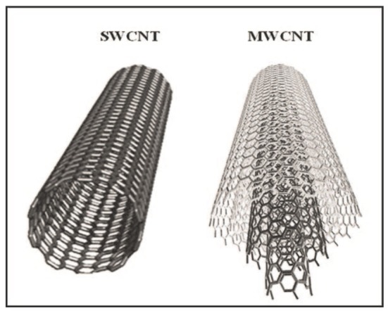
During the last decades, neuroscientists have increasingly exploited a variety of artificial, de-novo synthesized materials with controlled nano-sized features. For instance, a renewed interest in the development of prostheses or neural interfaces was driven by the availability of novel nanomaterials that enabled the fabrication of implantable bioelectronics interfaces with reduced side effects and increased integration with the target biological tissue. The peculiar physical-chemical properties of nanomaterials have also contributed to the engineering of novel imaging devices toward sophisticated experimental settings, to smart fabricated scaffolds and microelectrodes, or other tools ultimately aimed at a better understanding of neural tissue functions. In this review, we focus on nanomaterials and specifically on carbon-based nanomaterials, such as carbon nanotubes (CNTs) and graphene. While these materials raise potential safety concerns, they represent a tremendous technological opportunity for the restoration of neuronal functions. We then describe nanotools such as nanowires and nano-modified MEA for high-performance electrophysiological recording and stimulation of neuronal electrical activity. We finally focus on the fabrication of three-dimensional synthetic nanostructures, used as substrates to interface biological cells and tissues in vitro and in vivo.

Keywords: nanomaterials; nanoscience; nanotools; neuroengineering; neuroscience.


Figures

Figure 1
Figure 1
Schematic representation of a SWCNT, composed by just one graphene sheet, compared to a MWCNT, composed by more (three in this cartoon) graphene sheets (Choudhary and Gupta, 2011).
Figure 2
Figure 2
Neurons grown on a CNT substrates display (Left) increased spontaneous activity and firing (Reprinted with permission from Lovat et al., , American Chemical Society) and (Centre) increased GABAergic synaptogenesis. On the right a confocal reconstruction of a 3D-MWCNT scaffold (in gray) with neurons (in red) grown suspended within a pore, and glial cells (in green) acting as a support (Bosi et al., 2015). (A) Spontaneous synaptic currents recorded from control and CNTs substrates. (B) Current clamp recordings from hippocampal neurons grown on control and CNTs substrates.
Figure 3
Figure 3
SEM micrograph showing a peripheral neuronal fiber establishing intimate contacts (red arrows) with the CNT carpet, suggesting that also in the case of spinal explants the ability of CNTs to couple tight to neural membranes (Modified with the permission from Fabbro et al., , American Chemical Society).
Figure 4
Figure 4
Samples of (a) classic confocal and (b) STED imaging of fibroblasts. Scale bar: 2 μm. (Modified with the permission of Variola, , Published by the PCCP Owner Societies).
Figure 5
Figure 5
(a) SEM image of the nine silicon nanoneedles that constitutes the active region of a 3D-NEA. (b) SEM micrograph of a rat cortical neuron on top of an electrode pad; (c) example of stimulation and recording of rat cortical neurons showing that Action potentials (upper blue trace, measured by a patch pipette) could be reliably stimulated by voltage pulses applied to the nanoelectrodes (lower magenta trace; reprinted with permission from Alivisatos et al., , American Chemical Society).
Figure 6
Figure 6
Left: Zoomed-in view of a single electrode along with the platinum traces; Right: Effects of low frequency monopolar stimulation on the ER. Early responses (1–3 ms latency) recorded in the MG (top row) and TA (bottom row) bilaterally during low frequency (1 Hz) monopolar stimulation (3–6 V) at each electrode on the array. The height of each bar indicates the amplitude and the color indicates the latency of the response. The black box indicates a case where no response was recorded for that particular window (Modified with the permission from Gad et al., 2013).
Figure 7
Figure 7
Techniques employed in patterned scaffold generation. (A) Diagram showing the basis of the Electrospinning technique; (B) Schematics of the steps of microcontact printing, showing the creation of a PDMS mask and the deposition of biomolecules onto a substrate using the stamp (Cooper and Nadeau, 2009).
Figure 8
Figure 8
(Left) Human iN populations robustly express MAP2 in 2D and 3D conditions, while populations of unconverted, proliferative Ki67-expressing iPS cells persist in iN populations plated in 2D conditions. Scale bar, 100 mm (Carlson et al., 2016). (Right) snapshots of representative fields of neuronal cultures grown on 2D-PDMS (top) and 3D-PDMS (bottom) substrates, stained with the Oregon Green 488-BAPTA-1 AM. Scale bar: 50 μm. Repetitive Ca2+−events spontaneously recorded in hippocampal cultures of 9 DIV highlighted an higher frequency and synchronization of events in 3D cultures (Bosi et al., 2015).

Similar articles

Cited by

References

    1. Ahmad R., Mahmoudi T., Ahn M. S., Hahn Y. B. (2018). Recent advances in nanowires-based field-effect transistors for biological sensor applications. Biosens. Bioelectron. 100, 312–325. 10.1016/j.bios.2017.09.024 - DOI - PMC - PubMed
    1. Ajetunmobi A., Prina-Mello A., Volkov Y., Corvin A., Tropea D. (2014). Nanotechnologies for the study of the central nervous system. Prog. Neurobiol. 123, 18–36. 10.1016/j.pneurobio.2014.09.004 - DOI - PubMed
    1. Alivisatos A. P., Andrews A. M., Boyden E. S., Chun M., Church G. M., Deisseroth K., et al. . (2013). Nanotools for neuroscience and brain activity mapping. ACS Nano. 7, 1850–1866. 10.1021/nn4012847 - DOI - PMC - PubMed
    1. Aurand E. R., Usmani S., Medelin M., Scaini D., Bosi S., Rosselli F. B., et al. (2017). Nanostructures to engineer 3D neural-interfaces: directing axonal navigation toward successful bridging of spinal segments. Adv. Funct. Mat. 28:1700550 10.1002/adfm.201700550 - DOI
    1. Baldrighi M., Trusel M., Tonini R., Giordani S. (2016). Carbon nanomaterials interfacing with neurons: an in vivo perspective. Front. Neurosci. 10:250. 10.3389/fnins.2016.00250 - DOI - PMC - PubMed

LinkOut - more resources