. 2019 Jan 15:12:74.
doi: 10.3389/fnsys.2018.00074. eCollection 2018.

Abnormal Cortico-Cerebellar Functional Connectivity in Autism Spectrum Disorder

Affiliations

Abnormal Cortico-Cerebellar Functional Connectivity in Autism Spectrum Disorder

Taiane Coelho Ramos et al. Front Syst Neurosci. .

Abstract

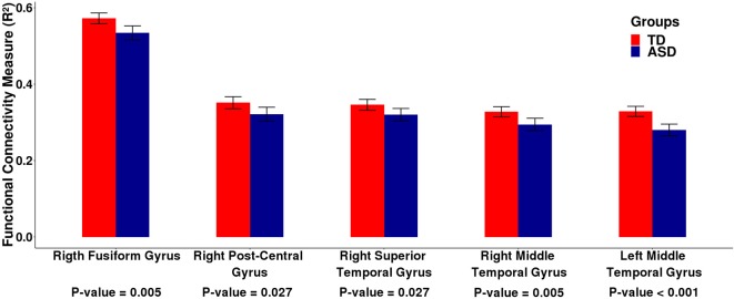
The cerebral cortex and the cerebellum are spatially remote areas that are connected by complex circuits that link both primary and associative areas. Previous studies have revealed abnormalities in autism spectrum disorder (ASD); however, it is not clear whether cortico-cerebellar connectivity is differentially manifested in the disorder. To explore this issue, we investigated differences in intrinsic cortico-cerebellar functional connectivity between individuals with typical development (TD) and those with ASD. To this end, we used functional magnetic resonance imaging (fMRI) of 708 subjects under a resting state protocol provided by the ABIDE I Consortium. We found that people with ASD had diminished functional connectivity between the cerebellum and the following cortical regions: (i) right fusiform gyrus, (ii) right postcentral gyrus, (iii) right superior temporal gyrus, (iv) right middle temporal gyrus, and (v) left middle temporal gyrus. All of these regions are involved in many cognitive systems that contribute to commonly affected functions in ASD. For right fusiform gyrus, right superior temporal gyrus, and left middle temporal gyrus, we reproduced the results in an independent cohort composed of 585 subjects of the ABIDE II Consortium. Our results points toward a consistent atypical cortico-cerebellar connectivity in ASD.

Keywords: ABIDE; ASD; autism spectrum disorders; cerebellum; cortico-cerebellar connectivity; functional connectivity; resting-state fMRI; underconnectivity.


Figures

Figure 1
Figure 1
Cortical ROIs with a differential association with the cerebellum between TD and ASD groups obtained by analyzing the ABIDE I dataset. Panels (A,B) represent coronal slices and two lateral views of the brain, respectively. The colors represent the cortical ROIs that differentially associated with the cerebellum between TD and ASD groups, namely, the right fusiform gyrus, right postcentral gyrus, right superior temporal gyrus, right middle temporal gyrus, and left middle temporal gyrus.
Figure 2
Figure 2
Barplots of the functional connectivity measure (R2) between the cortical ROI and the cerebellum obtained by analyzing the ABIDE I dataset. Each pair of bars represent the mean functional connectivity measurements between the cortical ROI and the cerebellum for the TD and ASD groups, respectively. The error bars represent 95% confidence interval. P-values were obtained by linear regression with the measure of functional connectivity (R2) as the response variable and the diagnostic group (TD or ASD) as the predictor variable with age, gender, and site as covariates. All p-values were corrected for multiple tests by FDR. Note that the mean functional connectivity measurement (R2) is significantly lower in the ASD group compared to the TD group, suggesting decreased functional cortico-cerebellar connectivity in the ASD group.
Figure 3
Figure 3
Barplots of the functional connectivity measure (R2) between the cortical ROI and the cerebellum for an independent dataset (ABIDE II). Each pair of bars represent the mean functional connectivity measurements between the cortical ROI and the cerebellum for the TD and ASD groups, respectively. The error bars represent 95% confidence interval. P-values were obtained by linear regression with the measure of functional connectivity (R2) as the response variable and the diagnostic group (TD or ASD) as the predictor variable with age, gender, and site as covariates. All p-values were corrected for multiple tests by FDR. Note that the median functional connectivity measurement (R2) is significantly lower in the ASD group compared to the TD group, suggesting decreased functional cortico-cerebellar connectivity in the ASD group. These data confirm the findings that were obtained by analyzing the ABIDE I dataset.

Similar articles

Cited by

References

    1. Alexander A. L., Lee J. E., Lazar M., Boudos R., DuBray M. B., Oakes T. R., et al. . (2007). Diffusion tensor imaging of the corpus callosum in autism. Neuroimage 34, 61–73. 10.1016/j.neuroimage.2006.08.032 - DOI - PubMed
    1. American Psychiatric Association (2013). Diagnostic and Statistical Manual of Mental Disorders (DSM-5®). Arlington, VA: American Psychiatric Pub.
    1. Barnea-Goraly N., Kwon H., Menon V., Eliez S., Lotspeich L., Reiss A. L. (2004). White matter structure in autism: preliminary evidence from diffusion tensor imaging. Biol. Psychiatry 55, 323–326. 10.1016/j.biopsych.2003.10.022 - DOI - PubMed
    1. Bauman M. L., Kemper T. L. (2005). Neuroanatomic observations of the brain in autism: a review and future directions. Int. J. Dev. Neurosci. 23, 183–187. 10.1016/j.ijdevneu.2004.09.006 - DOI - PubMed
    1. Baumann O., Borra R. J., Bower J. M., Cullen K. E., Habas C., Ivry R. B., et al. . (2015). Consensus paper: the role of the cerebellum in perceptual processes. Cerebellum 14, 197–220. 10.1007/s12311-014-0627-7 - DOI - PMC - PubMed

LinkOut - more resources