Review
. 2019 Jan 15:9:1834.
doi: 10.3389/fphys.2018.01834. eCollection 2018.

Effect of Estrogen on Musculoskeletal Performance and Injury Risk

Affiliations
Review

Effect of Estrogen on Musculoskeletal Performance and Injury Risk

Nkechinyere Chidi-Ogbolu et al. Front Physiol. .

Abstract

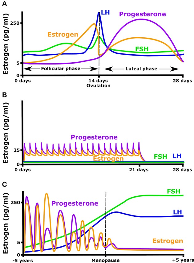
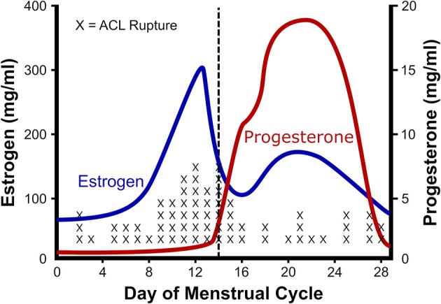
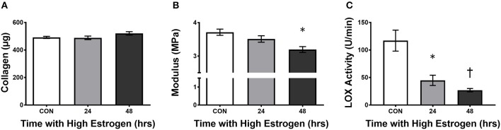
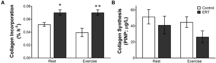
Estrogen has a dramatic effect on musculoskeletal function. Beyond the known relationship between estrogen and bone, it directly affects the structure and function of other musculoskeletal tissues such as muscle, tendon, and ligament. In these other musculoskeletal tissues, estrogen improves muscle mass and strength, and increases the collagen content of connective tissues. However, unlike bone and muscle where estrogen improves function, in tendons and ligaments estrogen decreases stiffness, and this directly affects performance and injury rates. High estrogen levels can decrease power and performance and make women more prone for catastrophic ligament injury. The goal of the current work is to review the research that forms the basis of our understanding how estrogen affects muscle, tendon, and ligament and how hormonal manipulation can be used to optimize performance and promote female participation in an active lifestyle at any age.

Keywords: ACL; estrogen; exercise; injury risk; ligament; muscle; tendon.


Figures

Figure 1
Figure 1
Hormonal fluctuation during (A) a normal menstrual cycle, (B) while taking an oral contraceptive (OC) containing both estrogen and progesterone, and (C) in the years before and after menopause.
Figure 2
Figure 2
Relationship between estrogen and ACL rupture in a normal cycle. The rate of anterior cruciate ligament (ACL) rupture in relation to female hormones throughout a standard menstrual cycle. Note that with the ovulatory rise in estrogen there is a concomitant rise in ACL ruptures. Adapted from Wojtys et al. (2002).
Figure 3
Figure 3
High estrogen decreasing engineered ligament stiffness due to inhibition of lysyl oxidase. (A) Collagen content, (B) tangent modulus, and (C) lysyl oxidase (LOX) activity in ligaments engineered from human ACL cells isolated from women following 24 or 48 h of treatment of the constructs with physiologically high (500 pg/ml) of estrogen. Note that even though there is a slight rise in collagen, the stiffness of the grafts decreases concomitant with an increase in estrogen in the media. *indicates different than control (p < 0.05), whereas indicates different than 24 h (p < 0.05). Adapted from Lee C. A. et al. (2015).
Figure 4
Figure 4
Differential measures of collagen incorporation and synthesis with estrogen replacement and exercise. The rate of (A) collagen incorporation of proline into the patellar tendon or (B) the appearance of the N-terminal propeptide of collagen I in post-menopausal women ± estrogen replacement therapy (ERT) and exercise. Note that with ERT collagen incorporation is higher in the same women where collagen synthesis is repressed. Further, exercise tends to decrease collagen incorporation and synthesis in controls, whereas ERT users show no effect on incorporation or a large drop in collagen synthesis. Symbols (*P < 0.01, **P < 0.001) show significance determined by unpaired t-test Control vs. ERT. These data suggest that there is a large methodological discrepancy between the two measures. Adapted from Hansen et al. (2009b).

Similar articles

Cited by

References

    1. Adachi N., Nawata K., Maeta M., Kurozawa Y. (2008). Relationship of the menstrual cycle phase to anterior cruciate ligament injuries in teenaged female athletes. Arch. Orthop. Trauma Surg. 128, 473–478. 10.1007/s00402-007-0461-1 - DOI - PubMed
    1. Arendt E., Dick R. (1995). Knee injury patterns among men and women in collegiate basketball and soccer: NCAA data and review of literature. Am. J. Sports Med. 23, 694–701. 10.1177/036354659502300611 - DOI - PubMed
    1. Baltgalvis K. A., Greising S. M., Warren G. L., Lowe D. A. (2010). Estrogen regulates estrogen receptors and antioxidant gene expression in mouse skeletal muscle. PLoS ONE 5:e10164. 10.1371/journal.pone.0010164 - DOI - PMC - PubMed
    1. Bamman M. M., Hill V. J., Adams G. R., Haddad F., Wetzstein C. J., Gower B. A., et al. . (2003). Gender differences in resistance-training-induced myofiber hypertrophy among older adults. J. Gerontol. Ser. A Biol. Sci. Med. Sci. 58, B108–B116. 10.1093/gerona/58.2.B108 - DOI - PubMed
    1. Barros R. P., Gustafsson J. Å. (2011). Estrogen receptors and the metabolic network. Cell Metabol. 14, 289–299. 10.1016/j.cmet.2011.08.005 - DOI - PubMed