. 2019 Jul;70(1):154-167.
doi: 10.1002/hep.30530. Epub 2019 Mar 20.

Keratin 23 Is a Peroxisome Proliferator-Activated Receptor Alpha-Dependent, MYC-Amplified Oncogene That Promotes Hepatocyte Proliferation

Affiliations

Keratin 23 Is a Peroxisome Proliferator-Activated Receptor Alpha-Dependent, MYC-Amplified Oncogene That Promotes Hepatocyte Proliferation

Donghwan Kim et al. Hepatology. 2019 Jul.

Abstract

Chronic activation of the nuclear receptor peroxisome proliferator-activated receptor alpha (PPARA) promotes MYC-linked hepatocellular carcinoma (HCC) in mice. Recent studies have shown that MYC can function as an amplifier of transcription where MYC does not act as an "on-off" switch for gene expression but rather accelerates transcription rates at active promoters by stimulating transcript elongation. Considering the possibility that MYC may amplify the expression of PPARA target genes to potentiate cell proliferation and liver cancer, gene expression was analyzed from livers of wild-type and liver-specific Myc knockout (MycΔHep ) mice treated with the PPARA agonist pirinixic acid. A subset of PPARA target genes was amplified in the presence of MYC, including keratin 23 (Krt23). The induction of Krt23 was significantly attenuated in MycΔHep mice and completely abolished in Ppara-null mice. Reporter gene assays and chromatin immunoprecipitation confirmed direct binding of both PPARA and MYC to sites within the Krt23 promoter. Forced expression of KRT23 in primary hepatocytes induced cell cycle-related genes. These data indicate that PPARA activation elevates MYC expression, which in turn potentiates the expression of select PPARA target genes involved in cell proliferation. Finally, KRT23 protein is highly elevated in human HCCs. Conclusion: These results revealed that MYC-mediated transcriptional potentiation of select PPARA target genes, such as Krt23, may remove rate-limiting constraints on hepatocyte growth and proliferation leading to liver cancer.


Conflict of interest statement

CONFLICT OF INTERESTS: The authors declare no conflict of interest.

Figures

FIG. 1.
FIG. 1.
MYC expression amplifies select PPARA target genes. (A) Liver microarray data from Myc-floxed (Mycfl/fl) and hepatocyte-specific Myc-null (MycΔHep) mice treated with Wy-14643. Heatmap showing representative MYC-amplified genes from microarray data. Upper panel depicts PPARA target gene mRNAs amplified by MYC. Lower panel shows PPARA targets unaffected by MYC status. (B) Myc and Krt23 expression in wild-type and MycΔHep mice treated with Wy-14643. (C) Western blotting analysis of KRT23 protein expression in liver from Mycfl/fl and MycΔHep mice treated with Wy-14643. (D) Densitometry analysis of KRT23 protein levels. (E) Quantification of Cxcr1, Fam40b, Fa2h, Krt23, Myc, and Otop1 mRNAs in wild-type, MycΔHep, and Ppara-null mice treated with Wy-14643. (F) Quantification of PPARA-dependent MYC-independent Acox1, Cyp4a10, and Ehhadh mRNAs in wild-type, MycΔHep, and Ppara-null mice treated with Wy-14643. Experiments were performed on at least five mice/group. Each data point represents the mean ± SD (ns, not significant; *, p < 0.05; ***, p < 0.001).
FIG. 2.
FIG. 2.
Liver-specific KRT23 response supports a role in PPARA-dependent hepatocyte proliferation. Wild-type (Ppara+/+) and Ppara-null (Ppara−/−) mice were treated with Wy-14643 for 48 hours. (A) qRT-PCR analysis of the PPARA target gene mRNA Cyp4a14 in different tissues. (B and C) Expression of Myc and Krt23 mRNA in Ppara+/+ and Ppara−/− mice with and without Wy-14643. (D) Western blotting of KRT23 in Wy-14643 treated Ppara+/+ and Ppara−/− mice. (E) Hematoxylin and eosin (H&E) staining of liver sections from control, Wy-14643-treated and Wy-14643 treated in MycΔHep mice. (F) Immunohistochemical staining of KRT23 in liver sections from control, Wy-14643 treated and Wy-14643 treated in MycΔHep mice. Representative images are shown. Scale bars represent 100 nm (200X). At least five mice were analyzed/genotype and treatment group. Each data point represents the mean ± SD (***, p < 0.001).
FIG. 3.
FIG. 3.
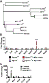
KRT23 is the principal keratin upregulated during PPARA-induced hepatocyte proliferation. (A) Dendrogram depicting homology between mouse KRT protein sequences. Diagnostic and prognostic markers denoted by ‘#’ and ‘*’, respectively. (B) Ppara+/+ and Ppara-null mice were treated with Wy-14643 for 48 hours then livers were collected and expression of Krt mRNA measured by qRT-PCR. (C) Analysis of Krt7, Krt8, Krt18, Krt19 and Krt23 mRNA in primary hepatocytes treated with AAV-KRT23. Experiments were performed on at least five mice/group. Each data point represents the mean ± SD (nd, not detected; *, p < 0.05; **, p < 0.01; ***, p < 0.001).
FIG. 4.
FIG. 4.
PPARA directly regulates Krt23 through promoter PPRE sites. (A) Schematic representation of predicted PPRE and Ebox sites found within the Krt23 promoter (−1KB). ChIP primer binding sites and reporter construct inserts are shown below. (B) Luciferase-based reporter assays confirmed functional PPREs sites found within the Krt23 promoter. (C) PPARA ChIP assays assessed PPRE binding in liver samples from Ppara+/+ and Ppara-null mice treated with Wy-14643. Experiments were performed with at least four replicates. Each data point represents the mean ± SD (ns, not significant; *, p < 0.05; **, p < 0.01; ***, p < 0.001).
FIG. 5.
FIG. 5.
MYC directly regulates Krt23 through promoter Ebox sites. (A) Schematic representation of KRT23 promoter and predicted Ebox binding sites. Reporter construct inserts are detailed below. (B) Luciferase-based reporter assays confirmed MYC transactivation activities in primary hepatocytes transfected with empty plasmid (Vector) or a MYC expression vector (MYC). (C) ChIP primer site locations within the KRT23 promoter used to confirm MYC Ebox binding. (D) MYC ChIP assays for determining promoter occupancy was examined using liver tissue from control and Wy-14643 treated mice. Experiments were performed with at least four replicates. Each data point represents the mean ± SD (***, p < 0.001).
FIG. 6.
FIG. 6.
Adenovirus expressing KRT23 induces G2/M-related gene expression in primary hepatocytes. (A) qRT-PCR analysis of G2/M-related genes Cdc25c, Cdk1, Ccnb1, Ccnb2, Ki67, and Pcna mRNA in primary hepatocytes infected with KRT23 expressing adenovirus (Ad-KRT23). Uninfected cells (Control) and cells infected with GFP expressing adenovirus (Ad-GFP) were used as negative controls. (B) Aurora kinase (Aurk) and Aurk-associated gene expression in KRT23 adenovirus infected primary hepatocytes. (C) Western blotting of KRT23, CDC25C, CCNB, and (D) AURKA protein expression in KRT23 adenovirus infected cells. ACTB expression was used as a loading control. (E) Immunoprecipitation with anti-KRT23 antibody followed by western blotting of 14–3-3ε in liver tissue from control or Wy-14643 treated mice. Each data point represents the mean ± SD (***, p < 0.001).
FIG. 7.
FIG. 7.
Aurora kinase inhibition attenuates MYC and KRT23 expression in the PPARA agonist-induced liver proliferation model. (A) qRT-PCR analysis of Krt23 and Myc mRNA in livers from Wy-14643 treated mice for 3 days or mice treated with Wy-14643 and MK-8745 (Wy + MK-8745) for 3 days. Vehicle treated mice were used as a negative control (Vehicle). (B) Western blot analysis of KRT23 and MYC in liver from Wy-14643 treated mice or treated with MK-8745 in mice. (C) Densitometric analysis of MYC and KRT23 protein expression. (D) Analysis of PPARA-dependent, MYC-amplified gene expression in response to AURK inhibition. (E) Proposed mechanism of action for KRT23 during PPARA-mediated hepatocyte proliferation. Experiments were performed on at least five mice/group. Each data point represents the mean ± SD (ns, not significant; *, p < 0.05; **, p < 0.01; ***, p < 0.001).
FIG. 8.
FIG. 8.
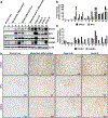
KRT23 expression correlates with human HCC progression. (A) Western blot analysis of KRT23, MYC, CCNB1, and AURKA protein expression in human liver biopsies. (B) Densitometric analysis of KRT23 and MYC protein expression. (C) Densitometric analysis of CCNB1 and AURKA protein expression. (D) Immunohistochemical staining of KRT23, MYC, and AURKA in from normal or cancerous human liver sections. Representative images are shown. Scale bars represent 20 µm (400X).

References

    1. Morimura K, Cheung C, Ward JM, Reddy JK, Gonzalez FJ. Differential susceptibility of mice humanized for peroxisome proliferator-activated receptor alpha to Wy-14,643-induced liver tumorigenesis. Carcinogenesis 2006;27:1074–1080. - PMC - PubMed
    1. Brocker CN, Yue J, Kim D, Qu A, Bonzo JA, Gonzalez FJ. Hepatocyte-specific PPARA expression exclusively promotes agonist-induced cell proliferation without influence from nonparenchymal cells. Am J Physiol Gastrointest Liver Physiol 2017;312:G283–G299. - PMC - PubMed
    1. Shah YM, Morimura K, Yang Q, Tanabe T, Takagi M, Gonzalez FJ. Peroxisome proliferator-activated receptor alpha regulates a microRNA-mediated signaling cascade responsible for hepatocellular proliferation. Mol Cell Biol 2007;27:4238–4247. - PMC - PubMed
    1. Qu A, Jiang C, Cai Y, Kim JH, Tanaka N, Ward JM, Shah YM, et al. Role of Myc in hepatocellular proliferation and hepatocarcinogenesis. J Hepatol 2014;60:331–338. - PMC - PubMed
    1. Wolf E, Lin CY, Eilers M, Levens DL. Taming of the beast: shaping Myc-dependent amplification. Trends Cell Biol 2015;25:241–248. - PMC - PubMed

Publication types