

White matter has impaired resting oxygen delivery in sickle cell patients
- PMID: 30697803
- PMCID: PMC6874897
- DOI: 10.1002/ajh.25423
White matter has impaired resting oxygen delivery in sickle cell patients
Abstract
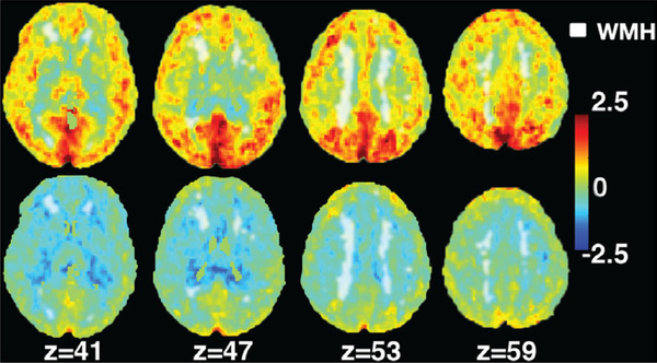
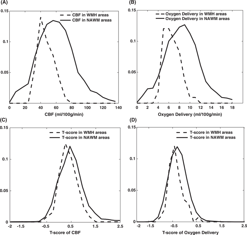
Although modern medical management has lowered overt stroke occurrence in patients with sickle cell disease (SCD), progressive white matter (WM) damage remains common. It is known that cerebral blood flow (CBF) increases to compensate for anemia, but sufficiency of cerebral oxygen delivery, especially in the WM, has not been systematically investigated. Cerebral perfusion was measured by arterial spin labeling in 32 SCD patients (age range: 10-42 years old, 14 males, 7 with HbSC, 25 HbSS) and 25 age and race-matched healthy controls (age range: 15-45 years old, 10 males, 12 with HbAS, 13 HbAA); 8/24 SCD patients were receiving regular blood transfusions and 14/24 non-transfused SCD patients were taking hydroxyurea. Imaging data from control subjects were used to calculate maps for CBF and oxygen delivery in SCD patients and their T-score maps. Whole brain CBF was increased in SCD patients with a mean T-score of 0.5 and correlated with lactate dehydrogenase (r2 = 0.58, P < 0.0001). When corrected for oxygen content and arterial saturation, whole brain and gray matter (GM) oxygen delivery were normal in SCD, but WM oxygen delivery was 35% lower than in controls. Age and hematocrit were the strongest predictors for WM CBF and oxygen delivery in patients with SCD. There was spatial co-localization between regions of low oxygen delivery and WM hyperintensities on T2 FLAIR imaging. To conclude, oxygen delivery is preserved in the GM of SCD patients, but is decreased throughout the WM, particularly in areas prone to WM silent strokes.
© 2019 Wiley Periodicals, Inc.
Conflict of interest statement
CONFLICT OF INTEREST
JCW receives research support in kind from Philips Healthcare. None of the other authors have conflicts relevant to the study.
Figures
References
-
- Ohene-Frempong K, Weiner SJ, Sleeper LA, et al. Cerebrovascular accidents in sickle cell disease: rates and risk factors. Blood. 1998;91 (1):288–294. - PubMed
-
- Miller ST, Macklin EA, Pegelow CH, et al. Silent infarction as a risk factor for overt stroke in children with sickle cell anemia: A report from the Cooperative Study of Sickle Cell Disease. J Pediatr. 2001;139(3):385–390. - PubMed
-
- Adams RJ, McKie VC, Hsu L, et al. Prevention of a first stroke by transfusions in children with sickle cell anemia and abnormal results on transcranial Doppler ultrasonography. N Engl J Med. 1998;339(1):5–11. - PubMed
Publication types
MeSH terms
Substances
Grants and funding
LinkOut - more resources
Full Text Sources
Medical