. 2019 Oct;107(7):2335-2342.
doi: 10.1002/jbm.b.34326. Epub 2019 Jan 29.

The optimal incubation time for in vitro hemocompatibility testing: Assessment using polymer reference materials under pulsatile flow with physiological wall shear stress conditions

Affiliations

The optimal incubation time for in vitro hemocompatibility testing: Assessment using polymer reference materials under pulsatile flow with physiological wall shear stress conditions

Sjoerd Leendert Johannes Blok et al. J Biomed Mater Res B Appl Biomater. 2019 Oct.

Abstract

During hemocompatibility testing, activation products may reach plateau values which can result in less distinction between hemocompatible and hemo-incompatible materials. Of concern is an underestimation of the blood activation caused by the biomaterial of interest, which may result in a false assessment of hemocompatibility. To elucidate the optimal incubation time for in vitro hemocompatibility testing, we used the Haemobile circulation model with human whole blood. Blood from healthy volunteers was in vitro incubated under pulsatile flow with physiological wall shear stress conditions at 37°C for 30, 60, 120, or 240 min. Test loops containing low-density polyethylene and polydimethylsiloxane served as low and high reference materials, that is, hemocompatible and hemo-incompatible biomaterials, respectively. In addition, empty loops served as a negative reference. Thrombogenicity, platelet function, inflammatory response, coagulation, and hemolysis between references and incubation times were compared. We found that thrombogenicity and platelet function were significantly affected by both the duration of incubation and the type of material. In particular, thrombogenicity and platelet function assessments were affected by incubation time. We found that an exposure time of 60 min was sufficient, and for almost all variables an optimal incubation time to discriminate between the low and high reference material. © 2019 The Authors. Journal of Biomedical Materials Research Part B: Applied Biomaterials published by Wiley Periodicals, Inc. J Biomed Mater Res Part B: Appl Biomater 107B: 2335-2342, 2019.

Keywords: biomaterials; blood-material interaction; cardiovascular device; hemocompatibility; hemocompatibility testing.


Figures

Figure 1
Figure 1
(a) Three‐dimensional model of the Haemobile. The black arrows indicate the back and forth movement of the round plateau carrying the stacked test loops. The test loops have a radius of 72.5 mm. The blue components represent the unidirectional check valves. (b) The angle of the round plateau in time, with the Haemobile programmed to the following settings: angle of rotation = 180°; clockwise angular velocity = 720°/s; anticlockwise angular velocity = 360°/s; angular acceleration/deceleration = 3600°/s2 (fixed setting). (c) Ensemble average of the measured flow (solid blue line) and typical coronary blood flow (dashed red line). (d) Calculated shear stress across the internal diameter of the test loop in time.
Figure 2
Figure 2
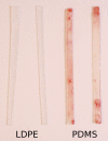
Macroscopic image of low‐density polyethylene (LDPE) and polydimethylsiloxane (PDMS) flat sheets after in vitro incubation under pulsatile flow with physiological wall shear stress conditions with human whole blood at 37°C for 1 h. LDPE barely showing thrombus formation; PDMS clearly showing vast red‐thrombus formation.
Figure 3
Figure 3
Scanning electron microscopy images of low‐density polyethylene (LDPE) and polydimethylsiloxane (PDMS) surfaces before and after in vitro incubation under pulsatile flow with physiological wall shear stress conditions with human whole blood at 37°C for 1 h. (a, b) 375× and 1500× magnification of LDPE before incubation, respectively. A clean, smooth surface is visible. (c, d) 420× and 1500× magnification of PDMS before incubation, respectively. A clean, rough surface is visible. (e, f) 375× and 6000× magnification of LDPE after incubation, respectively. An almost clean surface, showing only few adhered platelets is visible. (g, h) 385× and 1500× magnification of PDMS after incubation, respectively. The bare polymer surface is almost completely covered by vast thrombus layers, mostly consisting of fibrin and red blood cells.
Figure 4
Figure 4
(a) Platelet adhesion and (b) fibrin binding on low‐density polyethylene (LDPE) and polydimethylsiloxane (PDMS) after in vitro incubation under pulsatile flow with physiological wall shear stress conditions with human whole blood at 37°C for various incubation times. **p < 0.05 between indicated references, using an independent samples t test.
Figure 5
Figure 5
(a) Whole blood platelet count, (b) platelet adhesion to collagen, (c) surface‐bound P‐selectin expression, and (d) thromboxane B2 release in plasma after in vitro incubation under pulsatile flow with physiological wall shear stress conditions with human whole blood at 37°C for various incubation times using empty loops (Empty) as negative reference, low‐density polyethylene (LDPE) as low reference, and polydimethylsiloxane (PDMS) as high reference. *p < 0.05 compared with the baseline (0 min), using a paired samples t test. **p < 0.05 between indicated references, using an independent samples t test.
Figure 6
Figure 6
(a) Elastase release in plasma, (b) complement complex C5b‐9 formation in plasma, (c) thrombin–antithrombin III complex formation in plasma, and (d) free hemoglobin in plasma after in vitro incubation under pulsatile flow with physiological wall shear stress conditions with human whole blood at 37°C for various incubation times using empty loops (Empty) as negative reference, low‐density polyethylene (LDPE) as low reference, and polydimethylsiloxane (PDMS) as high reference. *p < 0.05 compared with the baseline (0 min), using a paired samples t test. **p < 0.05 between indicated references, using an independent samples t test.

References

    1. Blok SLJ, Engels GE, van Oeveren W. In vitro hemocompatibility testing: The importance of fresh blood. Biointerphases 2016;11:029802. - PubMed
    1. van Oeveren W. Obstacles in haemocompatibility testing. Scientifica (Cairo) 2013;2013:392584. - PMC - PubMed
    1. International Organization for Standardization . ISO 10993, Biological Evaluation of Medical Devices—Part 4: Selection of Tests for Interactions with Blood. Genève: ISO; 2017.
    1. Seyfert UT, Biehl V, Schenk J. In vitro hemocompatibility testing of biomaterials according to the ISO 10993‐4. Biomol Eng 2002;19:91–96. - PubMed
    1. Stevens KNJ, Aldenhoff YBJ, van der Veen FH, Maessen JG, Koole LH. Bioengineering of improved biomaterials coatings for extracorporeal circulation requires extended observation of blood‐biomaterial interaction under flow. J Biomed Biotechnol 2007;2007:1–10. - PMC - PubMed

LinkOut - more resources