. 2019 Sep 18;40(9):1099-1109.
doi: 10.1093/carcin/bgz012.

C/EBPβ suppresses keratinocyte autonomous type 1 IFN response and p53 to increase cell survival and susceptibility to UVB-induced skin cancer

Affiliations

C/EBPβ suppresses keratinocyte autonomous type 1 IFN response and p53 to increase cell survival and susceptibility to UVB-induced skin cancer

Hann W Tam et al. Carcinogenesis. .

Abstract

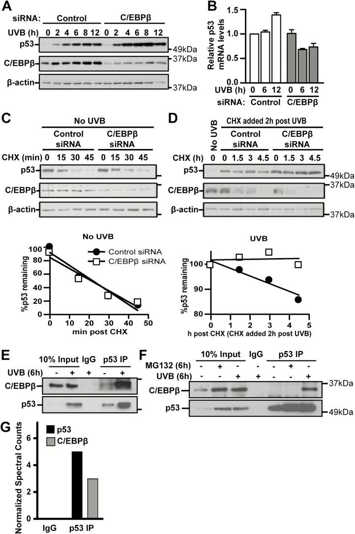
p53 is activated by DNA damage and oncogenic stimuli to regulate senescence, apoptosis and cell-cycle arrest, which are essential to prevent cancer. Here, we utilized UVB radiation, a potent inducer of DNA damage, p53, apoptosis and skin cancer to investigate the mechanism of CCAAT/enhancer binding protein-β (C/EBPβ) in regulating p53-mediated apoptosis in keratinocytes and to test whether the deletion of C/EBPβ in epidermis can protect mice from UVB-induced skin cancer. UVB-treatment of C/EBPβ skin conditional knockout (CKOβ) mice increased p53 protein levels in epidermis and enhanced p53-dependent apoptotic activity 3-fold compared with UVB-treated control mice. UVB increased C/EBPβ levels through a p53-dependent pathway and stimulated the formation of a C/EBPβ-p53 protein complex; knockdown of C/EBPβ increased p53 protein stability in keratinocytes. These results suggest a p53-C/EBPβ feedback loop, whereby C/EBPβ, a transcriptional target of a p53 pathway, functions as a survival factor by negatively regulating p53 apoptotic activity in response to DNA damage. RNAseq analysis of UVB-treated CKOβ epidermis unexpectedly revealed that type 1 interferon (IFN) pathway was the most highly enriched pathway. Numerous pro-apoptotic interferon stimulated genes were upregulated including some known to enhance p53 apoptosis. Our results indicate that p53 and IFN pathways function together in response to DNA damage to result in the activation of extrinsic apoptosis pathways and caspase 8 cleavage. Last, we observed CKOβ mice were resistant to UVB-induced skin cancer. Our results suggest that C/EBPβ represses apoptosis through keratinocyte autonomous suppression of the type 1 IFN response and p53 to increase cell survival and susceptibility to UVB-induced skin cancer.


Figures

Figure 1.
Figure 1.
Deletion of C/EBPβ in UVB-treated mouse epidermal keratinocytes results in elevated p53 protein levels and p53-dependent apoptosis in response to UVB. (A) K5Cre and CKOβ mice were exposed to 100 mJ/cm2 UVB and epidermis was collected 6 h post-UVB treatment. Epidermal protein lysates were prepared and immunoblot analysis for C/EBPβ, p53, p21 and β-actin was conducted. (B) K5Cre and CKOβ mice were exposed to 100 mJ/cm2 UVB and collected at the indicated times post UVB treatment. Mouse skin was processed and embedded in paraffin and skin sections were stained with anti-p53 antibody. Bar graph represent quantification of p53-stained-positive keratinocytes in untreated and UVB-treated K5Cre and CKOβ mouse epidermis. *denotes significantly different from UVB-treated K5Cre mice as determined by two-tailed Student t-test for paired data with the significance level set to P < 0.05. (C) K5Cre and CKOβ mice were exposed to 100 mJ/cm2 UVB and collected at 6 and 9 h post-UVB treatment. Mouse skin was processed and embedded in paraffin and skin sections were stained with anti-phospho-p53 Ser18 antibody. Bar graph represents quantification of phospho-p53 Ser18-stained-positive keratinocytes in untreated and UVB-treated K5Cre and CKOβ mouse epidermis. *denotes significantly different from UVB-treated K5Cre mice as determined by two-tailed Student t-test for paired data with the significance level set to P<0.05. (D) K5Cre and CKOβ mice were exposed to 100 mJ/cm2 UVB and skin was collected at the indicated times post-UVB treatment. Mouse skin was processed and embedded in paraffin and skin sections were stained with hematoxylin and eosin. Bar graph represents quantification of apoptotic cells in untreated and UVB-treated mouse epidermis as described in Materials and methods. *denotes significantly different from UVB-treated K5Cre mice as determined by two-tailed Student t-test for paired data with the significance level set to P < 0.05. (E) K5Cre, C/EBPβflox/flox and CKOβ mice were exposed to 100 mJ/cm2 UVB and skin was collected 6 h post-UVB treatment. Mouse skin was processed and embedded in paraffin and skin sections were stained with hematoxylin and eosin. Bar graph represents quantification of apoptotic cells in untreated and UVB-treated mouse epidermis as described in Materials and methods. *denotes significantly different from UVB-treated K5Cre mice as determined by two-tailed Student t-test for paired data with the significance level set to P < 0.05. (F) K5Cre, CKOβ, CKOp53 and DCKO mice were exposed to 100 mJ/cm2 UVB and epidermis was collected 6 h post-UVB treatment. Epidermal protein lysates were prepared and immunoblot analysis for C/EBPβ, p53 and β-actin was conducted. (G) K5Cre, CKOβ, and DCKO mice were exposed to 100 mJ/cm2 UVB and skin was collected 6 h post-UVB treatment. Mouse skin was processed and embedded in paraffin and skin sections were stained with hematoxylin and eosin. Bar graph represents quantification of apoptotic cells in untreated and UVB-treated mouse epidermis as described in Materials and methods. *denotes significantly different from UVB-treated K5Cre mice as determined by two-tailed Student t-test for paired data with the significance level set to P < 0.05. N = 3 for (B), (D), (E) and (G), and N = 5 for (C) mice/genotype/treatment. Data are expressed as the mean ± SD.
Figure 2.
Figure 2.
UVB treatment increases C/EBPβ mRNA and protein levels through a p53-dependent pathway. (A) BALB/MK2 mouse keratinocytes cells were treated with 10 mJ/cm2 of UVB at collected at the indicated times post-UVB treatment. Whole cell lysates were prepared and immunoblot analysis for C/EBPβ and β-actin was conducted. (B) BALB/MK2 mouse keratinocytes cells were treated with 10 mJ/cm2 of UVB at collected at the indicated times post-UVB treatment. Total RNA was isolated and qRT-PCR analysis of C/EBPβ mRNA levels (N = 3). Data represent mean ± SD. *denotes significantly different from untreated cells as determined by two-tailed Student t-test for paired data with the significance level set to P < 0.05. (C) BALB/MK2 mouse keratinocytes were treated with control or p53 siRNA. 40 h post-siRNA cells were exposed to 10 mJ/cm2 UVB radiation and collected 12 h post-UVB treatment. Whole cell lysates were prepared and immunoblot analysis for C/EBPβ, p53 and β-actin was conducted. (D) Wild-type SKH1 mice were exposed to 50 mJ/cm2 of UVB radiation of left untreated, 6 h post-UVB epidermal protein lysates were prepared and immunoblot analysis for C/EBPβ and β-actin was conducted. (E) Wild-type SKH1 mice were exposed to 50 mJ/cm2 of UVB radiation. Mouse skin was collected, processed and embedded in paraffin and skin sections were stained with anti-C/EBPβ antibody. Bar graph represents quantification of C/EBPβ-positive staining keratinocytes within the basal or suprabasal layer of wild-type SKH1 mice treated at the indicated times post-UVB treatment (N = 3). Data represent mean ± SD. *denotes significantly different from untreated K5Cre as determined by two-tailed student t-test for paired data with the significance level set to P < 0.05. (F) K5Cre and CKOp53 mice were exposed to 100 mJ/cm2 of UVB radiation, and skin was collected 6 h post-treatment. Mouse skin was processed and embedded in paraffin and skin sections were stained with anti-C/EBPβ antibody. Bar graph represents quantification of C/EBPβ-positive staining keratinocytes within the basal or suprabasal layer of mouse epidermis (N = 3). Data represent mean ± SD. *denotes significantly different from untreated K5Cre as determined by two-tailed Student t-test for paired data with the significance level set to P < 0.05. (G) Experimental model depicting C/EBPβ–p53 negative feedback loop in response to UVB radiation.
Figure 3.
Figure 3.
Deletion of C/EBPβ in mouse keratinocytes results in increase of p53 stability in response to UVB and UVB induces formation of C/EBPβ-p53 protein complex. (A) BALB/MK2 mouse keratinocytes were treated with control or C/EBPβ siRNA. 40 h post-siRNA cells were exposed to 10 mJ/cm2 UVB and collected at the indicated times post UVB treatment. Whole cell lysates were prepared and immunoblot analysis for C/EBPβ, p53 and β-actin was conducted. (B) BALB/MK2 mouse keratinocytes were treated with control or C/EBPβ siRNA, exposed to 10 mJ/cm2 UVB and collected 6 and 12 h post-UVB treatment. Total RNA was isolated and qRT-PCR analysis of p53 mRNA levels was conducted and normalized to Gapdh mRNA levels (N = 3). Data in bar graph are expressed as the mean ± SD. Evaluation by two-tailed Student t-test for paired data with the significance level set to P < 0.05 determined that changes in p53 mRNA levels were not significant. (C and D) BALB/MK2 mouse keratinocytes were treated with control or C/EBPβ siRNA; 40 h post-siRNA cells were treated with 25 µg/ml cyclohexamide (CHX) and collected at the indicated times post-CHX addition (C) or exposed to 10 mJ/cm2 UVB and 2 h post-UVB treated with 25 µg/ml CHX and collected at the indicated times post-CHX addition (D). Whole cell lysates were prepared an immunoblot analysis for C/EBPβ, p53 and β-actin was conducted. Densitometric analysis of p53 immunoblots was conducted and normalized to β-actin and the percent p53 remaining at each time was plotted (bottom). The data are representative of two independent experiments. (E) BALB/MK2 mouse keratinocytes were exposed to 10 mJ/cm2 UVB and collected 6 h post-UVB treatment. Whole cell lysates were prepared and p53 was immunoprecipitated using anti-p53 antibody (FL-393). Immunoblot analysis for p53 and C/EBPβ was conducted. (F) BALB/MK2 mouse keratinocytes were treated with 10 μM MG-132 or 10 mJ/cm2 UVB and collected 6 h post-treatment. Whole cell lysates were prepared and p53 was immunoprecipitated using anti-p53 antibody (FL-393). Immunoblot analysis for p53 and C/EBPβ was conducted. (G) BALB/MK2 mouse keratinocytes were exposed to 10 mJ/cm2 UVB and collected 6 h post-UVB treatment. Whole cell lysates were prepared and p53 was immunoprecipitated using anti-p53 antibody (FL-393). Immunoprecipitated proteins were analyzed by LC/MS/MS and plot of spectral counts for unique peptides for the proteins of interest were plotted.
Figure 4.
Figure 4.
UVB-treated CKOβ epidermis displays significant alterations in gene expression and enrichment of type 1 interferon and p53 signaling pathways. K5Cre and CKOβ mice were dosed with 100mJ/cm2 (N = 4). Total RNA was isolated from the epidermis at 6 h post-UVB treatment and was subjected to RNA sequencing. (A) Row-scaled heatmap showing total gene expression. All genes above white line at top are statistically decreased in UVB-treated CKOβ mice and all genes below, the white line on bottom are statistically increased in UVB-treated CKOβ mice. (B) Gene set enrichment analysis with top 25 statistically significant (FDR < 0.1, redundant gene sets removed), positively enriched pathways displayed. (C) Heatmap of genes in combined MSigDB interferon gene sets. (D) IFN response genes significantly altered (FDR ≤ 0.1) in CKOβ UVB-treated epidermis. Grayed genes represent genes overlapping with CKOβ epidermis and non-grayed genes (black) represents significantly altered genes unique to CKOβ UVB-treated epidermis. (E) Heatmap of genes in the MSigDB hallmark p53 pathway gene set. (F) Hallmark p53 genes significantly altered (FDR ≤ 0.1) in UVB-treated CKOβ epidermis. Grayed genes represent genes overlapping with CKOβ epidermis and non-grayed genes (black) represent significantly altered genes unique to CKOβ UVB-treated epidermis. (G) BALB/MK2 mouse keratinocytes were treated with a combination of control, C/EBPβ and INFAR1 siRNA; 40 h post-siRNA cells were exposed to 10 mJ/cm2 UVB and collected 6 and 18 h post-UVB treatment. Whole cell lysates were prepared and immunoblot analysis for C/EBPβ, p53 and α-tubulin was conducted. Densitometric analysis of p53 immunoblots was conducted and normalized to α-tubulin. Bar graph for p53 protein levels is representative of three independent experiments. Data are expressed as the mean ± SD. *denotes significantly different from control siRNA as determined by two-tailed Student t-test for paired data with the significance level set to P < 0.05. Knockdown of INAR1 was confirmed by TaqMAN reverse transcription–-PCR and was >70% knockdown. (H) K5Cre, CKOβ and DCKO mice were exposed to 100 mJ/cm2 UVB and skin was collected 6 h post-UVB treatment. Mouse skin was processed and embedded in paraffin and skin sections were stained with cleaved caspase 8 antibody. Bar graph represents quantification of cleaved caspase 8 positive staining keratinocytes in mouse epidermis (N = 3). Data represent mean ± SD. *denotes significantly different from untreated K5Cre as determined by two-tailed Student t-test for paired data with the significance level set to P < 0.05. (I) Representative picture of cleaved-caspase 8 stained cells in CKOβ mouse skin 6 h post-100 mJ/cm2 UVB (E, epidermis; D, dermis). Scale bar= 10 μm.
Figure 5.
Figure 5.
Deletion of C/EBPβ protects mice from UVB-induced skin tumorigenesis. K5Cre and CKOβ SKH1 mice were treated with 50 mJ/cm2 UVB five days/week for 27 weeks. (N=18 K5Cre and N=15 CKOβ). (A) Quantification of skin tumor incidence. (B) Quantification of skin tumor multiplicity. (C) Quantification of final tumor size. Brackets show mean and SEM of each genotype. * denotes significantly different from K5Cre tumors as determined by the Welch two sample t-test with the significance level set to P<0.05. (D) Classification of tumor types into four categories: I (papilloma), II (in situ carcinoma), III (microinvasive carcinoma), and IV (squamous cell carcinoma).

References

    1. Meek, D.W. et al. (2009) Posttranslational modification of p53: cooperative integrators of function. Cold Spring Harb. Perspect. Biol., 1, a000950. - PMC - PubMed
    1. Kastan, M.B. (2007) Wild-type p53: tumors can’t stand it. Cell, 128, 837–840. - PubMed
    1. Ramji, D.P. et al. (2002) CCAAT/enhancer-binding proteins: structure, function and regulation. Biochem. J., 365(Pt 3), 561–575. - PMC - PubMed
    1. House, J.S. et al. (2010) C/EBPalpha and C/EBPbeta are required for Sebocyte differentiation and stratified squamous differentiation in adult mouse skin. PLoS One, 5, e9837. - PMC - PubMed
    1. Tsukada, J. et al. (2011) The CCAAT/enhancer (C/EBP) family of basic-leucine zipper (bZIP) transcription factors is a multifaceted highly-regulated system for gene regulation. Cytokine, 54, 6–19. - PubMed