. 2019 Apr 1;121(4):1279-1288.
doi: 10.1152/jn.00481.2018. Epub 2019 Jan 30.

Age-related reweighting of visual and vestibular cues for vertical perception

Affiliations

Age-related reweighting of visual and vestibular cues for vertical perception

Bart B G T Alberts et al. J Neurophysiol. .

Abstract

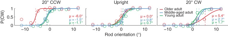
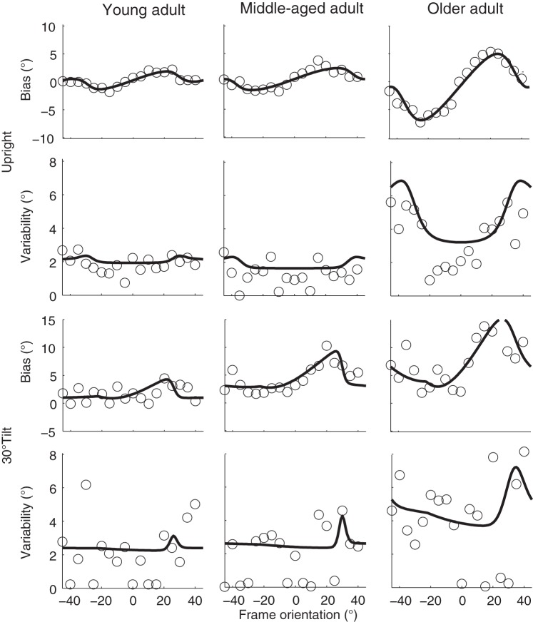
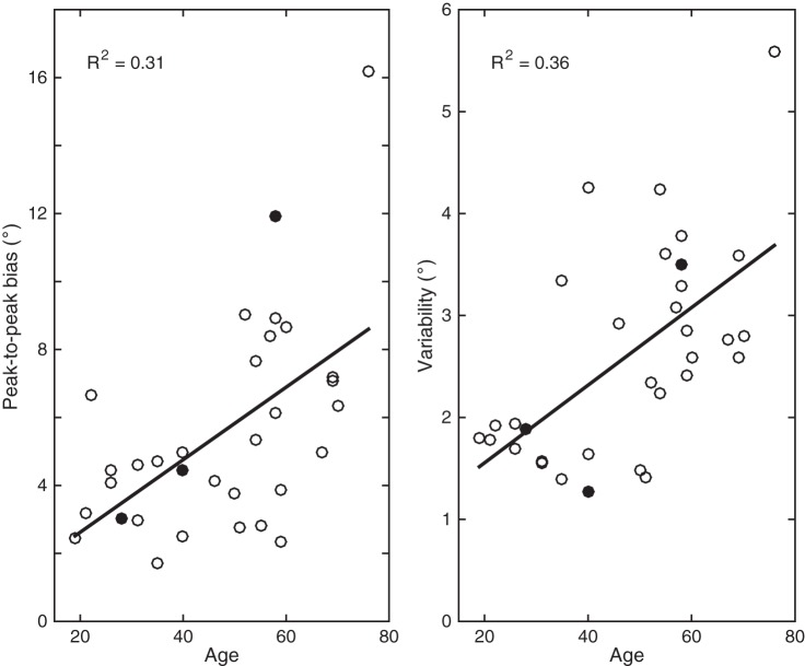
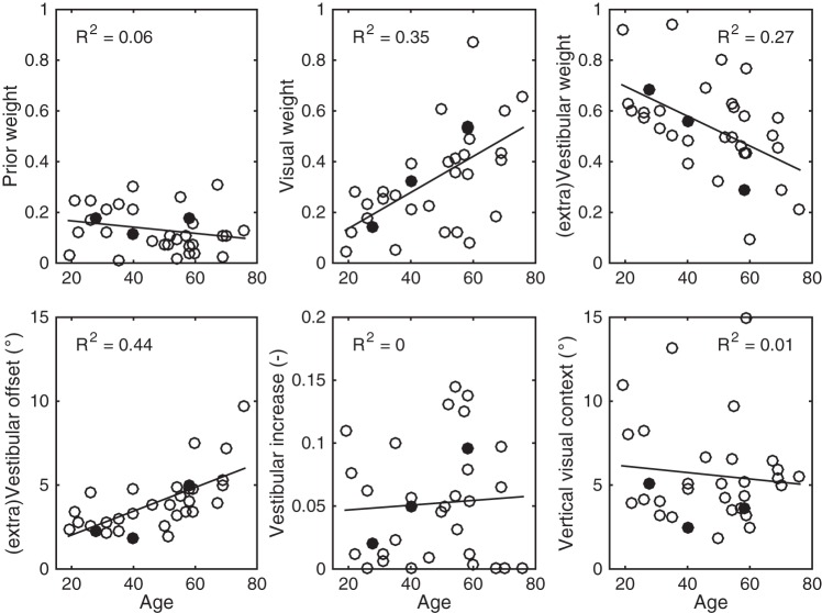
As we age, the acuity of our sensory organs declines, which may affect our lifestyle. Sensory deterioration in the vestibular system is typically bilateral and gradual, and could lead to problems with balance and spatial orientation. To compensate for the sensory deterioration, it has been suggested that the brain reweights the sensory information sources according to their relative noise characteristics. For rehabilitation and training programs, it is important to understand the consequences of this reweighting, preferably at the individual subject level. We psychometrically examined the age-dependent reweighting of visual and vestibular cues used in spatial orientation in a group of 32 subjects (age range: 19-76 yr). We asked subjects to indicate the orientation of a line (clockwise or counterclockwise relative to the gravitational vertical) presented within an oriented square visual frame when seated upright or with their head tilted 30° relative to the body. Results show that subjects' vertical perception is biased by the orientation of the visual frame. Both the magnitude of this bias and response variability become larger with increasing age. Deducing the underlying sensory noise characteristics, using Bayesian inference, suggests an age-dependent reweighting of sensory information, with an increasing weight of the visual contextual information. Further scrutiny of the model suggests that this shift in sensory weights is the result of an increase in the noise of the vestibular signal. Our approach quantifies how noise properties of visual and vestibular systems change over the life span, which helps to understand the aging process at the neurocomputational level. NEW & NOTEWORTHY Perception of visual vertical involves a weighted fusion of visual and vestibular tilt cues. Using a Bayesian approach and experimental psychophysics, we quantify how this fusion process changes with age. We show that, with age, the vestibular information is down-weighted whereas the visual weight is increased. This shift in sensory reweighting is primarily due to an age-related increase of the noise of vestibular signals.

Keywords: aging; internal models; rod-frame illusion; sensory reweighting; spatial orientation; verticality perception.


Conflict of interest statement

No conflicts of interest, financial or otherwise, are declared by the authors.

Figures

Fig. 1.
Fig. 1.
A: experimental procedure of the rod-and-frame task. After presentation of a square frame for 250 ms, a rod is briefly (33 ms) flashed within the frame. When the rod disappears, the square remains visible until the subject responds whether the rod was oriented clockwise or counterclockwise from upright. A 500-ms black screen is presented before the start of a new trial. B: schematic representation of the optimal integration model for verticality perception, integrating global visual frame-on-retina (θR), vestibular head-in-space (HS), and prior knowledge (HP) information into an optimal head-in-space estimate. All sensory signals are assumed to be accurate but contaminated with Gaussian noise, represented by σver and σhor for the visual, αHS and βHS for the vestibular, and σHP for the prior knowledge information. The optimal head-in-space estimate is then combined with local visual information about the rod-on-retina and eye-in-head information (magnitude AOCR) to determine the optimal rod-in-space orientation.
Fig. 2.
Fig. 2.
Probability of clockwise (CW) responses [P(CW)] plotted against rod orientation when the frame is displayed 20° counterclockwise (CCW), upright, or 20° CW for a young adult (green circles), middle-aged adult (blue circles), and older adult (red circles). Solid lines plotted through the data represent the psychometric functions, quantifying the bias (μ; dashed lines) and response variability of the subject.
Fig. 3.
Fig. 3.
Bias and variability plotted against frame orientation for the 3 representative subjects in the upright (top two rows) and 30° tilt condition (bottom two rows). Solid lines represent the best-fit of the Bayesian optimal integration model of Fig. 1B.
Fig. 4.
Fig. 4.
Peak-to-peak bias and response variability in the head-upright condition plotted against age. Peak-to-peak biases are calculated by fitting a sinusoid through the bias patterns. Solid lines represent the linear regression analysis, which is reported by the R2 correlation measure in each panel. Data points for the 3 exemplar subjects are closed circles.
Fig. 5.
Fig. 5.
A–C: for all subjects, prior knowledge (A), visual context (B), and vestibular weights (C) are plotted against age. Solid lines represent the linear regression analysis, which is reported by the R2 measure in each panel. D–F: analyses indicating whether the effects shown in A–C are the result of a correlation with age in the additive vestibular offset (D), multiplicative vestibular increase in noise (E), or vertical visual context (F). Data points for the 3 exemplar subjects are closed circles.

References

    1. Alberts BB, de Brouwer AJ, Selen LP, Medendorp WP. A Bayesian account of visual-vestibular interactions in the rod-and-frame task. eNeuro 3: ENEURO.0093-16.2016, 2016a. doi:10.1523/ENEURO.0093-16.2016. - DOI - PMC - PubMed
    1. Alberts BB, Selen LP, Bertolini G, Straumann D, Medendorp WP, Tarnutzer AA. Dissociating vestibular and somatosensory contributions to spatial orientation. J Neurophysiol 116: 30–40, 2016b. doi:10.1152/jn.00056.2016. - DOI - PMC - PubMed
    1. Alberts BB, Selen LP, Verhagen WI, Pennings RJ, Medendorp WP. Bayesian quantification of sensory reweighting in a familial bilateral vestibular disorder (DFNA9). J Neurophysiol 119: 1209–1221, 2018. doi:10.1152/jn.00082.2017. - DOI - PMC - PubMed
    1. Anson E, Jeka J. Perspectives on aging vestibular function. Front Neurol 6: 269, 2016. doi:10.3389/fneur.2015.00269. - DOI - PMC - PubMed
    1. Baezner H, Blahak C, Poggesi A, Pantoni L, Inzitari D, Chabriat H, Erkinjuntti T, Fazekas F, Ferro JM, Langhorne P, O’Brien J, Scheltens P, Visser MC, Wahlund LO, Waldemar G, Wallin A, Hennerici MG; LADIS Study Group . Association of gait and balance disorders with age-related white matter changes: the LADIS study. Neurology 70: 935–942, 2008. doi:10.1212/01.wnl.0000305959.46197.e6. - DOI - PubMed

Publication types