. 2019 Mar;160(3):712-723.
doi: 10.1097/j.pain.0000000000001450.

Islet-cell autoantigen 69 mediates the antihyperalgesic effects of electroacupuncture on inflammatory pain by regulating spinal glutamate receptor subunit 2 phosphorylation through protein interacting with C-kinase 1 in mice

Affiliations

Islet-cell autoantigen 69 mediates the antihyperalgesic effects of electroacupuncture on inflammatory pain by regulating spinal glutamate receptor subunit 2 phosphorylation through protein interacting with C-kinase 1 in mice

Kunyuan Han et al. Pain. 2019 Mar.

Abstract

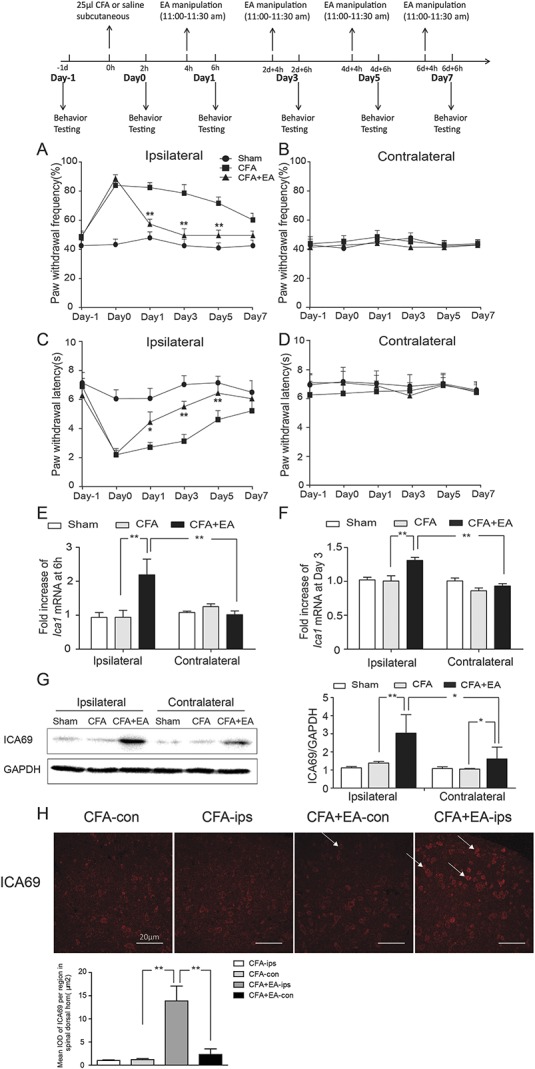
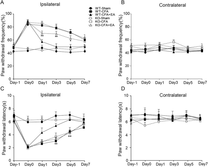
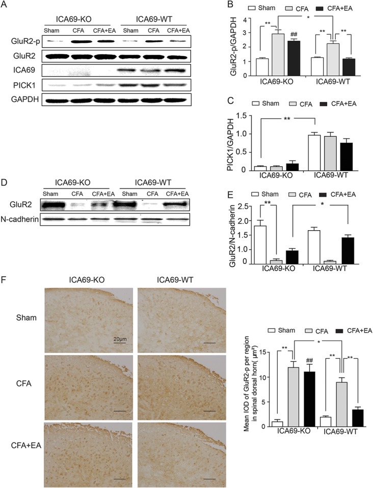
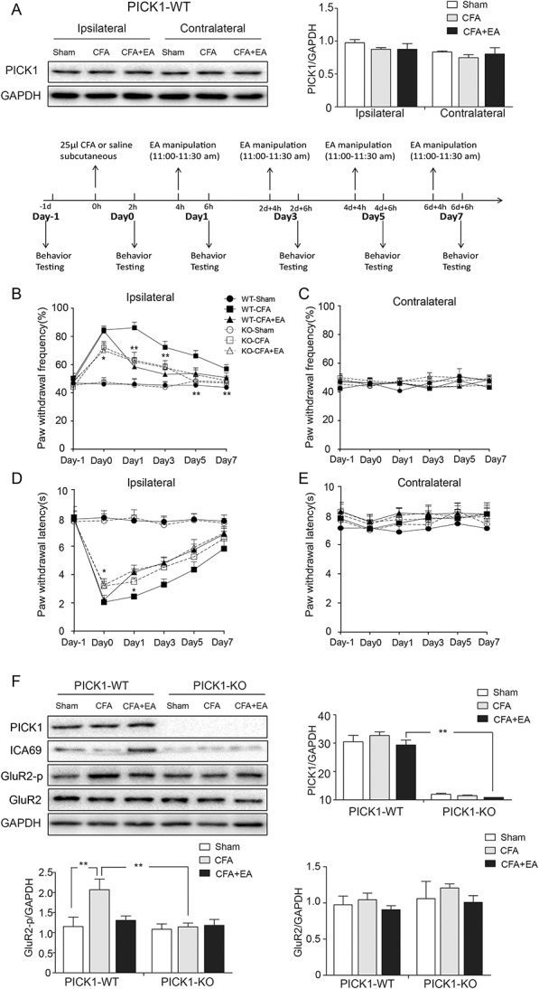
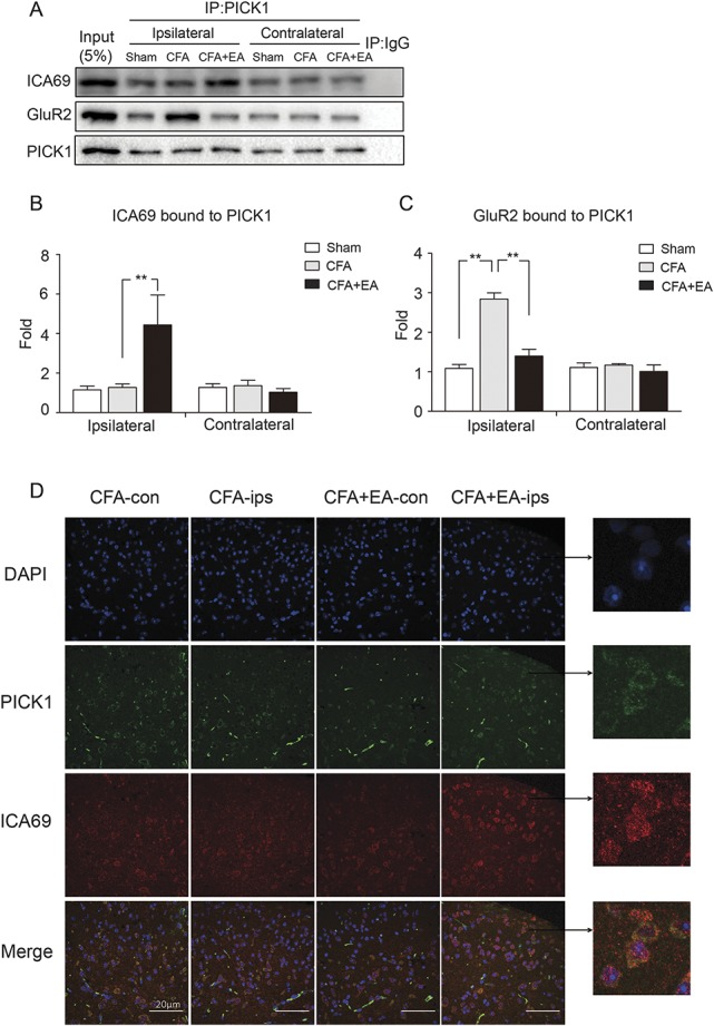
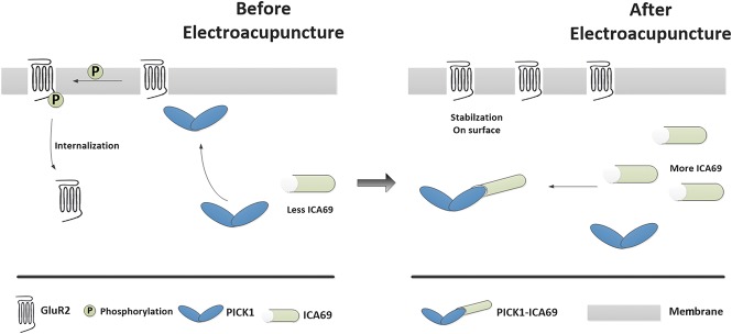
Electroacupuncture (EA) is widely used in clinical settings to reduce inflammatory pain. Islet-cell autoantigen 69 (ICA69) has been reported to regulate long-lasting hyperalgesia in mice. ICA69 knockout led to reduced protein interacting with C-kinase 1 (PICK1) expression and increased glutamate receptor subunit 2 (GluR2) phosphorylation at Ser880 in spinal dorsal horn. In this study, we evaluated the role of ICA69 in the antihyperalgesic effects of EA and the underlying mechanism through regulation of GluR2 and PICK1 in spinal dorsal horn. Hyperalgesia was induced in mice with subcutaneous plantar injection of complete Freund adjuvant (CFA) to cause inflammatory pain. Electroacupuncture was then applied for 30 minutes every other day after CFA injection. When compared with CFA group, paw withdrawal frequency of CFA+EA group was significantly decreased. Remarkable increases in Ica1 mRNA expression and ICA69 protein levels on the ipsilateral side were detected in the CFA+EA group. ICA69 expression reached the peak value around day 3. More importantly, ICA69 deletion impaired the antihyperalgesic effects of EA on GluR2-p, but PICK1 deletion could not. Injecting ICA69 peptide into the intrathecal space of ICA69-knockout mice mimicked the effects of EA analgesic and inhibited GluR2-p. Electroacupuncture had no effects on the total protein of PICK1 and GluR2. And, EA could increase the formation of ICA69-PICK1 complexes and decrease the amount of PICK1-GluR2 complexes. Our findings indicate that ICA69 mediates the antihyperalgesic effects of EA on CFA-induced inflammatory pain by regulating spinal GluR2 through PICK1 in mice.


Conflict of interest statement

Sponsorships or competing interests that may be relevant to content are disclosed at the end of this article.

Figures

Figure 1.
Figure 1.
Effects of EA on CFA-induced hyperalgesia and the content of ICA69 expression. (A) Reduction in paw withdrawal frequency of the CFA+EA group compared with the CFA group on the ipsilateral side (n = 13; *P < 0.05); this reduction was particularly obvious at day 1, day 3, and day 5 (n = 13; *P < 0.05, **P < 0.01). (B) No difference among the 3 groups on the contralateral side (n = 13; P > 0.05). (C) Increase in paw withdrawal latency of the CFA+EA group compared with the CFA group on the ipsilateral side; this effect was most obvious at day 3 and day 5 (n = 13; *P < 0.05, **P < 0.01). (D) No difference among the 3 groups on the contralateral side (n = 13; P > 0.05). (E and F) Effects of EA on the expression of Ica1 mRNA in SDH at 6 hours and 3 days after CFA injection. There was an increase of the Ica1 mRNA level in the CFA+EA group compared with the CFA group on the ipsilateral side (n = 9, **P < 0.01), and an increase of the ipsilateral side compared with the contralateral side in the CFA+EA group (n = 9, **P < 0.01). (G) The ICA69 protein content increased in the CFA+EA group compared with the CFA group on the ipsilateral side (n = 9, **P < 0.01); there was an increase of the ipsilateral side compared with the contralateral side in the CFA+EA group (n = 9, *P < 0.05). (H) Immunofluorescence results of ICA69 in SDH in the 4 indicated groups: the red staining represents the ICA69 protein, and the white arrow points to the positive cells in the SDH area. Scale bars = 20 µm. Quantification of ICA69 per µm2 in SDH (n = 3; group values are indicated by mean ± SEM; **P < 0.01, *P < 0.05). CFA, complete Freund adjuvant; EA, electroacupuncture; SDH, spinal dorsal horn.
Figure 2.
Figure 2.
Effects of EA on CFA-induced hyperalgesia in ICA69-WT and ICA69-KO mice. (A) An increase in paw withdrawal frequency of the KO-CFA+EA group compared with the WT-CFA+EA group on the ipsilateral side (n = 13; *P < 0.05); this effect was obvious at day 1, day 3, and day 5 (n = 13; *P < 0.05, **P < 0.01). There was no difference between the KO-CFA+EA and the KO-CFA group (n = 13; P > 0.05), and there was no difference between KO-CFA and WT-CFA groups (n = 13; P > 0.05). (B) There was no difference among the 6 groups on the contralateral side (n = 13; P > 0.05). (C) Reduction in paw withdrawal latency of the KO-CFA+EA group compared with the WT-CFA+EA group on the ipsilateral side (n = 13; *P < 0.05); this effect was particularly obvious at day 3 and day 5 (n = 13; *P < 0.05, **P < 0.01). There was no difference between the KO-CFA+EA group and the KO-CFA group (n = 13; P > 0.05), and there was no difference between KO-CFA and WT-CFA groups (n = 13; P > 0.05). (D) No difference among the 6 groups was detected on the contralateral side (n = 13; P > 0.05). CFA, complete Freund adjuvant; EA, electroacupuncture; KO, knockout; WT, wild-type.
Figure 3.
Figure 3.
Effects of EA on the protein levels of GluR2-p, ICA69, PICK1, GluR2 (total), and GluR2 (membrane) in ICA69-WT and ICA69-KO mice. (A) Representative Western blot bands of GluR2-p, GluR2 (total), ICA69, and PICK1. Quantitative analyses of GluR2-p (##KO-CFA+EA vs WT-CFA+EA, P < 0.001) (B), PICK1 (C), and GluR2 (membrane) (E) (n = 3, group values are indicated by mean ± SEM; **P < 0.01; *P < 0.05). (D) Representative Western blot bands of GluR2 (membrane); (n = 3, group values are indicated by mean ± SEM; **P < 0.01; *P < 0.05). (F) Immunohistochemical results of GluR2-p in SDH in the 6 indicated groups: the tawny staining represents the GluR2-p protein. Scale bar = 20 μm. Quantification of GluR2-p per µm2 in SDH (n = 3; ##KO-CFA+EA vs WT-CFA+EA, P < 0.001; group values are indicated by mean ± SEM; **P < 0.01; *P < 0.05). CFA, complete Freund adjuvant; EA, electroacupuncture; IOD, integrated optical density; KO, knockout; SDH, spinal dorsal horn; WT, wild-type.
Figure 4.
Figure 4.
Effects of EA on CFA-induced hyperalgesia and the protein levels of PICK1, ICA69, GluR2-p, and GluR2 in PICK1-WT and PICK1-KO mice. (A) Western blot bands of PICK1 and quantitative analyses of PICK1 on both sides in PICK1-WT mice. (B) Reduction in paw withdrawal frequency of the KO-CFA group compared with the WT-CFA group on the ipsilateral side (n = 13; *P < 0.05); the effect was particularly obvious at day 0, day 1, day 3, day 5, and day 7 (n = 13; *P < 0.05, **P < 0.01). There was no difference between KO-CFA+EA and KO-CFA groups (n = 13; P > 0.05), and there was no difference between KO-CFA and WT-CFA+EA groups (n = 13; P > 0.05). (C) No difference among the 6 groups on the contralateral side (n = 13; P > 0.05). (D) Increase in paw withdrawal latency of the KO-CFA group compared with the WT-CFA group on the ipsilateral side (n = 13; *P < 0.05); the effect was particularly obvious at day 0 and day 1 (n = 13; *P < 0.05). There was no difference between KO-CFA+EA and KO-CFA groups (n = 13; P > 0.05), and there was no difference between KO-CFA and WT-CFA+EA groups (n = 13; P > 0.05). (E) There was no difference among the 6 groups on the contralateral side (n = 13; P > 0.05). (F) Representative Western blot bands of PICK1, ICA69, GluR2-p, and GluR2 in PICK1-WT and PICK1-KO mice, and quantitative analyses of these bands (n = 3, group values are indicated by mean ± SEM; **P < 0.01). CFA, complete Freund adjuvant; EA, electroacupuncture; KO, knockout; WT, wild-type.
Figure 5.
Figure 5.
Effects of EA on the formation of endogenous ICA69-PICK1 and PICK1-GluR2 complexes. (A) Endogenous protein was collected from SDH. Representative Western blot bands of ICA69, GluR2, and PICK1 in WT mice by co-IP. (B and C) Quantitative analyses of ICA69 and GluR2 to evaluate the ability of binding to PICK1. Increased ICA69 and reduced GluR2 interactions were observed with PICK1 in the CFA+EA group (n = 3, group values are indicated by mean ± SEM; **P < 0.01). (D) Electroacupuncture caused an increase in colocalization between endogenous ICA69 (red) and PICK1 (green) on the ipsilateral side in SDH. CFA, complete Freund adjuvant; EA, electroacupuncture; SDH, spinal dorsal horn; WT, wild-type.
Figure 6.
Figure 6.
Proposed mechanism of the antihyperalgesic effects of EA stimulation. Before EA, there is a relatively low amount of ICA69 protein in SDH, and it does not abundantly combine with PICK1 protein in the cytoplasm; the medial PICK1 protein is therefore free to transport to the cell membrane, resulting in GluR2 phosphorylation. Once GluR2 is phosphorylated into GluR2-p, endogenous GluR2 will move from the cell membrane into the cytoplasm. AMPA receptors lacking GluR2 become permeable to Ca2+, resulting in cellular inflammation that leads to hyperalgesia. By contrast, EA treatment increases the amount of ICA69 proteins, forming abundant complexes with PICK1, thereby stranding PICK1 in the cytoplasm. This prevents GluR2 phosphorylation, and therefore AMPA receptors can maintain the impermeability of the cell membrane to Ca2+, ultimately producing an anti-inflammatory pain effect. EA, electroacupuncture.

Similar articles

Cited by

References

    1. Aoyama K, Matsumura N, Watabe M, Nakaki T. Oxidative stress on EAAC1 is involved in MPTP-induced glutathione depletion and motor dysfunction. Eur J Neurosci 2008;27:20–30. - PubMed
    1. Arvan P, Pietropaolo M, Ostrov D, Rhodes CJ. Islet autoantigens: structure, function, localization, and regulation. Cold Spring Harb Perspect Med 2012;2:a007658. - PMC - PubMed
    1. Atianjoh FE, Yaster M, Zhao X, Takamiya K, Xia J, Gauda EB, Huganir RL, Tao YX. Spinal cord protein interacting with C kinase 1 is required for the maintenance of complete Freund's adjuvant-induced inflammatory pain but not for incision-induced post-operative pain. PAIN 2010;151:226–34. - PMC - PubMed
    1. Barnes PM, Barbara B, Nahin RL. Complementary and alternative medicine use among adults and children: United States, 2007. Natl Health Stat Rep 2008;12:1–23. - PubMed
    1. Berman BM, Lao L, Langenberg P, Lee WL, Gilpin AM, Hochberg MC. Effectiveness of acupuncture as adjunctive therapy in osteoarthritis of the knee: a randomized, controlled trial. Ann Intern Med 2004;141:901–10. - PubMed

MeSH terms