. 2019 Jan 30;15(1):e1007578.
doi: 10.1371/journal.ppat.1007578. eCollection 2019 Jan.

Oncogenic KSHV-encoded interferon regulatory factor upregulates HMGB2 and CMPK1 expression to promote cell invasion by disrupting a complex lncRNA-OIP5-AS1/miR-218-5p network

Affiliations

Oncogenic KSHV-encoded interferon regulatory factor upregulates HMGB2 and CMPK1 expression to promote cell invasion by disrupting a complex lncRNA-OIP5-AS1/miR-218-5p network

Wan Li et al. PLoS Pathog. .

Erratum in

Abstract

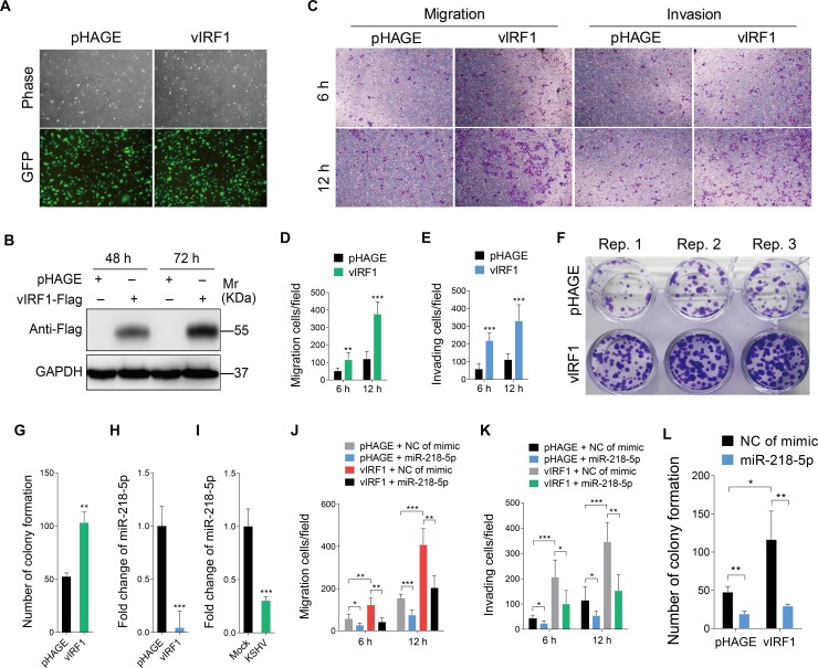
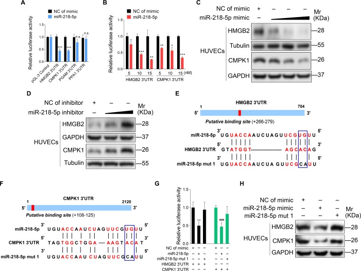
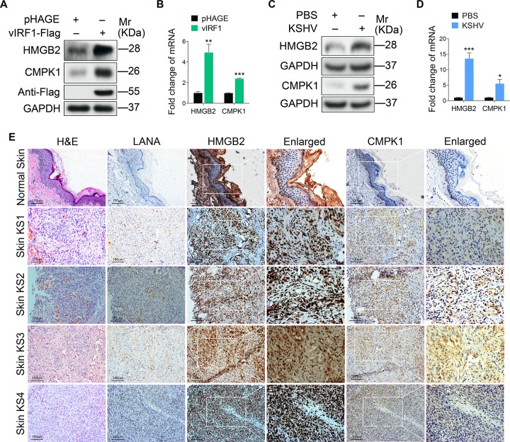
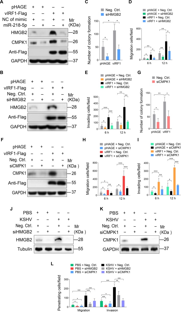
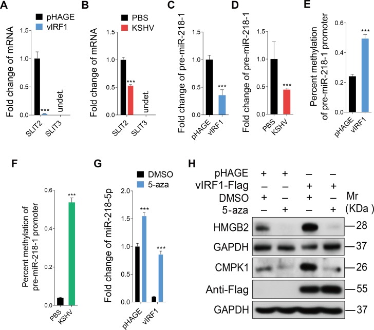
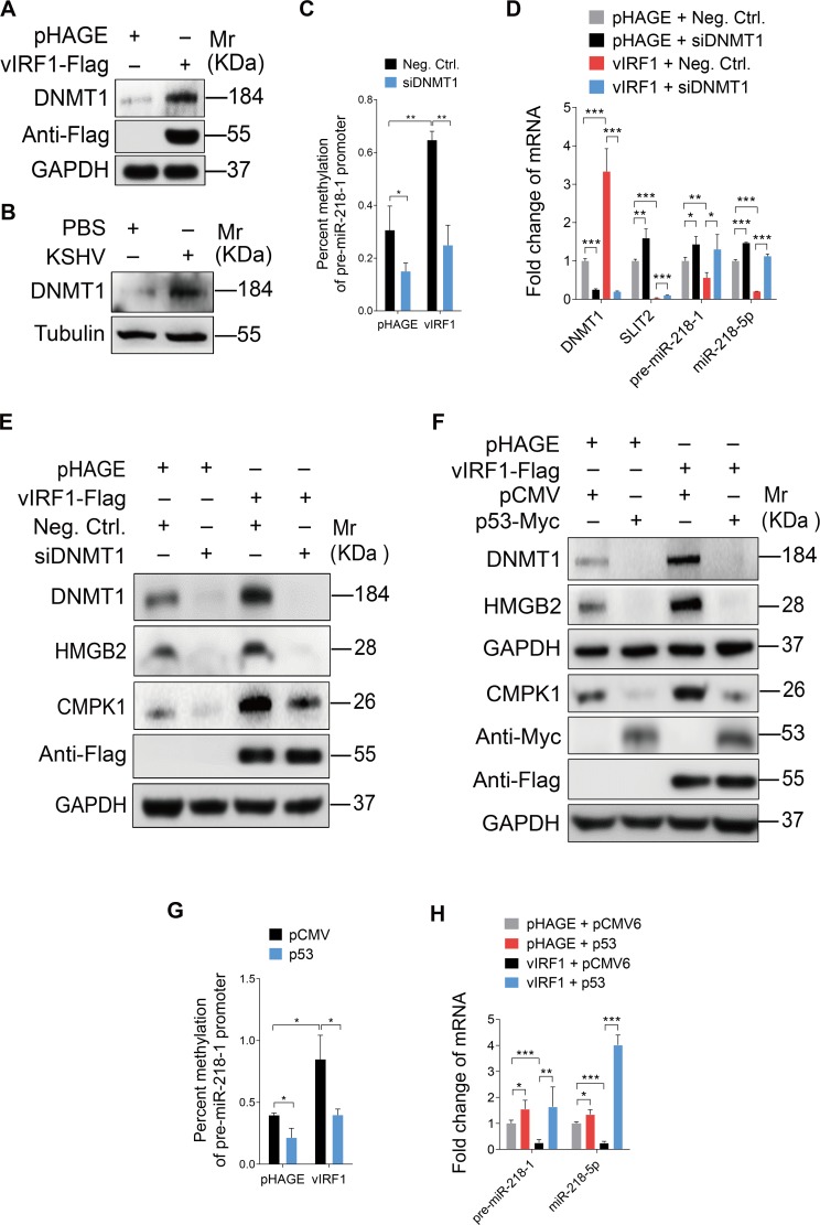
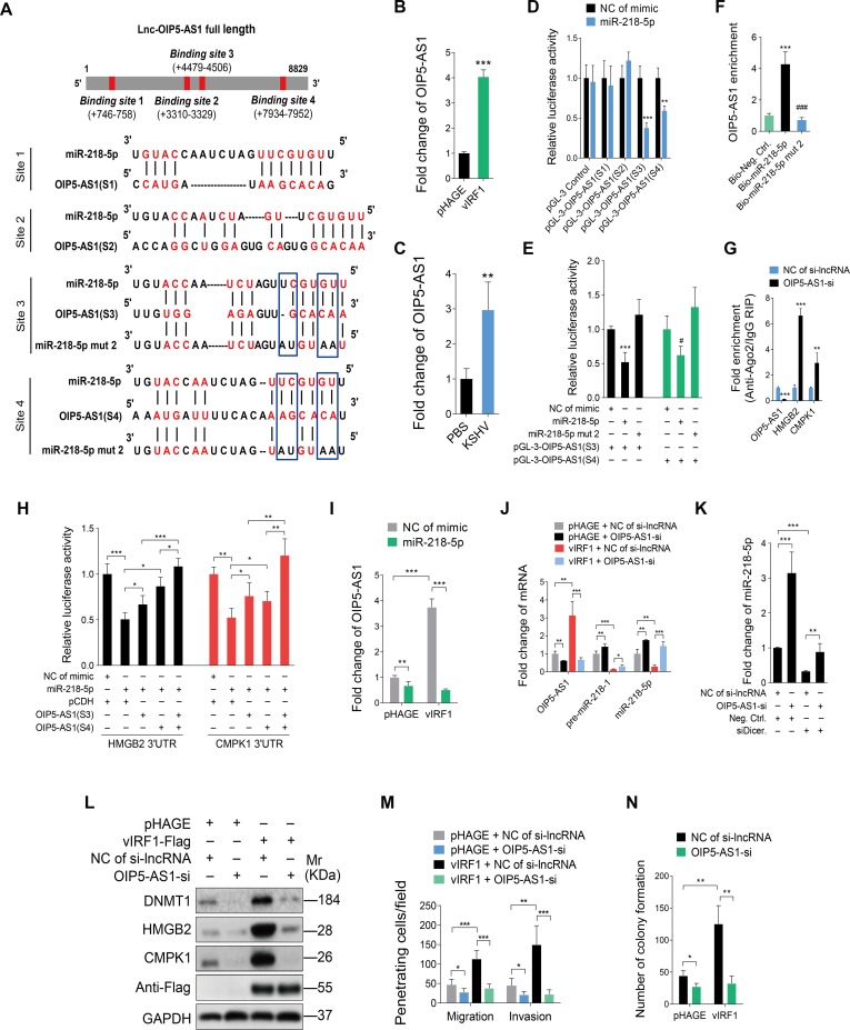
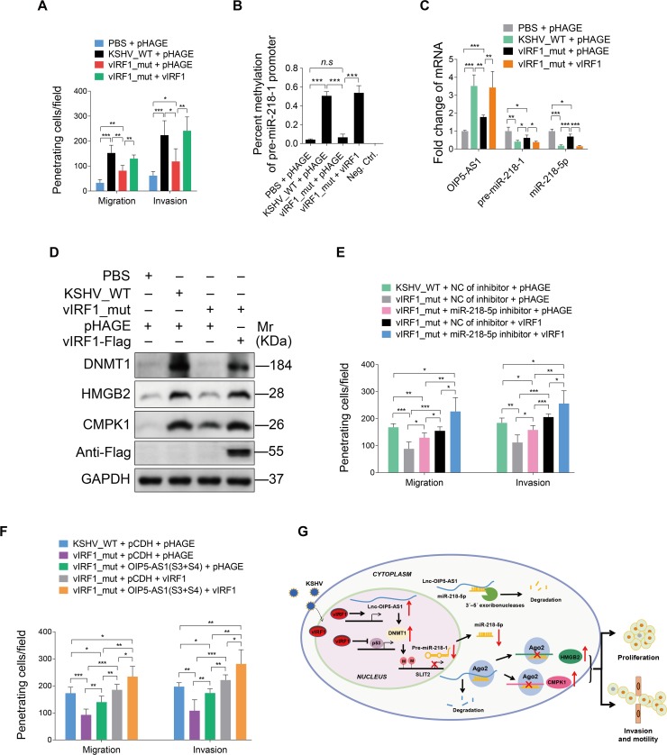
Kaposi's sarcoma (KS), a highly disseminated tumor of hyperproliferative spindle endothelial cells, is the most common AIDS-associated malignancy caused by infection of Kaposi's sarcoma-associated herpesvirus (KSHV). KSHV-encoded viral interferon regulatory factor 1 (vIRF1) is a viral oncogene but its role in KSHV-induced tumor invasiveness and motility remains unknown. Here, we report that vIRF1 promotes endothelial cell migration, invasion and proliferation by down-regulating miR-218-5p to relieve its suppression of downstream targets high mobility group box 2 (HMGB2) and cytidine/uridine monophosphate kinase 1 (CMPK1). Mechanistically, vIRF1 inhibits p53 function to increase the expression of DNA methyltransferase 1 (DNMT1) and DNA methylation of the promoter of pre-miR-218-1, a precursor of miR-218-5p, and increases the expression of a long non-coding RNA OIP5 antisense RNA 1 (lnc-OIP5-AS1), which acts as a competing endogenous RNA (ceRNA) of miR-218-5p to inhibit its function and reduce its stability. Moreover, lnc-OIP5-AS1 increases DNA methylation of the pre-miR-218-1 promoter. Finally, deletion of vIRF1 from the KSHV genome reduces the level of lnc-OIP5-AS1, increases the level of miR-218-5p, and inhibits KSHV-induced invasion. Together, these results define a novel complex lnc-OIP5-AS1/miR-218-5p network hijacked by vIRF1 to promote invasiveness and motility of KSHV-induced tumors.


Conflict of interest statement

The authors have declared that no competing interests exist.

Figures

Fig 1
Fig 1. Overexpression of vIRF1 negatively regulates miR-218-5p expression and enhances endothelial cell motility, invasion and proliferation.
(A). Representational images of HUVECs transduced with vIRF1 (vIRF1) or control lentivirus (pHAGE). Magnification, ×100. (B). Western blotting analysis for vIRF1 protein in HUVECs infected with vIRF1 (vIRF1) or control lentivirus (pHAGE) for 48 h and 72 h, respectively. The antibody against the Flag-tag was used to detect vIRF1. GAPDH was used as loading control. (C). Migration and invasion analysis of HUVECs expressing vIRF1 (vIRF1) or control lentivirus (pHAGE) at 6 h and 12 h. (D) and (E). Quantification of cell migration and invasion described in (C), respectively. Results are from three independent experiments, each with quintuple technical replicates, were performed. (F). Plate colony formation assay of HUVECs transduced with vIRF1 (vIRF1) or control lentivirus (pHAGE). (G). Quantification of cell colony formation described in (F). Results are from three independent experiments, each with cubic technical replicates, were performed. (H) and (I). qPCR showing miR-218-5p expression in vIRF1-transduced and KSHV-infected cells, respectively. (J). Migration analysis of vIRF1-infected HUVECs transfected with miR-218-5p mimic for 48 h. (K). Invasion analysis of vIRF1-expressing HUVECs treated as in (J). (L). Colony formation assay of HUVECs treated as in (J). The quantified results represent mean ± SD. * P < 0.05, ** P < 0.01, and *** P < 0.001, Student's t-test.
Fig 2
Fig 2. miR-218-5p directly targets HMGB2 and CMPK1 3'UTR.
(A). Luciferase activity in HEK293T cells cotransfected with miR-218-5p mimic (miR-218-5p; 10 nM) or its control (NC of mimic; 10 nM) and the reporter constructs for 48 h. (B). Luciferase activity in HEK293T cells cotransfected with increasing amounts of miR-218-5p mimic (miR-218-5p; 5, 10, and 15 nM) or its control (NC of mimic), and the pGL-3-HMGB2 3'UTR reporter or pGL-3-CMPK1 3'UTR reporter for 48 h. (C). Western blotting of HMGB2 and CMPK1 expression in HUVECs transfected with increasing amounts of miR-218-5p mimic (10, 20 and 40 nM) or its control for 48 h. (D). Western blotting of HMGB2 and CMPK1 expression in HUVECs transfected with a miR-218-5p inhibitor for 48 h. (E). Putative binding site of miR-218-5p in the 3'UTR region of HMGB2 and mutagenesis of target site in miR-218-5p. (F). Putative binding site of miR-218-5p in the 3'UTR region of CMPK1 and mutagenesis of target site in miR-218-5p. (G). Luciferase activity in HEK293T cells cotransfected with miR-218-5p mimic (miR-218-5p; 10 nM), miR-218-5p mutant mimic (miR-218-5p mut 1; 10 nM) or a negative control (NC of mimic; 10 nM), and the HMGB2 3'UTR or CMPK1 3'UTR reporter construct for 48 h. (H). Western-blotting of HMGB2 and CMPK1 expression in HUVECs transfected with a negative control mimic (NC of mimic; 20 nM), miR-218-5p mimic (miR-218-5p mimic; 20 nM) or miR-218-5p mutant mimic (miR-218-5p mut 1; 20 nM) for 48 h, respectively. The quantified results represent mean ± SD. Results are from three independent experiments, each with quadruple technical replicates, were performed. * P <0.05, ** P <0.01, *** P < 0.001, and ### P < 0.001, Student's t-test. n.s, not significant.
Fig 3
Fig 3. HMGB2 and CMPK1 expression is increased in vIRF1-transduced and KSHV-infected HUVECs, and KS lesion samples.
(A). Western-blotting of HMGB2 and CMPK1 expression in vIRF1-transduced HUVECs. (B). qPCR showing HMGB2 and CMPK1 mRNA transcription in vIRF1-transduced HUVECs. (C). Western-blotting of HMGB2 and CMPK1 expression in KSHV-infected HUVECs. (D). qPCR showing HMGB2 and CMPK1 mRNA transcription in KSHV-infected HUVECs. (E). H&E staining, and immunohistochemical staining of KSHV LANA, HMGB2 and CMPK1 in normal skin, skin KS of patient #1 (Skin KS1), skin KS of patient #2 (Skin KS2), skin KS of patient #3 (Skin KS3) and skin KS patient #4 (Skin KS4). Magnification, ×200, ×400. The quantified results represent mean ± SD. Results are from three independent experiments, each with quadruple technical replicates, were performed. * P < 0.05, ** P < 0.01, and *** P < 0.001, Student's t-test.
Fig 4
Fig 4. miR-218-5p directly targets HMGB2 and CMPK1 to mediate vIRF1-induced cell motility, invasion and proliferation.
(A). Western-blotting of HMGB2 and CMPK1 expression in vIRF1-infected HUVECs transfected with a negative control mimic (NC of mimic; 20 nM), miR-218-5p mimic (miR-218-5p; 20 nM). (B). Western-blotting of HMGB2 expression in vIRF1-expressing HUVECs transfected with a mixture of siRNAs targeting HMGB2 (siHMGB2). (C). Plate colony formation assay of HUVECs treated as in (B). (D). Migration analysis of HUVECs treated as in (B). (E). Invasion analysis of HUVECs treated as in (B). (F). Western-blotting of CMPK1 expression in vIRF1-expressing HUVECs transfected with a mixture of siRNAs targeting CMPK1 (siCMPK1). (G). Plate colony formation assay of HUVECs treated as in (F). (H). Migration analysis of HUVECs treated as in (F). (I). Invasion analysis of HUVECs treated as in (F). (J). Western-blotting of HMGB2 expression in KSHV-infected HUVECs transfected with a mixture of siRNAs targeting HMGB2 (siHMGB2). (K). Western-blotting of CMPK1 expression in KSHV-infected HUVECs transfected with a mixture of siRNAs targeting CMPK1 (siCMPK1). (L). Migration and invasion analyses of HUVECs treated as in (J) and (K) at 6 h. The quantified results represent mean ± SD. Results are from three independent experiments, each with cubic (colony formation) or quadruple (migration and invasion) technical replicates, were performed. * P < 0.05, ** P < 0.01, and *** P < 0.001, Student's t-test.
Fig 5
Fig 5. vIRF1 suppresses miR-218-5p via hypermethylation of the pre-miR-218-1 promoter.
(A). qPCR showing SLIT2 and SLIT3 expression in vIRF1-transduced HUVECs. (B). qPCR showing SLIT2 and SLIT3 expression in KSHV-infected HUVECs. (C). qPCR showing pre-miR-218-1 expression in vIRF1-transduced HUVECs. (D). qPCR showing pre-miR-218-1 expression in KSHV-infected HUVECs (E). Methylation-specific PCR showing DNA methylation of in the pre-miR-218-1 promoter in vIRF1-transduced HUVECs. (F). Methylation-specific PCR showing DNA methylation of the pre-miR-218-1 promoter in KSHV-infected cells. (G). qPCR showing miR-218-5p expression in vIRF1-transduced HUVECs treated with 5-aza. (H). Western-blotting of HMGB2 and CMPK1 expression in vIRF1-transduced HUVECs treated with 5-aza. The Western blots were ran with the same samples but in two different gels, with each of them calibrated by independent GAPDH panels. The quantified results represent mean ± SD. *** P < 0.001, Student's t-test. undet., undetermined.
Fig 6
Fig 6. vIRF1 suppresses miR-218-5p via hypermethylation of the pre-miR-218-1 promoter by inhibiting p53 to increase DNMT1 expression.
(A). Western-blotting of DNMT1 expression in vIRF1-transduced HUVECs. (B). Western-blotting of DNMT1 in KSHV-infected HUVECs. (C). Methylation-specific PCR showing DNA methylation of the pre-miR-218-1 promoter in vIRF1-expressing HUVECs transfected with a mixture of siRNAs targeting DNMT1 (siDNMT1). (D). qPCR showing DNMT1, SLIT2, pre-miR-218-1 and miR-218-5p expression in vIRF1-expressing HUVECs transfected with a mixture of siRNAs targeting DNMT1 (siDNMT1). (E). Western-blotting of DNMT1, HMGB2 and CMPK1 expression in vIRF1-expressing HUVECs transfected with a mixture of siRNAs targeting DNMT1 (siDNMT1). (F). Western-blotting of DNMT1, HMGB2 and CMPK1 expression in vIRF1-expressing HUVECs transfected with pCMV6-Entry-C-Myc-p53 construct. The Western blots were ran with the same samples but in two different gels, with each of them calibrated by independent GAPDH panels. (G). Methylation-specific PCR showing DNA methylation of the pre-miR-218-1 promoter in vIRF1-expressing HUVECs transfected with pCMV6-Entry-C-Myc-p53 construct. (H). qPCR showing pre-miR-218-1 and miR-218-5p expression in vIRF1-expressing HUVECs transfected with pCMV6-Entry-C-Myc-p53 construct. The quantified results represent mean ± SD. * P < 0.05, ** P < 0.01, and *** P < 0.001, Student's t-test.
Fig 7
Fig 7. The crosstalk between miR-218-5p and lncRNA-OIP5-AS1 contributes to vIRF1-induced cell motility, invasion and proliferation.
(A). Putative binding site of miR-218-5p in lnc-OIP5-AS1 and mutagenesis of target sites in miR-218-5p. (B). qPCR showing lncRNA-OIP5-AS1 expression in vIRF1-transduced HUVECs. (C). qPCR showing lnc-OIP5-AS1 expression in KSHV-infected HUVECs. (D). Luciferase activity in HEK293T cells cotransfected with miR-218-5p mimic (miR-218-5p; 20 nM) or a negative control (NC of mimic; 20 nM), together with the pGL-3-OIP5-AS1(S1), pGL-3-OIP5-AS1(S2), pGL-3-OIP5-AS1(S3) or pGL-3-OIP5-AS1(S4) reporter construct for 48 h. (E). Luciferase activity in HEK293T cells cotransfected with the miR-218-5p mimic (miR-218-5p; 20 nM), miR-218-5p mutant mimic (miR-218-5p mut 2; 20 nM) or a negative control (NC of mimic; 20 nM) together with the pGL-3-OIP5-AS1(S3), or pGL-3-OIP5-AS1(S4) reporter construct for 48 h. (F). RNA pull-down analysis was performed with biotin-labeled miRNA and its control including bio-Neg. Ctrl., bio-miR-218-5p and bio-miR-218-5p mut 2. Specific primers were used to detect the enrichment of lnc-OIP5-AS1. (G). RIP results showing the enrichment of binding of lnc-OIP5-AS1, HMGB2 and CMPK1 with miR-218-5p based on Ago2 pull down assay. (H). Luciferase activity in HEK293T cells cotransfected with miR-218-5p mimic (miR-218-5p; 20 nM) or a negative control (NC of mimic; 20 nM) together with pGL3-HMGB2 3'UTR reporter or pGL3-CMPK1 3'UTR reporter with and without lnc-OIP5-AS1(S3) or lnc-OIP5-AS1(S4) fragment. (I). qPCR showing lnc-OIP5-AS1 expression in vIRF1-expressing HUVECs transfected with miR-218-5p mimic. (J). qPCR showing lnc-OIP5-AS1, pre-miR-218-1 and miR-218-5p expression in vIRF1-expressing HUVECs with lnc-OIP5-AS1 silencing. (K). qPCR showing miR-218-5p expression in lnc-OIP5-AS1 silenced HUVECs with Dicer knockdown. (L). Western-blotting of DNMT1, HMGB2 and CMPK1 expression in vIRF1 cells with lnc-OIP5-AS1 silencing. (M). Migration and invasion analyses of vIRF1 cells with lnc-OIP5-AS1 silencing at 6 h. (N). Plate colony formation assay of vIRF1-transduced HUVECs with lnc-OIP5-AS1 silencing. The quantified results represent mean ± SD. * P < 0.05, ** P < 0.01, *** P < 0.001, and ### P < 0.001, Student's t-test.
Fig 8
Fig 8. Loss of vIRF1 reduces cell motility, invasion and proliferation induced by KSHV.
(A). Migration and invasion analyses of HUVECs treated with PBS (PBS) or infected with wild-type KSHV (KSHV_WT) or vIRF1 mutant virus (vIRF1_mut) followed by transduction with lentiviral vIRF1 at MOI 2 at 6 hpi. (B). Methylation-specific PCR showing DNA methylation of HUVECs treated as in (A). (C). Expression of lnc-OIP5-AS1, pre-miR-218-1 and miR-218-5p expression detected by qPCR in HUVECs treated as in (A). (D). Western-blotting of DNMT1, HMGB2 and CMPK1 expression in HUVECs treated as in (A). (E). Migration and invasion analyses of wild-type KSHV (KSHV_WT) cells, vIRF1 mutant cells (vIRF1_mut) or vIRF1-transduced mutant cells followed by transduction with a specific miR-218-5p inhibitor at 6 hpi. (F). Migration and invasion analyses of wild-type KSHV (KSHV_WT) cells, vIRF1 mutant cells (vIRF1_mut) or vIRF1-transduced mutant cells followed by transduction with lentiviral lnc-OIP5-AS1 (S3) construct and lentiviral lnc-OIP5-AS1 (S4) construct at 6 h. (G). A hypothetical model of the mechanism of how vIRF1 facilitates endothelial cell motility, invasion and proliferation. The quantified results represent mean ± SD. * P < 0.05, ** P < 0.01, and *** P < 0.001, Student's t-test. n.s, not significant.

Similar articles

Cited by

References

    1. Chang Y, Cesarman E, Pessin MS, Lee F, Culpepper J, Knowles DM, et al. Identification of herpesvirus-like DNA sequences in AIDS-associated Kaposi's sarcoma. Science. 1994;266(5192):1865–9. Epub 1994/12/16. . - PubMed
    1. Moore PS, Chang Y. Molecular virology of Kaposi's sarcoma-associated herpesvirus. Philos Trans R Soc Lond B Biol Sci. 2001;356(1408):499–516. Epub 2001/04/21. 10.1098/rstb.2000.0777 ; PubMed Central PMCID: PMCPmc1088441. - DOI - PMC - PubMed
    1. Grundhoff A, Ganem D. Inefficient establishment of KSHV latency suggests an additional role for continued lytic replication in Kaposi sarcoma pathogenesis. J Clin Invest. 2004;113(1):124–36. Epub 2004/01/01. 10.1172/JCI200417803 ; PubMed Central PMCID: PMCPmc300762. - DOI - PMC - PubMed
    1. Mesri EA, Cesarman E, Boshoff C. Kaposi's sarcoma and its associated herpesvirus. Nat Rev Cancer. 2010;10(10):707–19. 10.1038/nrc2888 - DOI - PMC - PubMed
    1. Baresova P, Pitha PM, Lubyova B. Distinct roles of Kaposi's sarcoma-associated herpesvirus-encoded viral interferon regulatory factors in inflammatory response and cancer. J Virol. 2013;87(17):9398–410. 10.1128/JVI.03315-12 - DOI - PMC - PubMed

Publication types

MeSH terms