. 2019 Jan 30;14(1):e0211542.
doi: 10.1371/journal.pone.0211542. eCollection 2019.

New insights of the local immune response against both fertile and infertile hydatid cysts

Affiliations

New insights of the local immune response against both fertile and infertile hydatid cysts

Christian Hidalgo et al. PLoS One. .

Abstract

Background: Cystic echinococcosis is caused by the metacestode of the zoonotic flatworm Echinococcus granulosus. Within the viscera of the intermediate host, the metacestode grows as a unilocular cyst known as hydatid cyst. This cyst is comprised of two layers of parasite origin: germinal and laminated layers, and one of host origin: the adventitial layer, that encapsulates the parasite. This adventitial layer is composed of collagen fibers, epithelioid cells, eosinophils and lymphocytes. To establish itself inside the host, the germinal layer produces the laminated layer, and to continue its life cycle, generates protoscoleces. Some cysts are unable to produce protoscoleces, and are defined as infertile cysts. The molecular mechanisms involved in cyst fertility are not clear, however, the host immune response could play a crucial role.

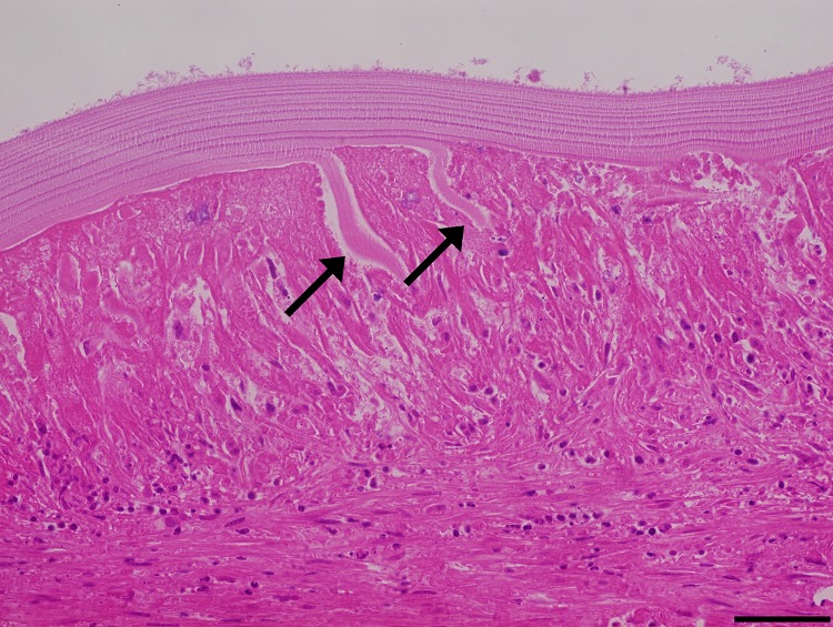
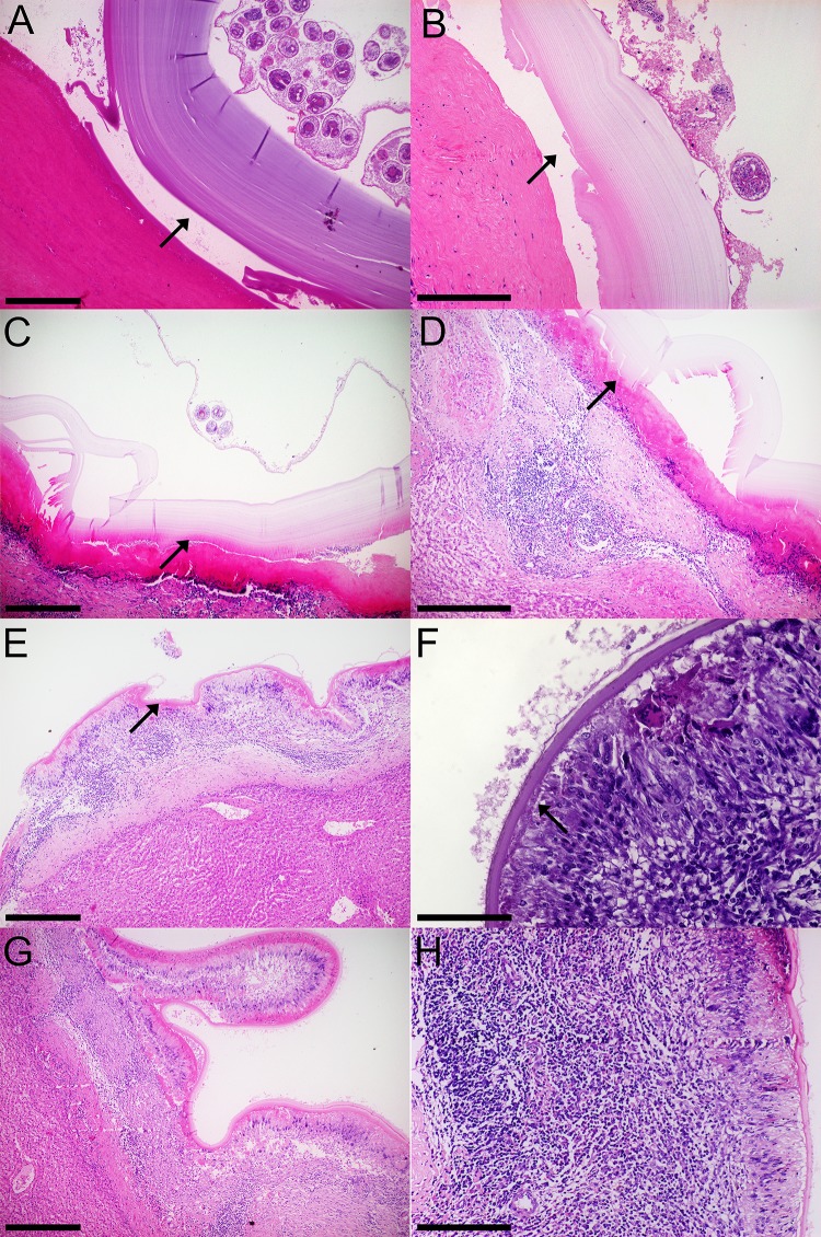
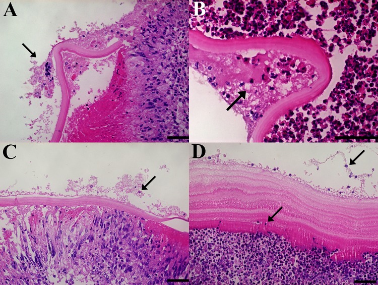
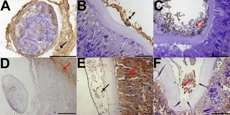
Methodology/principal findings: We collected hydatid cysts from both liver and lungs of slaughtered cattle, and histological sections of fertile, infertile and small hydatid cysts were stained with haematoxylin-eosin. A common feature observed in infertile cysts was the disorganization of the laminated layer by the infiltration of host immune cells. These infiltrating cells eventually destroy parts of laminated layer. Immunohistochemical analysis of both parasite and host antigens, identify these cells as cattle macrophages and are present inside the cysts associated to germinal layer.

Conclusions/significance: This is the first report that indicates to cell from immune system present in adventitial layer of infertile bovine hydatid cysts could disrupt the laminated layer, infiltrating and probably causing the infertility of cyst.


Conflict of interest statement

The authors have declared that no competing interests exist.

Figures

Fig 1
Fig 1. Laminated layer thickness (μm) according to hydatid cyst location and fertility.
Data is shown as mean +/- standard deviation. Fertile, lung: n = 4. Fertile, liver: n = 5. Infertile, lung: n = 83. Infertile, liver: n = 36. Small, lung: n = 10. Small, liver: n = 6. The only statistical differences were between fertile and infertile/small cysts, regardless of organ localization.
Fig 2
Fig 2. Laminated layer disorganization.
The different layers that compose the laminated layer disorganize (arrows), while the adventitial layer inflammatory cells infiltrate between them. Size bar: 50 μm.
Fig 3
Fig 3. Histological characteristics of fertile, infertile and small bovine hydatid cysts from both lungs and livers.
A, B: Fertile hydatid cyst with high viability protoscoleces (PSC) are characterized by a germinal layer with PSC, followed by a thick laminated layer and an adventitial layer composed of collagen fibers that detach from the laminated layer (arrows). C, D: Fertile hydatid cysts with low viability PSC feature the same germinal and laminated layer characteristic of high viability fertile hydatid cysts, but have an adventitial layer composed of inflammatory cells that are tightly attached to the laminated layer (arrows). E, F: Infertile hydatid cysts have little to none germinal layer cells with a thin laminated layer, the adventitial layer is composed palisading foamy macrophages, multinucleated giant cells and lymphoid follicles. This inflammatory infiltrate is tightly attached to the laminated layer (arrows). G, H: Small hydatid cysts (i.e. <1 cm) feature the same histological characteristics as infertile cysts. Size bar: A: 400 μm, B: 200 μm, C: 400 μm, D: 200 μm, E: 400 μm, F: 100 μm, G: 400 μm, H: 200 μm.
Fig 4
Fig 4. Inflammatory cell score between fertile (A), infertile (B) and small (C) bovine hydatid cysts from both lungs and liver.
Cell score ranged from 0 to 3 and was divided between low (0 to 1) and high (2 to 3) scores. Data is presented as a percentage of total samples. ALI = Adventitial layer inflammation; GMC = Multinucleated giant cells. Fertile n = 9, Infertile n = 129, Small n = 16.
Fig 5
Fig 5. Cells with big nuclei within the germinal layer of both infertile and small hydatid cysts.
Histological sections of cyst wall samples with the germinal, laminated and adventitial layers in succession. The arrow points at cells with bigger nuclei than parasite cells, either inside the laminated layer or within the germinal layer. Stained with H&E. Size bar: 50 μm.
Fig 6
Fig 6. Immunohistochemical analysis of both fertile and infertile bovine hydatid cysts with anti-Echinococcus granulosus aldolase (A, B, C) and anti-macrophages antibodies (D, E, F).
A: Fertile hydatid cyst, black arrow points at positive parasite cells inside the protoscolex. B: Infertile hydatid cyst without host cell infiltration, black arrow points at positive parasite cells along the germinal layer. C: Infertile hydatid cyst with host cell infiltration, red arrows points at host cells nucleus, which are negative to Echinococcus granulosus aldolase antibody detection. D Fertile hydatid cyst, red arrow points at positive macrophage while Echinococcus granulosus cells are negative E: Infertile hydatid cysts without host cell infiltration, red arrows points at positive macrophages while black arrow points at negative germinal layer cells. F: Infertile hydatid cyst with host cell infiltration, red arrow points at positive macrophages within both the germinal layer and the adventitial layer. Size bar: A, B, C, D, E: 50 μm. F: 100 μm.

References

    1. Aziz A, Zhang W, Li J, Loukas A, McManus DP, Mulvenna J. Proteomic characterisation of Echinococcus granulosus hydatid cyst fluid from sheep, cattle and humans. J Proteomics. 2011;74(9):1560–72. 10.1016/j.jprot.2011.02.021 . - DOI - PubMed
    1. Moro P, Schantz PM. Cystic echinococcosis in the Americas. Parasitol Int. 2006;55 Suppl:S181–6. 10.1016/j.parint.2005.11.048 . - DOI - PubMed
    1. Eckert J, Deplazes P. Biological, epidemiological, and clinical aspects of echinococcosis, a zoonosis of increasing concern. Clin Microbiol Rev. 2004;17(1):107–35. 10.1128/CMR.17.1.107-135.2004 . - DOI - PMC - PubMed
    1. Aboelhadid SM, El-Dakhly KM, Yanai T, Fukushi H, Hassanin KM. Molecular characterization of Echinococcus granulosus in Egyptian donkeys. Vet Parasitol. 2013;193(1–3):292–6. 10.1016/j.vetpar.2012.11.019 . - DOI - PubMed
    1. Jawad RA, Sulbi IM, Jameel YJ. Epidemiological study of the prevalence of hydatidosis in ruminants at the Holy City of Karbala, Iraq. Ann Parasitol. 2018;64(3):211–5. . - PubMed

Publication types