. 2019 Jan 29;20(3):575.
doi: 10.3390/ijms20030575.

The Role of the CopA Copper Efflux System in Acinetobacter baumannii Virulence

Affiliations

The Role of the CopA Copper Efflux System in Acinetobacter baumannii Virulence

Saleh F Alquethamy et al. Int J Mol Sci. .

Abstract

Acinetobacter baumannii has emerged as one of the leading causative agents of nosocomial infections. Due to its high level of intrinsic and adapted antibiotic resistance, treatment failure rates are high, which allows this opportunistic pathogen to thrive during infection in immune-compromised patients. A. baumannii can cause infections within a broad range of host niches, with pneumonia and bacteraemia being associated with the greatest levels of morbidity and mortality. Although its resistance to antibiotics is widely studied, our understanding of the mechanisms required for dealing with environmental stresses related to virulence and hospital persistence, such as copper toxicity, is limited. Here, we performed an in silico analysis of the A. baumannii copper resistome, examining its regulation under copper stress. Using comparative analyses of bacterial P-type ATPases, we propose that A. baumannii encodes a member of a novel subgroup of P1B-1 ATPases. Analyses of three putative inner membrane copper efflux systems identified the P1B-1 ATPase CopA as the primary mediator of cytoplasmic copper resistance in A. baumannii. Using a murine model of A. baumannii pneumonia, we reveal that CopA contributes to the virulence of A. baumannii. Collectively, this study advances our understanding of how A. baumannii deals with environmental copper toxicity, and it provides novel insights into how A. baumannii combats adversities encountered as part of the host immune defence.

Keywords: P-type ATPases; P1B-1; bacterial; metal ions; virulence.


Conflict of interest statement

The authors declare no conflict of interest.

Figures
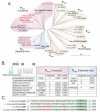
Figure 1
Figure 1
The P1B-1 ATPases display significant sequence variation. (A) Phylogenetic analysis of the P-type ATPases, including members of the P1B-1 (classical, FixI/CopA2, and histidine-rich), P1B-2, P1B-3, P1B-4, and P1B-5 subgroups; ADP1, Acinetobacter baylyi; Aea, Aquifex aeolicus; Afu, Archaeoglobus fulgidus; Ajunii, Acinetobacter junii; Alwoffii, Acinetobacter lwoffii; Anosoc, Acinetobacter nosocomialis; Apittii, Acinetobacter pittii; Atown, Acinetobacter towneri; Bme, Brucella melitensis; Bsu, Bacillus subtilis; Cgl, Corynebacterium glutamicum; Crf, Citrobacter freundii; Eco, Escherichia coli; Ehi, Enterococcus hirae; Hpy, Helicobacter pylori; Kpn, Klebsiella pneumoniae; Lla, Lactococcus lactis; LpCopA, Legionella pneumophila; Mlo, Mesorhizobium loti; Pae, Pseudomonas aeruginosa; Pat, Pectobacterium atrosepticum; Sco, Streptomyces coelicolor; Sen, Salmonella enterica; Sme, Sinorhizobium meliloti; Sth, Symbiobacterium thermophilum; Ssp, Synechocystis sp.; Sty, Salmonella enterica Typhy. All P-type ATPases from Acinetobacter are depicted in red and those functionally characterised are in a larger font. (B) Cartoon model of a P1B-1 ATPase is depicted with its eight transmembrane domains. The model serves to highlight the position of the N-terminal metal-binding domain (in green). The table presents data of the N-terminal metal-binding domain of the classical and histidine-rich P1B-1 ATPases only, showing the total number of histidine residues and the presence of the CxxC motifs. (C) The 40 amino acids at the N-terminal side of the conserved “TCPMHPEIR” domain (dark green shading) of the four histidine-rich P1B-1 ATPases are displayed (light green shading). The first transmembrane domain (TM1) starts 35 or 36 amino acids (light green shading) towards the C-terminal end from the “TCPMHPEIR” domain. All histidine residues are displayed in bold and red.
Figure 2
Figure 2
CopA is a contributor to copper resistance in A. baumannii. The growth of wild-type (WT) AB5075_UW cells (black) and the copA::T26 (burgundy), cueA::T26 (blue) and czcX::T26 (orange) mutants was determined by measuring the optical density at 600 nm (OD600) under (A) untreated conditions or (B) following supplementation of 1 mM CuSO4. The cultures were analysed at 37 °C with shaking (600 rpm), with measurements taken every 30 min. The data represent the mean (± standard error of the mean [SEM]) of at least biological triplicates.
Figure 3
Figure 3
The role of CopA in A. baumannii metal ion homeostasis. (A) To examine the total cellular copper levels, mid-log phase cells, with or without 200 µM CuSO4, were analysed by ICP-MS, with the data represented as the weight of metal (µg) per dry weight of cell material (g). (B) Cellular copper was also examined by measuring coppersensor-1 fluorescence by flow cytometry (fluorescent units corrected to unlabelled cells) in cells grown with or without 200 µM CuSO4. To examine the total cellular (C) zinc and (D) iron levels, mid-log phase cells, with or without 200 µM CuSO4, were analysed by ICP-MS, with the data represented as the weight of metal (µg) per dry weight of cell material (g). The data in all analyses represent the mean (±SEM) of at least biological triplicates. Statistical analyses were performed with a one-way ANOVA (* p < 0.05; **** p < 0.0001).
Figure 4
Figure 4
The effect of copper on oxidative stress tolerance. The growth of (A) wild-type AB5075_UW cells and (B) the copA::T26 mutant was determined by measuring the optical density at 600 nm (OD600) under untreated conditions or following supplementation of 400 µM CuSO4 and/or 60 µM paraquat (PQ). The cultures were analysed at 37 °C with shaking (600 rpm), with measurements taken every 30 min. The data represent the mean (± SEM) of at least biological triplicates.
Figure 5
Figure 5
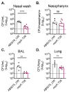
CopA aids in A. baumannii murine colonisation. Outbred Swiss mice (5-week-old females), were intranasally challenged with 2 × 10⁸ colony-forming units (CFUs) of strains AB5075_UW (black dots) or its copA::T26 derivative (burgundy dots). After 24 h, mice were euthanised and the A. baumannii cells were enumerated in (A) the nasal wash, (B) the nasopharynx, (C) the bronchoalveolar lavage (BAL), and (D) the lungs. The data represent the geometric mean with the dotted line indicating the limit of detection. Statistical analyses were performed with a Student’s t-test (ns = not significant, ** p < 0.01; **** p < 0.0001).

References

    1. Dijkshoorn L., Nemec A., Seifert H. An increasing threat in hospitals: Multidrug-resistant Acinetobacter baumannii. Nat. Rev. Microbiol. 2007;5:939–951. doi: 10.1038/nrmicro1789. - DOI - PubMed
    1. Dexter C., Murray G.L., Paulsen I.T., Peleg A.Y. Community-acquired Acinetobacter baumannii: Clinical characteristics, epidemiology and pathogenesis. Expert Rev. Anti-Infect. Ther. 2015;13:567–573. doi: 10.1586/14787210.2015.1025055. - DOI - PubMed
    1. WHO . WHO Publishes List of Bacteria for which New Antibiotics Are Urgently Needed. WHO Media Centre; Geneva, Switzerland: 2017.
    1. Eijkelkamp B.A., Stroeher U.H., Hassan K.A., Papadimitrious M.S., Paulsen I.T., Brown M.H. Adherence and motility characteristics of clinical Acinetobacter baumannii isolates. FEMS Microbiol. Lett. 2011;323:44–51. doi: 10.1111/j.1574-6968.2011.02362.x. - DOI - PubMed
    1. Giles S.K., Stroeher U.H., Eijkelkamp B.A., Brown M.H. Identification of genes essential for pellicle formation in Acinetobacter baumannii. BMC Microbiol. 2015;15:116. doi: 10.1186/s12866-015-0440-6. - DOI - PMC - PubMed

MeSH terms