

The Paroxysmal Depolarization Shift: Reconsidering Its Role in Epilepsy, Epileptogenesis and Beyond
- PMID: 30699993
- PMCID: PMC6387313
- DOI: 10.3390/ijms20030577
The Paroxysmal Depolarization Shift: Reconsidering Its Role in Epilepsy, Epileptogenesis and Beyond
Abstract
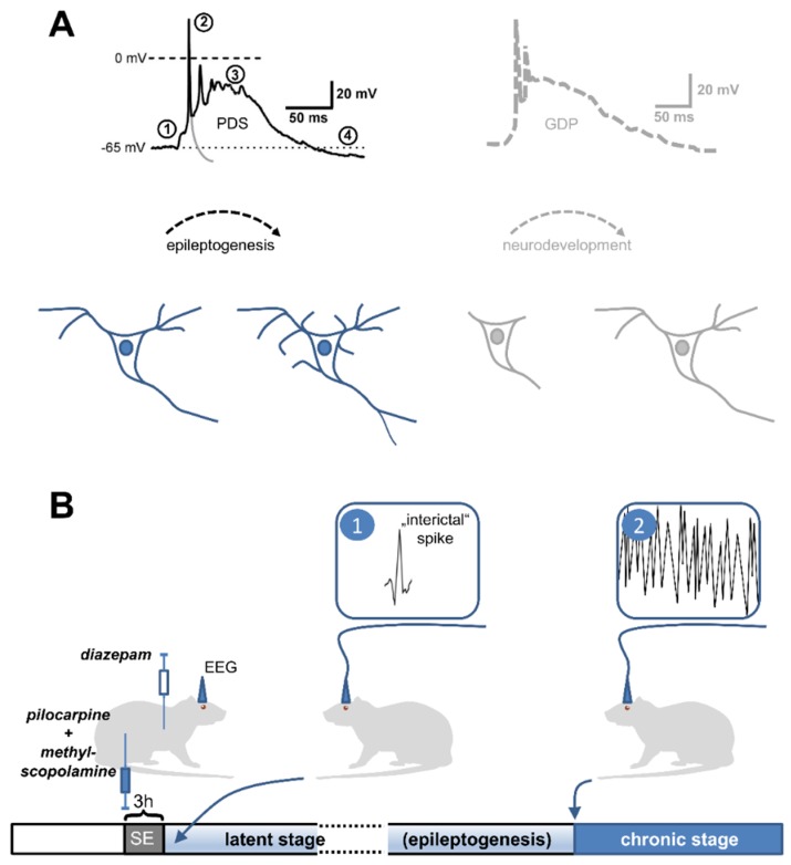
Paroxysmal depolarization shifts (PDS) have been described by epileptologists for the first time several decades ago, but controversy still exists to date regarding their role in epilepsy. In addition to the initial view of a lack of such a role, seemingly opposing hypotheses on epileptogenic and anti-ictogenic effects of PDS have emerged. Hence, PDS may provide novel targets for epilepsy therapy. Evidence for the roles of PDS has often been obtained from investigations of the multi-unit correlate of PDS, an electrographic spike termed "interictal" because of its occurrence during seizure-free periods of epilepsy patients. Meanwhile, interictal spikes have been found to be associated with neuronal diseases other than epilepsy, e.g., Alzheimer's disease, which may indicate a broader implication of PDS in neuropathologies. In this article, we give an introduction to PDS and review evidence that links PDS to pro- as well as anti-epileptic mechanisms, and to other types of neuronal dysfunction. The perturbation of neuronal membrane voltage and of intracellular Ca2+ that comes with PDS offers many conceivable pathomechanisms of neuronal dysfunction. Out of these, the operation of L-type voltage-gated calcium channels, which play a major role in coupling excitation to long-lasting neuronal changes, is addressed in detail.
Keywords: Alzheimer’s disease; L-type voltage-gated calcium channels; dendrites; electrophysiology; giant depolarizing potentials; hippocampal neurons; neuronal dysfunction; neuronal remodelling; seizures.
Conflict of interest statement
The authors declare no conflict of interest.
Figures
Similar articles
-
The paroxysmal depolarization shift in epilepsy research.Int J Biochem Cell Biol. 2019 Feb;107:77-81. doi: 10.1016/j.biocel.2018.12.006. Epub 2018 Dec 14. Int J Biochem Cell Biol. 2019. PMID: 30557621 Free PMC article. Review.
-
On the Origin of Paroxysmal Depolarization Shifts: The Contribution of Cav1.x Channels as the Common Denominator of a Polymorphous Neuronal Discharge Pattern.Neuroscience. 2021 Aug 1;468:265-281. doi: 10.1016/j.neuroscience.2021.05.011. Epub 2021 May 18. Neuroscience. 2021. PMID: 34015369
-
Cav 1.3 channels play a crucial role in the formation of paroxysmal depolarization shifts in cultured hippocampal neurons.Epilepsia. 2017 May;58(5):858-871. doi: 10.1111/epi.13719. Epub 2017 Mar 11. Epilepsia. 2017. PMID: 28295232 Free PMC article.
-
Inter-ictal- and ictal-like epileptic discharges in the dendritic tree of neocortical pyramidal neurons.J Neurophysiol. 2002 Dec;88(6):2954-62. doi: 10.1152/jn.00525.2001. J Neurophysiol. 2002. PMID: 12466421
-
The Paroxysmal Depolarizing Shift: The First Cellular Marker of Focal Epileptogenesis.In: Noebels JL, Avoli M, Rogawski MA, Vezzani A, Delgado-Escueta AV, editors. Jasper's Basic Mechanisms of the Epilepsies. 5th edition. New York: Oxford University Press; 2024. Chapter 1. In: Noebels JL, Avoli M, Rogawski MA, Vezzani A, Delgado-Escueta AV, editors. Jasper's Basic Mechanisms of the Epilepsies. 5th edition. New York: Oxford University Press; 2024. Chapter 1. PMID: 39637141 Free Books & Documents. Review.
Cited by
-
An ictogenic marker in the mesial temporal epilepsy and its temporal evolutionary features.Front Neurol. 2025 Jan 10;15:1510108. doi: 10.3389/fneur.2024.1510108. eCollection 2024. Front Neurol. 2025. PMID: 39866518 Free PMC article.
-
Novel test strategies for in vitro seizure liability assessment.Expert Opin Drug Metab Toxicol. 2021 Aug;17(8):923-936. doi: 10.1080/17425255.2021.1876026. Epub 2021 Feb 17. Expert Opin Drug Metab Toxicol. 2021. PMID: 33595380 Free PMC article. Review.
-
Utilizing machine learning algorithms to predict subject genetic mutation class from in silico models of neuronal networks.BMC Med Inform Decis Mak. 2022 Nov 9;22(1):290. doi: 10.1186/s12911-022-02038-7. BMC Med Inform Decis Mak. 2022. PMID: 36352381 Free PMC article.
-
Mitochondrial Glutamine Metabolism Drives Epileptogenesis in Primary Hippocampal Neurons.J Neurosci. 2025 May 21;45(21):e0110252025. doi: 10.1523/JNEUROSCI.0110-25.2025. J Neurosci. 2025. PMID: 40228896
-
Extract of Euterpe oleracea Martius Stone Presents Anticonvulsive Activity via the GABAA Receptor.Front Cell Neurosci. 2022 May 11;16:872743. doi: 10.3389/fncel.2022.872743. eCollection 2022. Front Cell Neurosci. 2022. PMID: 35634465 Free PMC article.
References
Publication types
MeSH terms
Substances
Grants and funding
LinkOut - more resources
Full Text Sources
Medical
Miscellaneous
