

Sirtuin 6 overexpression relieves sepsis-induced acute kidney injury by promoting autophagy
- PMID: 30700227
- PMCID: PMC6422466
- DOI: 10.1080/15384101.2019.1568746
Sirtuin 6 overexpression relieves sepsis-induced acute kidney injury by promoting autophagy
Abstract
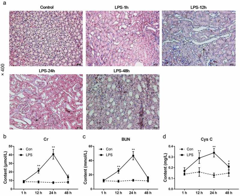
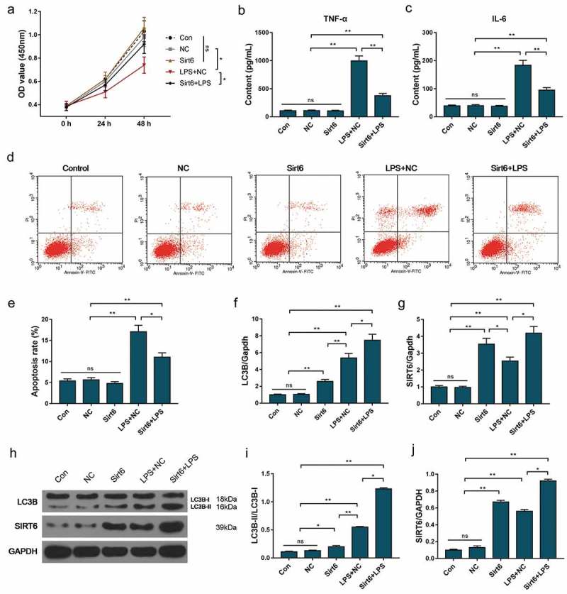
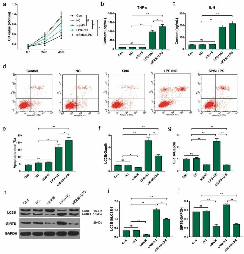
Sirtuin 6 (SIRT6) has the function of regulating autophagy. The aim of this study was to investigate the mechanism through which SIRT6 relieved acute kidney injury (AKI) caused by sepsis. The AKI model was established with lipopolysaccharides (LPS) using mice. Hematoxylin-eosin (HE) staining and streptavidin-perosidase (SP) staining was used to observe kidney tissue and test SIRT6 and LC3B proteins in kidney. Enzyme-linked immunosorbent assay (ELISA) was performed to detected the tumor necrosis factor-α (TNF-α) and interleukin-6 (IL-6) concentrations. Cell counting kit-8 (CCK-8) assay and flow cytometry were carried out to test the cell viability and apoptosis rate respectively. Protein and mRNA were determined by Western blot and quantitative real-time polymerase chain reaction (qRT-PCR). AKI induced by LPS had self-repairing ability. At 12 h after modeling, the expression levels of TNF-α, IL-6, SIRT6 and LC3B-II/LC3B-I were first significantly increased and were then significantly decreased at 48 h after modeling. LPS inhibited the growth of HK-2 cells and promoted the expressions of TNF-α, IL-6, SIRT6 and LC3B. Overexpression of SIRT6 down-regulated the secretion of TNF-α and IL-6 induced by LPS. SIRT6 overexpression inhibited apoptosis induced by LPS and promoted autophagy in HK-2 cells. Silencing of the SIRT6 gene not only promoted the secretion of TNF-α and IL-6 by HK-2 cells, but also promoted apoptosis and reduced autophagy. LPS up-regulated the expression of SIRT6 gene in HK-2 cells. Overexpression of the SIRT6 gene could inhibit apoptosis and induce autophagy, which might be involved in repairing kidney damage caused by LPS.
Keywords: Sirtuin 6; acute kidney injury; apoptosis; autophagy.
Figures


Similar articles
-
[Scutellarin alleviates lipopolysaccharide-induced renal injury via mediating cysteine-rich protein 61-connective tissue growth factor-nephroblastoma overexpressed gene 1 expression to inhibit nuclear factor-κB signaling pathway].Zhonghua Wei Zhong Bing Ji Jiu Yi Xue. 2022 Apr;34(4):400-406. doi: 10.3760/cma.j.cn121430-20210401-00767. Zhonghua Wei Zhong Bing Ji Jiu Yi Xue. 2022. PMID: 35692206 Chinese.
-
[Protective effect and mechanism of Ribociclib on sepsis induced-acute kidney injury].Zhonghua Wei Zhong Bing Ji Jiu Yi Xue. 2020 Feb;32(2):204-209. doi: 10.3760/cma.j.cn121430-20200108-00038. Zhonghua Wei Zhong Bing Ji Jiu Yi Xue. 2020. PMID: 32275007 Chinese.
-
The Effect of Overexpression of the Enhancer of Zeste Homolog 1 (EZH1) Gene on Aristolochic Acid-Induced Injury in HK-2 Human Kidney Proximal Tubule Cells In Vitro.Med Sci Monit. 2019 Jan 28;25:801-810. doi: 10.12659/MSM.911611. Med Sci Monit. 2019. PMID: 30688289 Free PMC article.
-
Epigenetic dysregulation of autophagy in sepsis-induced acute kidney injury: the underlying mechanisms for renoprotection.Front Immunol. 2023 May 5;14:1180866. doi: 10.3389/fimmu.2023.1180866. eCollection 2023. Front Immunol. 2023. PMID: 37215112 Free PMC article. Review.
-
Sirtuins and their influence on autophagy.J Cell Biochem. 2024 Nov;125(11):e30377. doi: 10.1002/jcb.30377. Epub 2023 Feb 6. J Cell Biochem. 2024. PMID: 36745668 Review.
Cited by
-
Polydatin Attenuates Cisplatin-Induced Acute Kidney Injury via SIRT6-Mediated Autophagy Activation.Oxid Med Cell Longev. 2022 Sep 16;2022:9035547. doi: 10.1155/2022/9035547. eCollection 2022. Oxid Med Cell Longev. 2022. PMID: 36160707 Free PMC article.
-
Sepsis-induced AKI: From pathogenesis to therapeutic approaches.Front Pharmacol. 2022 Sep 15;13:981578. doi: 10.3389/fphar.2022.981578. eCollection 2022. Front Pharmacol. 2022. PMID: 36188562 Free PMC article. Review.
-
Circ_001653 alleviates sepsis associated-acute kidney injury by recruiting BUD13 to regulate KEAP1/NRF2/HO-1 signaling pathway.J Inflamm (Lond). 2024 Sep 17;21(1):37. doi: 10.1186/s12950-024-00409-7. J Inflamm (Lond). 2024. PMID: 39289683 Free PMC article.
-
USP10 alleviates sepsis-induced acute kidney injury by regulating Sirt6-mediated Nrf2/ARE signaling pathway.J Inflamm (Lond). 2021 Aug 19;18(1):25. doi: 10.1186/s12950-021-00291-7. J Inflamm (Lond). 2021. PMID: 34412625 Free PMC article.
-
Sirtuin 6 is a key contributor to gender differences in acute kidney injury.Cell Death Discov. 2023 Apr 25;9(1):134. doi: 10.1038/s41420-023-01432-y. Cell Death Discov. 2023. PMID: 37185276 Free PMC article.
References
-
- Dellinger RP, Levy MM, Rhodes A, et al. Surviving sepsis campaign: international guidelines for management of severe sepsis and septic shock: 2012. Crit Care Med. 2013. February;41(2):580–637. PubMed PMID: 23353941; eng. - PubMed
-
- Rhodes A, Evans LE, Alhazzani W, et al. Surviving Sepsis campaign: international guidelines for management of sepsis and septic shock: 2016. Crit Care Med. 2017. March;45(3):486–552. PubMed PMID: 28098591; eng. - PubMed
-
- Sossdorf M, Otto GP, Menge K, et al. Potential effect of physiotherapeutic treatment on mortality rate in patients with severe sepsis and septic shock: a retrospective cohort analysis. J Crit Care. 2013. December;28(6):954–958. PubMed PMID: 23958242; eng. - PubMed
-
- Poukkanen M, Wilkman E, Vaara ST, et al. Hemodynamic variables and progression of acute kidney injury in critically ill patients with severe sepsis: data from the prospective observational FINNAKI study. Crit Care. 2013. December 13;17(6):R295 PubMed PMID: 24330815; PubMed Central PMCID: PMCPMC4056430. eng. - PMC - PubMed
Publication types
MeSH terms
Substances
LinkOut - more resources
Full Text Sources
Medical