

Zebrafish duox mutations provide a model for human congenital hypothyroidism
- PMID: 30700401
- PMCID: PMC6398463
- DOI: 10.1242/bio.037655
Zebrafish duox mutations provide a model for human congenital hypothyroidism
Abstract
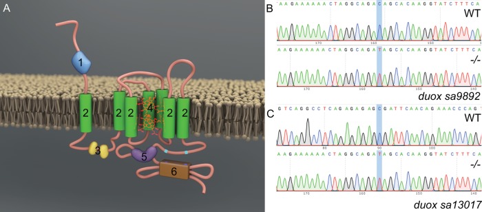
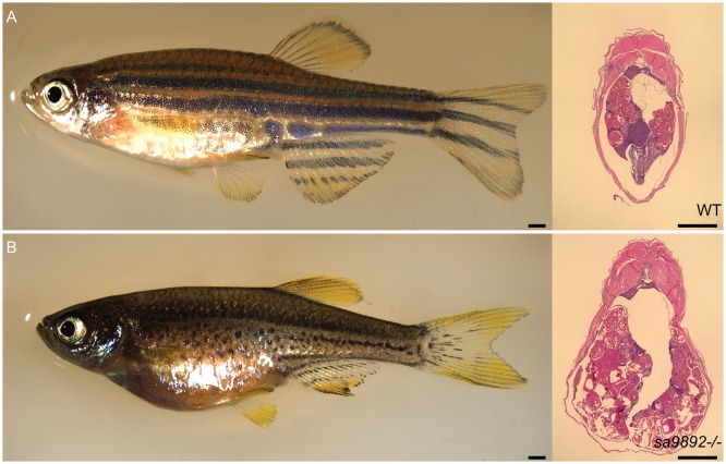
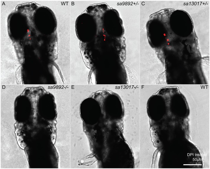
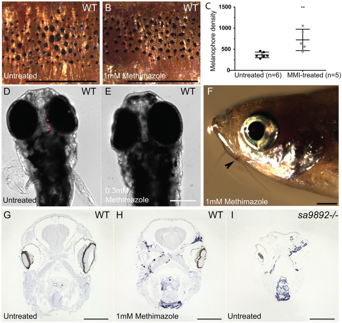
Thyroid dyshormonogenesis is a leading cause of congenital hypothyroidism, a highly prevalent but treatable condition. Thyroid hormone (TH) synthesis is dependent on the formation of reactive oxygen species (ROS). In humans, the primary sources for ROS production during thyroid hormone synthesis are the NADPH oxidases DUOX1 and DUOX2. Indeed, mutations in DUOX1 and DUOX2 have been linked with congenital hypothyroidism. Unlike humans, zebrafish has a single orthologue for DUOX1 and DUOX2 In this study, we investigated the phenotypes associated with two nonsense mutant alleles, sa9892 and sa13017, of the single duox gene in zebrafish. Both alleles gave rise to readily observable phenotypes reminiscent of congenital hypothyroidism, from the larval stages through to adulthood. By using various methods to examine external and internal phenotypes, we discovered a strong correlation between TH synthesis and duox function, beginning from an early larval stage, when T4 levels are already noticeably absent in the mutants. Loss of T4 production resulted in growth retardation, pigmentation defects, ragged fins, thyroid hyperplasia/external goiter and infertility. Remarkably, all of these defects associated with chronic congenital hypothyroidism could be rescued with T4 treatment, even when initiated when the fish had already reached adulthood. Our work suggests that these zebrafish duox mutants may provide a powerful model to understand the aetiology of untreated and treated congenital hypothyroidism even in advanced stages of development.This article has an associated First Person interview with the first author of the paper.
Keywords: Congenital hypothyroidism; Growth retardation; Infertility; Thyroid.
© 2019. Published by The Company of Biologists Ltd.
Conflict of interest statement
Competing interestsThe authors declare no competing or financial interests.
Figures


Similar articles
-
Genetic Manipulation on Zebrafish duox Recapitulate the Clinical Manifestations of Congenital Hypothyroidism.Endocrinology. 2021 Aug 1;162(8):bqab101. doi: 10.1210/endocr/bqab101. Endocrinology. 2021. PMID: 34019632
-
DUOX Defects and Their Roles in Congenital Hypothyroidism.Methods Mol Biol. 2019;1982:667-693. doi: 10.1007/978-1-4939-9424-3_37. Methods Mol Biol. 2019. PMID: 31172499 Review.
-
New phenotypes in thyroid dyshormonogenesis: hypothyroidism due to DUOX2 mutations.Endocr Dev. 2007;10:99-117. doi: 10.1159/000106822. Endocr Dev. 2007. PMID: 17684392 Review.
-
Inhibition of the thyroid hormonogenic H2O2 production by Duox/DuoxA in zebrafish reveals VAS2870 as a new goitrogenic compound.Mol Cell Endocrinol. 2020 Jan 15;500:110635. doi: 10.1016/j.mce.2019.110635. Epub 2019 Oct 31. Mol Cell Endocrinol. 2020. PMID: 31678421
-
Conformation of the N-Terminal Ectodomain Elicits Different Effects on DUOX Function: A Potential Impact on Congenital Hypothyroidism Caused by a H2O2 Production Defect.Thyroid. 2018 Aug;28(8):1052-1062. doi: 10.1089/thy.2017.0596. Epub 2018 Jul 24. Thyroid. 2018. PMID: 29845893
Cited by
-
Thyroid hormone regulates proximodistal patterning in fin rays.Proc Natl Acad Sci U S A. 2023 May 23;120(21):e2219770120. doi: 10.1073/pnas.2219770120. Epub 2023 May 15. Proc Natl Acad Sci U S A. 2023. PMID: 37186843 Free PMC article.
-
Duox is the primary NADPH oxidase responsible for ROS production during adult caudal fin regeneration in zebrafish.iScience. 2023 Feb 4;26(3):106147. doi: 10.1016/j.isci.2023.106147. eCollection 2023 Mar 17. iScience. 2023. PMID: 36843843 Free PMC article.
-
Effect of Thyroperoxidase and Deiodinase Inhibition on Anterior Swim Bladder Inflation in the Zebrafish.Environ Sci Technol. 2020 May 19;54(10):6213-6223. doi: 10.1021/acs.est.9b07204. Epub 2020 Apr 29. Environ Sci Technol. 2020. PMID: 32320227 Free PMC article.
-
The RNA helicase Ddx52 functions as a growth switch in juvenile zebrafish.Development. 2021 Aug 1;148(15):dev199578. doi: 10.1242/dev.199578. Epub 2021 Jul 29. Development. 2021. PMID: 34323273 Free PMC article.
-
Thyroid hormones regulate the formation and environmental plasticity of white bars in clownfishes.Proc Natl Acad Sci U S A. 2021 Jun 8;118(23):e2101634118. doi: 10.1073/pnas.2101634118. Proc Natl Acad Sci U S A. 2021. PMID: 34031155 Free PMC article.
References
Grants and funding
LinkOut - more resources
Full Text Sources
Molecular Biology Databases
