. 2019 Feb 11;8(2):bio036590.
doi: 10.1242/bio.036590.

Microtubule stabilizer reveals requirement of Ca2+-dependent conformational changes of microtubules for rapid coiling of haptonema in haptophyte algae

Affiliations

Microtubule stabilizer reveals requirement of Ca2+-dependent conformational changes of microtubules for rapid coiling of haptonema in haptophyte algae

Mami Nomura et al. Biol Open. .

Abstract

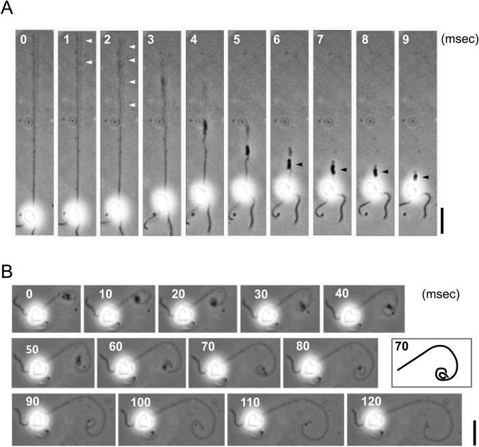
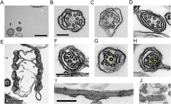
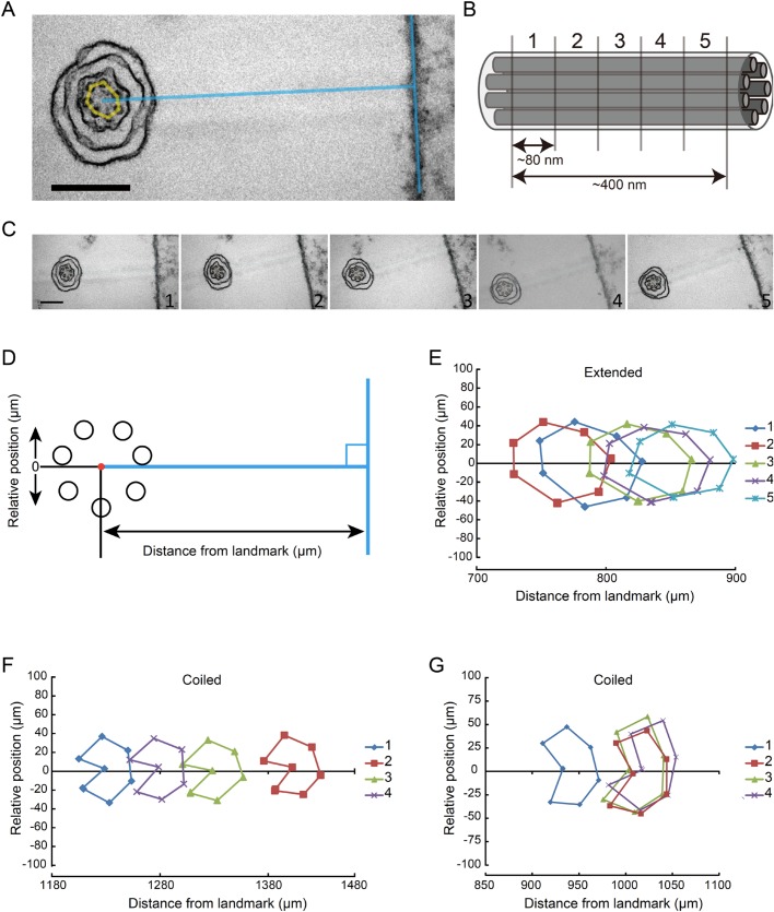
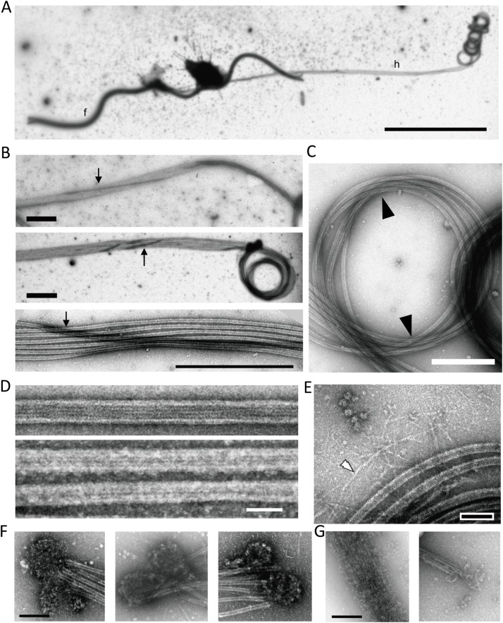
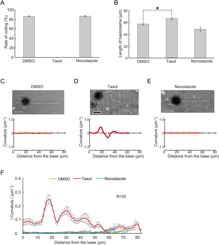
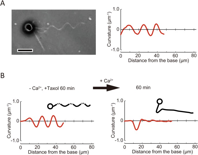
A haptonema is an elongated microtubule-based motile organelle uniquely present in haptophytes. The most notable and rapid movement of a haptonema is 'coiling', which occurs within a few milliseconds following mechanical stimulation in an unknown motor-independent mechanism. Here, we analyzed the coiling process in detail by high-speed filming and showed that haptonema coiling was initiated by left-handed twisting of the haptonema, followed by writhing to form a helix from the distal tip. On recovery from a mechanical stimulus, the helix slowly uncoiled from the proximal region. Electron microscopy showed that the seven microtubules in a haptonema were arranged mostly in parallel but that one of the microtubules often wound around the others in the extended state. A microtubule stabilizer, paclitaxel, inhibited coiling and induced right-handed twisting of the haptonema in the absence of Ca2+, suggesting changes in the mechanical properties of microtubules. Addition of Ca2+ resulted in the conversion of haptonematal twist into the planar bends near the proximal region. These results indicate that switching microtubule conformation, possibly with the aid of Ca2+-binding microtubule-associated proteins is responsible for rapid haptonematal coiling.

Keywords: Chrysochromulina; Curvature; Haptonema; Haptophyte; Microtubule; Paclitaxel.


Conflict of interest statement

Competing interestsThe authors declare no competing or financial interests.

Figures

Fig. 1.
Fig. 1.
High-speed analysis of haptonematal coiling and uncoiling. (A) Coiling process of a haptonema. High speed images recorded at 1000 fps. Gentle helices that initially formed in the distal half of a haptonema are indicated by white arrowheads. Stacked coils are indicated by black arrowheads. Scale bar: 20 µm. (B) Uncoiling process of a haptonema. High speed images recorded at 200 fps. Scale bar: 20 µm. The inset represents the trace of the haptonema at 70 ms for clarification of the coiling direction.
Fig. 2.
Fig. 2.
Thin-section images of extended and coiled haptonemata. (A) Extended haptonema at low magnification. Scale bar: 500 nm. (B–D) High magnification images of an extended haptonema. Scale bar: 100 nm. (E) Coiled haptonema at low magnification. Scale bar: 500 nm. (F–H) High magnification images of a coiled haptonema. Dashed yellow circles show a microtubule invaginated into the center. Scale bar: 100 nm. (I,J) Longitudinal images of extended (I) and coiled (J) haptonemata. Scale bar: 500 nm.
Fig. 3.
Fig. 3.
Arrangement of microtubules along a haptonema. (A) Extended haptonema adjacent to the poly-lysine-BSA coated Epon surface (landmark). To position each microtubule, a line was drawn from the center of the microtubule bundle at right angles to the surface. Scale bar: 200 nm. (B) Approximately 80 nm sequential sections were made. For example, five sections covered a 400 nm length of a haptonema along the longitudinal axis. (C) Example of sequential images of an extended haptonema. Scale bar: 200 nm. (D) Definition of the distance from the landmark and the relative position of each microtubule. (E) Typical plot of microtubule positions in five sequential images of an extended haptonema. (F) Typical plot of microtubule positions in five sequential images of a coiled haptonema. (G) Plot showing a helical arrangement of the seven microtubules in a coiled haptonema. Four out of 24 sets of sequential images of the coiled haptonema showed this pattern.
Fig. 4.
Fig. 4.
Negative stain images of extended and coiled haptonemata. (A) Negative stain image of a haptophyte. Because of demembranation, most parts of the cell body are removed. f, flagellum; h, haptonema. A long haptonema is observed on the right side with the tip coiled. Scale bar: 10 µm. (B) Three images showing that one, occasionally two microtubules (arrows), wind around the other microtubules in the extended region. Scale bar: 1 µm. (C) Coiled region. Microtubules are mostly parallel to each other, but there seems to be a twist with microtubules crossed over at the two opposite positions of the coil (arrowhead). Scale bar: 500 nm. (D) Microtubules polymerized from purified brain tubulin (upper panel) and microtubules in a demembranated haptonema (lower panel). Scale bar: 50 nm. (E) Image showing filamentous structures (white arrowhead) emanating from the microtubule bundle. Bar, 100 nm. (F) Mass of small particles observed at the distal tip of a haptonema. They are frequently observed in a pair. Scale bar: 200 nm. (G) Depolymerization of microtubules occasionally observed in demembranated haptonemata. The depolymerized microtubules are still bundled probably because of MAPs. The morphology of the small particles in F is similar to depolymerized tubulin dimers. Scale bar: 200 nm.
Fig. 5.
Fig. 5.
Effects of drugs that affect microtubules on haptonematal coiling. Haptophytes were suspended in artificial sea water containing 9.18 mM CaCl2 in the presence of drugs. After 60 min, haptonematal coiling was induced by tapping of the microscopic stage. (A) Rates of coiling in the presence of taxol (20 μM) or nocodazole (20 μM). Error bars show the standard deviation. N=5. (B) Length of haptonemata at 60 min after treatment with each drug. Error bars show the standard deviation. N=5. The asterisk represents that the difference is significant at P<0.01 (Student's t-test). (C–E) Effects of each drug on the curvature along the axis of a haptonema. The curvature was measured as described in Fig. S5, from the phase-contrast images shown above each plot. Scale bar: 10 µm. (F) Absolute values of the curvature at 60 min after incubation, showing the extent of bending along the haptonema. Error bars show the standard deviation. N=20.
Fig. 6.
Fig. 6.
Effects of Ca2+ on taxol-induced haptonematal bending. (A) A phase contrast image of a haptophyte treated with taxol (20 μM) in the absence of Ca2+ (CFSW+EGTA+BAPTA-AM) showing a helical configuration of the haptonema (left). The plot to the right shows the curvature along the haptonema (see Fig. S5). The curvature of the haptonema was calculated from the microscopic image, which is a two-dimensionally projected image. The curvature of a helical structure calculated in this way shows oscillation with a repeat distance equivalent to that of the helix, and a shift in the pattern represents propagation of the wave form. Scale bar: 10 µm. (B) Ca2+-dependent propagation of a taxol-induced haptonematal helix. The helix is also converted to planar bending with the maximum curvature of bend propagating to the proximal region. Trace images of haptophytes are shown as insets.

Similar articles

Cited by

  • A Novel Single-Domain Na+-Selective Voltage-Gated Channel in Photosynthetic Eukaryotes.
    Helliwell KE, Chrachri A, Koester JA, Wharam S, Taylor AR, Wheeler GL, Brownlee C. Helliwell KE, et al. Plant Physiol. 2020 Dec;184(4):1674-1683. doi: 10.1104/pp.20.00889. Epub 2020 Oct 1. Plant Physiol. 2020. PMID: 33004614 Free PMC article.
  • Tree of motility - A proposed history of motility systems in the tree of life.
    Miyata M, Robinson RC, Uyeda TQP, Fukumori Y, Fukushima SI, Haruta S, Homma M, Inaba K, Ito M, Kaito C, Kato K, Kenri T, Kinosita Y, Kojima S, Minamino T, Mori H, Nakamura S, Nakane D, Nakayama K, Nishiyama M, Shibata S, Shimabukuro K, Tamakoshi M, Taoka A, Tashiro Y, Tulum I, Wada H, Wakabayashi KI. Miyata M, et al. Genes Cells. 2020 Jan;25(1):6-21. doi: 10.1111/gtc.12737. Genes Cells. 2020. PMID: 31957229 Free PMC article. Review.
  • Origins of eukaryotic excitability.
    Wan KY, Jékely G. Wan KY, et al. Philos Trans R Soc Lond B Biol Sci. 2021 Mar 15;376(1820):20190758. doi: 10.1098/rstb.2019.0758. Epub 2021 Jan 25. Philos Trans R Soc Lond B Biol Sci. 2021. PMID: 33487111 Free PMC article. Review.

References

    1. Amos W. B., Routledge L. M. and Yew F. F. (1975). Calcium-binding proteins in a Vorticellid contractile organelle. J. Cell Sci. 19, 203-213. - PubMed
    1. Andersen R. A. (2004). Biology and systematics of heterokont and haptophyte algae. Am. J. Bot. 91, 1508-1522. 10.3732/ajb.91.10.1508 - DOI - PubMed
    1. Anderson R. A. and Kawachi M. (2005). Traditional microalgae isolation techniques. In Algal Culturing Techniques (ed. Anderson R. A.), pp. 83-100. Amsterdam: Elsevier Academic Press.
    1. Arnal I. and Wade R. H. (1995). How does taxol stabilize microtubules? Curr. Biol. 5, 900-908. 10.1016/S0960-9822(95)00180-1 - DOI - PubMed
    1. Asai H., Ochiai T., Fukui K., Watanabe M. and Kano F. (1978). Improved preparation and cooperative calcium contraction of glycerinated Vorticella. J. Biochem. (Tokyo) 83, 795-798. 10.1093/oxfordjournals.jbchem.a131974 - DOI - PubMed