

Cavernous sinus thrombosis due to ipsilateral sphenoid sinusitis
- PMID: 30700458
- PMCID: PMC6352844
- DOI: 10.1136/bcr-2018-227302
Cavernous sinus thrombosis due to ipsilateral sphenoid sinusitis
Abstract
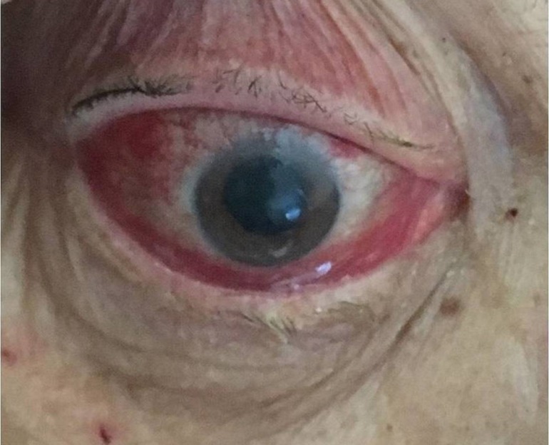
We report a case of septic thrombosis of the right cavernous sinus in a diabetic woman in her late 70's due to ipsilateral sphenoid sinusitis. The diagnosis was delayed and made only after the abrupt and dramatic appearance of the manifestations of sinus thrombosis. The patient developed, among the other symptoms, right peripheral facial palsy, which is a very rare manifestation in cavernous sinus thrombosis (CST). She was treated with broad-spectrum antibiotics and enoxaparin. The day of the scheduled drainage of sphenoid sinus-24 hours after the initiation of anticoagulation-she developed fatal subarachnoid haemorrhage. Our case demonstrates the difficulty of timely diagnosis of acute sphenoid sinusitis which has emerged as the most common primary infectious source potentially leading in CST. It also underscores the uncertainty concerning the use of anticoagulation in cerebral sinus thrombosis of infectious origin.
Keywords: cranial nerves; ear, nose and throat/otolaryngology; infection (neurology); neuroimaging.
© BMJ Publishing Group Limited 2019. No commercial re-use. See rights and permissions. Published by BMJ.
Conflict of interest statement
Competing interests: None declared.
Figures


Similar articles
-
Cavernous sinus and jugular thromboses, base of skull osteomyelitis and cranial nerve palsies: catastrophic complications of sphenoid sinusitis.BMJ Case Rep. 2023 Feb 2;16(2):e253496. doi: 10.1136/bcr-2022-253496. BMJ Case Rep. 2023. PMID: 36731941 Free PMC article.
-
Acute sphenoid sinusitis leading to contralateral cavernous sinus thrombosis: a case report.J Laryngol Otol. 2013 Aug;127(8):814-6. doi: 10.1017/S0022215113001527. Epub 2013 Jul 24. J Laryngol Otol. 2013. PMID: 23883649 Review.
-
A review of eight cases of cavernous sinus thrombosis secondary to sphenoid sinusitis, including a12-year-old girl at the present department.Infect Dis (Lond). 2017 Sep;49(9):641-646. doi: 10.1080/23744235.2017.1331465. Epub 2017 May 23. Infect Dis (Lond). 2017. PMID: 28535728 Review.
-
Cavernous sinus thrombosis caused by contralateral sphenoid sinusitis: a case report.Head Face Med. 2013 Mar 13;9:9. doi: 10.1186/1746-160X-9-9. Head Face Med. 2013. PMID: 23497466 Free PMC article.
-
Cavernous sinus thrombosis secondary to contralateral sphenoid sinusitis: a diagnostic challenge.J Laryngol Otol. 2010 Aug;124(8):928-30. doi: 10.1017/S0022215110000113. Epub 2010 Mar 5. J Laryngol Otol. 2010. PMID: 20202275
Cited by
-
Understanding Infection-Induced Thrombosis: Lessons Learned From Animal Models.Front Immunol. 2019 Nov 5;10:2569. doi: 10.3389/fimmu.2019.02569. eCollection 2019. Front Immunol. 2019. PMID: 31749809 Free PMC article. Review.
-
Isolated Fungal Sphenoid Sinusitis With Cavernous Sinus Thrombophlebitis: A Case Report.Cureus. 2022 May 16;14(5):e25034. doi: 10.7759/cureus.25034. eCollection 2022 May. Cureus. 2022. PMID: 35719783 Free PMC article.
-
A Rare Intersection of Septic Cavernous Sinus Thrombosis and Subarachnoid Hemorrhage: Insights from the Case of a 70-Year-Old Patient.Medicina (Kaunas). 2024 Feb 1;60(2):253. doi: 10.3390/medicina60020253. Medicina (Kaunas). 2024. PMID: 38399541 Free PMC article.
-
Bridging the Gap between Ophthalmology and Emergency Medicine in Community-Based Emergency Departments (EDs): A Neuro-Ophthalmology Guide for ED Practitioners.Clin Pract. 2021 Dec 2;11(4):919-932. doi: 10.3390/clinpract11040106. Clin Pract. 2021. PMID: 34940005 Free PMC article.
-
Case report: 18F-FDG PET confirmed pupil-sparing third nerve palsy heralding aseptic cavernous sinus embolism in patient with chest malignancy.Front Surg. 2022 Aug 31;9:893651. doi: 10.3389/fsurg.2022.893651. eCollection 2022. Front Surg. 2022. PMID: 36117807 Free PMC article.
References
Publication types
MeSH terms
LinkOut - more resources
Full Text Sources