. 2019 Apr;211(4):1315-1330.
doi: 10.1534/genetics.119.301970. Epub 2019 Jan 30.

Necessity and Contingency in Developmental Genetic Screens: EGF, Wnt, and Semaphorin Pathways in Vulval Induction of the Nematode Oscheius tipulae

Affiliations

Necessity and Contingency in Developmental Genetic Screens: EGF, Wnt, and Semaphorin Pathways in Vulval Induction of the Nematode Oscheius tipulae

Amhed M Vargas-Velazquez et al. Genetics. 2019 Apr.

Abstract

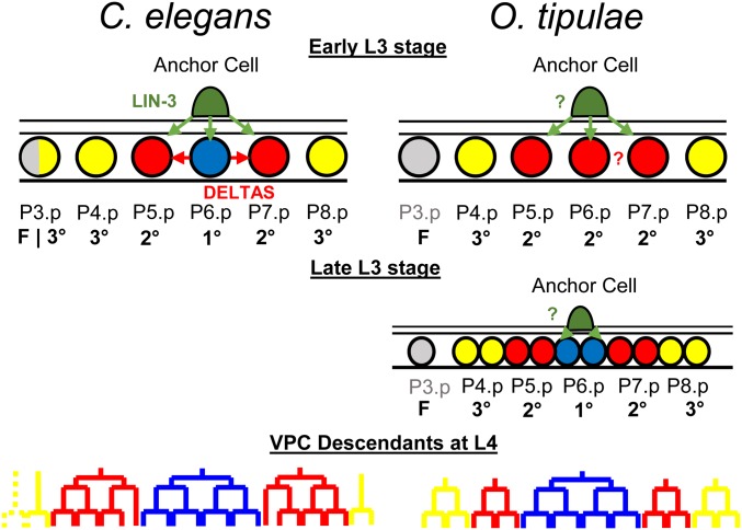
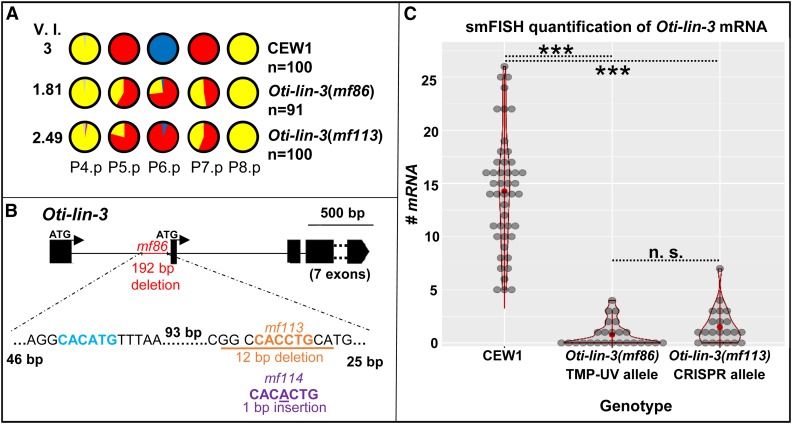
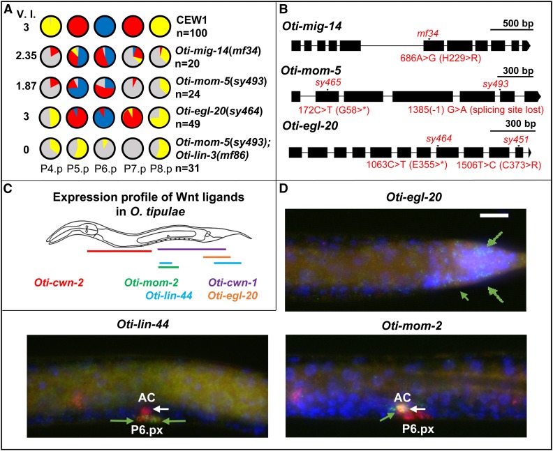
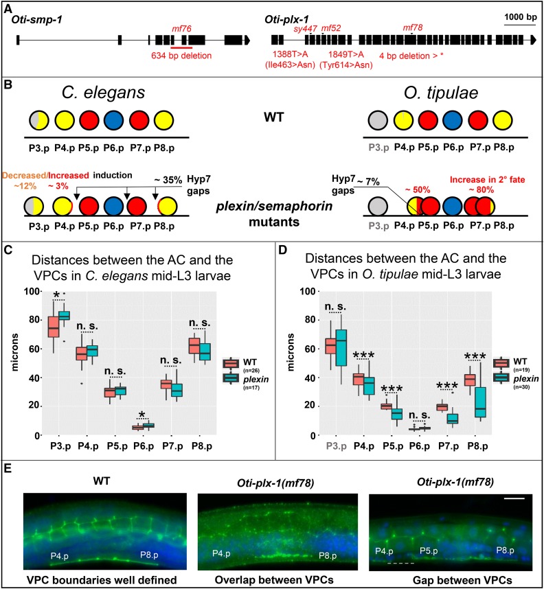
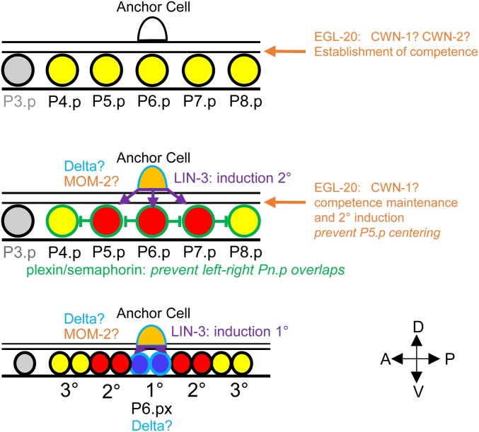
Genetic screens in the nematode Caenorhabditis elegans identified the EGF/Ras and Notch pathways as central for vulval precursor cell fate patterning. Schematically, the anchor cell secretes EGF, inducing the P6.p cell to a primary (1°) vulval fate; P6.p in turn induces its neighbors to a secondary (2°) fate through Delta-Notch signaling and represses Ras signaling. In the nematode Oscheius tipulae, the anchor cell successively induces 2° then 1° vulval fates. Here, we report on the molecular identification of mutations affecting vulval induction in O. tipulae A single Induction Vulvaless mutation was found, which we identify as a cis-regulatory deletion in a tissue-specific enhancer of the O. tipulae lin-3 homolog, confirmed by clustered regularly interspaced short palindromic repeats/Cas9 mutation. In contrast to this predictable Vulvaless mutation, mutations resulting in an excess of 2° fates unexpectedly correspond to the plexin/semaphorin pathway. Hyperinduction of P4.p and P8.p in these mutants likely results from mispositioning of these cells due to a lack of contact inhibition. The third signaling pathway found by forward genetics in O. tipulae is the Wnt pathway; a decrease in Wnt pathway activity results in loss of vulval precursor competence and induction, and 1° fate miscentering on P5.p. Our results suggest that the EGF and Wnt pathways have qualitatively similar activities in vulval induction in C. elegans and O. tipulae, albeit with quantitative differences in the effects of mutation. Thus, the derived induction process in C. elegans with an early induction of the 1° fate appeared during evolution, after the recruitment of the EGF pathway for vulval induction.

Keywords: C. elegans; O. tipulae; Wnt; evolution of development; genetic screens; lin-3/EGF; plexin; semaphorin.


Figures

Figure 1
Figure 1
Vulval cell fate patterning in C. elegans and O. tipulae. In the third larval stage (L3) of C. elegans, a cell from the somatic gonad known as the anchor cell (AC) produces an EGF-like inductive signal (LIN-3, green arrows) that activates the Ras pathway in the central vulval precursor cells (VPCs). High Ras signaling promotes the primary (1°) fate (blue circle) in P6.p, which, in turn, produces Deltas (red arrows) that induce a secondary (2°) fate (red circle), and represses the 1° fate in P5.p and P7.p. Both fates prevent the formation of nonspecialized epidermis [tertiary (3°) fate, yellow circles]. Only VPCs with 1° or 2° fates will give rise to the cells that will form the vulva (bottom). P3.p is not competent to acquire a vulval cell fate (gray) in O. tipulae. Unlike in C. elegans, the AC of O. tipulae has been shown to be required to induce the 1° fate in P6.p descendants after VPC division. While a similar vulval cell fate pattern is conserved between the two nematodes, the cell division patterns of the 2° and 3° fates are different.
Figure 2
Figure 2
Cis-regulatory lesions in Oti-lin-3/EGF cause hypoinduction of primary (1°) and secondary (2°) vulval cell fates. (A) P(4–8) cell fates in the wild-type CEW1 O. tipulae reference strain and mutants for lin-3/EGF. The pie diagrams represent the percentage of cell fates over individuals. Yellow, red, and blue are for the tertiary (3°), 2°, and 1° fates, respectively. Gray denotes an undivided cell fused to the hypodermis. The Vulva Index (V.I.) is calculated as the average number of cells acquiring a vulval cell fate in a set of animals. The quantifications of Oti-lin-3(mf86) are from Dichtel-Danjoy and Félix (2004b). (B) Position of the deletions in the trimethylpsoralene-ultraviolet (TMP-UV) and clustered regularly interspaced short palindromic repeat (CRISPR) alleles. As in C. elegans, the lin-3 gene of O. tipulae is predicted to have two alternative ATGs, with the anchor cell cis-regulatory element upstream of the second ATG. However, unlike C. elegans, O. tipulae has a single E-box “CACCTG” binding site and no nuclear hormone receptor (NHR) binding site. Note that the seven exons following the second ATG were excluded from the diagram. (C) Distributions of Oti-lin-3 mRNA number in the anchor cell of wild-type CEW1 and lin-3 cis-regulatory mutants, as quantified by single-molecule FISH. n.s., not significant. ***: p<0.001.
Figure 3
Figure 3
O. tipulae mutants in Wnt signaling display defects in competence and centering of the 1° fate on P5.p. (A) Pie diagrams representing the percentage of P(4–8).p cells acquiring one of the four possible cell fates (blue, red, yellow, and gray for the 1°, 2°, 3°, or 4° fates, respectively) for animals of different genotypes. The V.I. is calculated as the average number of cells acquiring a vulval cell fate in a set of animals. The quantifications in Oti-mig-14(mf34) and Oti-mom-5(sy493) animals are from Louvet-Vallée et al. (2003), and that of Oti-egl-20(sy464) from Dichtel et al. (2001). (B) Position of different mutations in genes encoding Wnt pathway components. A star designates a stop codon. (C) Diagram of Wnt ligand expression profiles in O. tipulae at mid-L3 stage. smFISH images of Oti-cwn-1 and Oti-cwn-2 are found in Figure S3. (D) smFISH images of Oti-egl-20, Oti-lin-44, and Oti-mom-2 Wnt ligands after P6.p division at the L3 stage. mRNAs are visible as green dots. The animals were also labeled with DAPI (in blue, labeling nuclei) and fluorophore probes for Oti-lag-2/Δ (in red, labeling the anchor cell, P6.p descendants, and distal tip cells outside the field of view). Oti-egl-20 is visible only in the posterior part of the animal (green arrows). Oti-mom-2 mRNAs (green arrow) are found in the anchor cell (white arrow), while Oti-lin-44 mRNAs (green arrows) appear in P6.p daughters (as well as sex myoblast precursors outside the focal plane). All the images are set to the same scale. The size of the white bar is 10 μm. Anterior is to the left in all images and the ventral side is down. 1°, primary; 2°, secondary; 3°, tertiary; AC, anchor cell; smFISH, small-molecule FISH; V.I., Vulva Index.
Figure 4
Figure 4
O. tipulae plexin/semaphorin mutants present defects in vulval induction and closer VPC cells. (A) Gene models of Oti-smp-1 and Oti-plx-1 with their respective mutations in O. tipulae. (B) Schematic depiction of the phenotypic effects of plexin/semaphorin mutants in C. elegans and O. tipulae on the induction and localization of Pn.p cells. Quantifications can be found in Table S4. In C. elegans, the percentages shown are the highest found along alleles, namely those in plx-1(ev724) mutants. Arrows show the most common localization of intercellular spaces (gaps) between VPCs. Each VPC diagram (circle) is colored according to the frequency of its acquired fate (yellow, red, and blue for the 3°, 2°, and 1° fates, respectively, and gray for undivided). Data from Dichtel-Danjoy and Félix (2004b). (C) Normalized distances between the AC and the VPCs in C. elegans WT and plx-1(ev724) animals at the mid-L3 stage after DU division. Only the distances between the AC and P3.p, and P6.p, are significantly larger in plx-1 mutants compared to WT (Wilcoxon rank sum test, P < 0.05). (D) Normalized distances between the AC and the VPCs in O. tipulae WT and Oti-plx-1(mf78) animals at the mid-L3 stage after DU division. Distances between each of P(4–8).p and the AC, except for P6.p, are all significantly smaller in Oti-plx-1(mf78) mutants relative to WT (Wilcoxon rank sum test, with P-values < 10−4). (E) Immunostaining of cell junctions with MH27 antibody (in green), with DAPI staining in blue. The central panel shows overlapping VPCs, while the right panel shows a rare instance of a gap (dotted line) in Oti-plx-1(mf78) animals. All the images are set to the same scale. Bar, 10 μm. Anterior is to the left in all images, and the ventral side is down. AC, anchor cell; n.s., not significant; VPC, vulval precursor cell; WT, wild-type; *: p<0.05; ***: p<0.001.
Figure 5
Figure 5
Expression of signaling molecules and vulval cell fate patterning in O. tipulae. The VPCs are color-coded according to their fate as in previous figures. Their boundary is color-coded according to the signaling molecules that they express (at least as mRNAs): LIN-3 in purple, Wnts in orange, plexin in green, and Delta in light blue. A question mark indicates that the effect of removing this signal is not known. Note that in addition, the sex myoblasts left and right of the AC express lin-44/Wnt. 1°, primary; 2°, secondary; 3°, tertiary; A, anterior; AC, anchor cell; D, dorsal; P, posterior; V, ventral; VPC, vulval precursor cell.

References

    1. Arribere J. A., Bell R. T., Fu B. X., Artiles K. L., Hartman P. S., et al. , 2014. Efficient marker-free recovery of custom genetic modifications with CRISPR/Cas9 in Caenorhabditis elegans. Genetics 198: 837–846. 10.1534/genetics.114.169730 - DOI - PMC - PubMed
    1. Bänziger C., Soldini D., Schutt C., Zipperlen P., Hausmann G., et al. , 2006. Wntless, a conserved membrane protein dedicated to the secretion of Wnt proteins from signaling cells. Cell 125: 509–522. 10.1016/j.cell.2006.02.049 - DOI - PubMed
    1. Barkoulas M., van Zon J. S., Milloz J., van Oudenaarden A., Félix M. A., 2013. Robustness and epistasis in the C. elegans vulval signaling network revealed by pathway dosage modulation. Dev. Cell 24: 64–75. 10.1016/j.devcel.2012.12.001 - DOI - PubMed
    1. Barkoulas M., Vargas Velazquez A. M., Peluffo A. E., Félix M. A., 2016. Evolution of new cis-regulatory motifs required for cell-specific gene expression in Caenorhabditis. PLoS Genet. 12: e1006278 10.1371/journal.pgen.1006278 - DOI - PMC - PubMed
    1. Besnard F., Koutsovoulos G., Dieudonné S., Blaxter M., Félix M.-A., 2017. Toward universal forward genetics: using a draft genome sequence of the nematode Oscheius tipulae to identify mutations affecting vulva development. Genetics 206: 1747–1761. 10.1534/genetics.117.203521 - DOI - PMC - PubMed

Publication types