. 2019 Mar 29;294(13):4981-4996.
doi: 10.1074/jbc.RA118.005634. Epub 2019 Jan 30.

Myocardial-specific ablation of Jumonji and AT-rich interaction domain-containing 2 (Jarid2) leads to dilated cardiomyopathy in mice

Affiliations

Myocardial-specific ablation of Jumonji and AT-rich interaction domain-containing 2 (Jarid2) leads to dilated cardiomyopathy in mice

Eunjin Cho et al. J Biol Chem. .

Abstract

Cardiomyopathy is a common myocardial disease that can lead to sudden death. However, molecular mechanisms underlying cardiomyopathy remain unclear. Jumonji and AT-rich interaction domain-containing 2 (Jarid2) is necessary for embryonic heart development, but functions of Jarid2 after birth remain to be elucidated. Here, we report that myocardial-specific deletion of Jarid2 using αMHC::Cre mice (Jarid2αMHC) causes dilated cardiomyopathy (DCM) and premature death 6-9 months after birth. To determine functions of Jarid2 in the adult heart and DCM, we analyzed gene expression in the heart at postnatal day (p)10 (neonatal) and 7 months (DCM). Pathway analyses revealed that dysregulated genes in Jarid2αMHC hearts at p10, prior to cardiomyopathy, represented heart development and muscle contraction pathways. At 7 months, down-regulated genes in Jarid2αMHC hearts were enriched in metabolic process and ion channel activity pathways and up-regulated genes in extracellular matrix components. In normal hearts, expression levels of contractile genes were increased from p10 to 7 months but were not sufficiently increased in Jarid2αMHC hearts. Moreover, Jarid2 was also necessary to repress fetal contractile genes such as TroponinI1, slow skeletal type (Tnni1) and Actin alpha 2, smooth muscle (Acta2) in neonatal stages through ErbB2-receptor tyrosine kinase 4 (ErbB4) signaling. Interestingly, Ankyrin repeat domain 1 (Ankrd1) and Neuregulin 1 (Nrg1), whose expression levels are known to be increased in the failing heart, were already elevated in Jarid2αMHC hearts within 1 month of birth. Thus, we demonstrate that ablation of Jarid2 in cardiomyocytes results in DCM and suggest that Jarid2 plays important roles in cardiomyocyte maturation during neonatal stages.

Keywords: Cardiomyocyte maturation; Dilated cardiomyopathy; Jarid2; Jumonji family; cardiomyocyte; cardiomyopathy; gene expression; gene regulation; heart failure; muscle contraction.


Conflict of interest statement

The authors declare that they have no conflicts of interest with the contents of this article

Figures

Figure 1.
Figure 1.
Conditional deletion of Jarid2 and age-dependent expression levels of Jarid2. A, genomic DNAs were isolated from the tail and the heart at 3 months, and PCR was performed with primers detecting floxed Jarid2 (1054 bp) or floxed out Jarid2 (354 bp) band. B, qRT-PCR was performed on p10 hearts to determine Jarid2 expression levels. The expression levels were normalized to control (Ctrl) levels (n = 3). C, immunostaining was performed on p10 hearts with Jarid2 antibody (brown). Scale bar, 100 μm. D, Jarid2 protein levels were detected by Western blotting on p10 hearts. The GAPDH was used as a loading control. E, LacZ staining was performed on the frozen sections of Jarid2 heterozygous hearts (27) at different stages. Scale bar, 100 μm. F, Jarid2 protein levels of WT hearts were detected by Western blotting on embryonic (e) and postnatal hearts. The GAPDH was used as a loading control. m, month(s).
Figure 2.
Figure 2.
Jarid2αMHC mice died at 6–9 months of age. A, Kaplan–Meier survival curves of control (Ctrl) and Jarid2αMHC mice were assessed by log-rank test. p < 0.001 (n = 10). B, gross morphology of Jarid2αMHC hearts at 7 months. Scale bar, 1 mm. C–E, the frontal (top panels) or transverse midline (bottom panels) heart sections were stained with H&E (C), and frontal sections for WGA (D), or PicroSirius red (E) staining on control and Jarid2αMHC hearts at 7 months. Scale bar, 1 mm. F, cardiomyocyte surface CSA in the left ventricular wall of 7-month hearts was measured by WGA staining using ImageJ software (n = 3). G, expression levels of hypertrophy marker genes were measured by qRT-PCR on control and Jarid2αMHC hearts at 3 or 7 months and normalized to control levels (n = 3–5). m, month(s).
Figure 3.
Figure 3.
Jarid2αMHC mice developed dilated cardiomyopathy. Cardiac structural and functional parameters were evaluated by echocardiography at 3 or 7 months of age. Jarid2αMHC mice showed increased LV inner diameters at diastole (LVID;d, A) and systole (LVID;s, B) at 7 months, and LV mass to body weight ratio (C). Heart function was measured by ejection fraction (EF, D), fractional shortening (F.S., E), and cardiac output (F). The values are means ± S.E. (n = 9–10). Ctrl, control; m, month(s).
Figure 4.
Figure 4.
Jarid2αMHC hearts revealed normal phenotypes at p10. A, whole heart images of control (Ctrl) and Jarid2αMHC mice at p10. Scale bar, 1 mm. B, H&E staining was performed on p10 hearts on frontal (top) and transverse (bottom) sections. Scale bar, 500 μm. C, CSA was measured by WGA staining at p10 (n = 6). The values are means ± S.E. D, expression of hypertrophy marker genes was evaluated by qRT-PCR on p10 hearts (n = 3–5).
Figure 5.
Figure 5.
Analyses of gene expression profiling in Jarid2αMHCversus control hearts at p10. A, volcano plots showed differentially expressed genes by EBSeq analyses. 61 genes were dysregulated at a statistically significant level (PPDE > 0.95), which was magnified in the right panel. Gray lines indicate ±1-fold change. Each gene is indicated by a dot. B, Venn diagram demonstrates numbers of DE genes by EBSeq and DESeq2. C, GO term analysis was performed with all 72 DE genes to determine significant BP and CC. The x axis indicates log-transformed FDR adjusted (adj) p value, and dotted lines indicate adjusted p value of <0.05. Colors reflect z scores. D, heat map indicates genes involved in organ morphogenesis, ion transmembrane transport, heart development, and muscle contraction. Increased genes are indicated in red, intermediate genes are in black, and decreased genes are in green.
Figure 6.
Figure 6.
Analyses of gene expression profiling in Jarid2αMHCversus control hearts at 7 months. A, volcano plots showed DE genes by EBSeq. 1044 genes were statistically significant (PPDE > 0.95), indicated by dotted line, and magnified in the right panel. All genes are indicated by dots. B, Venn diagram demonstrates numbers of differentially expressed genes by EBSeq and DESeq2. 1005 genes were common DE genes in EBSeq and DESeq2. C, GO term analysis was performed with 1005 genes. The top ten BPs and top five CCs and MFs are indicated (FDR adjusted (adj) p value of <0.05). D, GO term analysis was performed with 319 DE genes that showed greater than 2-fold changes. All significant categories were shown for down-regulated genes, and the top three significant categories were shown for up-regulated genes (FDR adjusted p value < 0.05). KEGG, Kyoto Encyclopedia of Genes and Genomes.
Figure 7.
Figure 7.
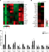
Jarid2 was required for myocardial maturation. A, the heat map shows statistically significantly dysregulated genes in Jarid2αMHC at both p10 and 7 months (m). Only 12 genes were identified as dysregulated genes among 72 genes and 2375 genes at p10 and 7 months, respectively. B, the heat map consists of 16 genes identified by time-dependent differential gene expression. 16 genes were statistically significant in our analysis, and the fold changes from p10 to 7 months are presented by colors. C, qRT-PCR was performed to determine expression levels of DE genes in p10 and 7-month hearts (n = 3–5). Ctrl, control.
Figure 8.
Figure 8.
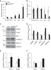
ErbB4 was up-regulated in Jarid2αMHC hearts at p10. A and B, Nrg1 (A) and ErbB4 (B) expression levels were determined by qRT-PCR on the heart at different ages. The expression levels in Jarid2αMHC hearts were normalized to each control (Ctrl) level (n = 3–5). C, Western blotting was performed to determine ErbB4, AKT, and ERK pathways on p10 hearts. D, the graph showed the protein levels of ErbB4, AKT, and ERK by standardization to GAPDH and normalization to control levels. E and F, qRT-PCR was performed on isolated cardiomyocytes from new born hearts, which were incubated in vehicle (E) or the ErbB inhibitor AG1478 (5μm) (F) (n = 3–5).
Figure 9.
Figure 9.
Summary of Jarid2 function in the postnatal heart. Jarid2 plays important roles in cardiomyocyte maturation during neonatal stages and inhibiting DCM development. HF, heart failure.

Similar articles

Cited by

References

    1. Ziaeian B., and Fonarow G. C. (2016) Epidemiology and aetiology of heart failure. Nat. Rev. Cardiol. 13, 368–378 10.1038/nrcardio.2016.25 - DOI - PMC - PubMed
    1. McNally E. M., Golbus J. R., and Puckelwartz M. J. (2013) Genetic mutations and mechanisms in dilated cardiomyopathy. J. Clin. Invest. 123, 19–26 10.1172/JCI62862 - DOI - PMC - PubMed
    1. Hall D. D., Ponce J. M., Chen B., Spitler K. M., Alexia A., Oudit G. Y., Song L. S., and Grueter C. E. (2017) Ectopic expression of Cdk8 induces eccentric hypertrophy and heart failure. JCI Insight 2, 92476 - PMC - PubMed
    1. van Berlo J. H., Maillet M., and Molkentin J. D. (2013) Signaling effectors underlying pathologic growth and remodeling of the heart. J. Clin. Invest. 123, 37–45 10.1172/JCI62839 - DOI - PMC - PubMed
    1. Ujfalusi Z., Vera C. D., Mijailovich S. M., Svicevic M., Yu E. C., Kawana M., Ruppel K. M., Spudich J. A., Geeves M. A., and Leinwand L. A. (2018) Dilated cardiomyopathy myosin mutants have reduced force-generating capacity. J. Biol. Chem. 293, 9017–9029 10.1074/jbc.RA118.001938 - DOI - PMC - PubMed

Publication types

MeSH terms