. 2019 Mar 21;93(7):e02084-18.
doi: 10.1128/JVI.02084-18. Print 2019 Apr 1.

Rare Detection of Antiviral Functions of Polyclonal IgA Isolated from Plasma and Breast Milk Compartments in Women Chronically Infected with HIV-1

Affiliations

Rare Detection of Antiviral Functions of Polyclonal IgA Isolated from Plasma and Breast Milk Compartments in Women Chronically Infected with HIV-1

Matthew Zirui Tay et al. J Virol. .

Abstract

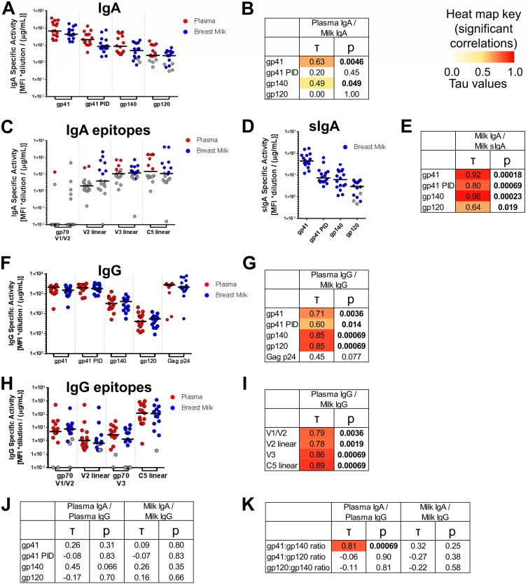
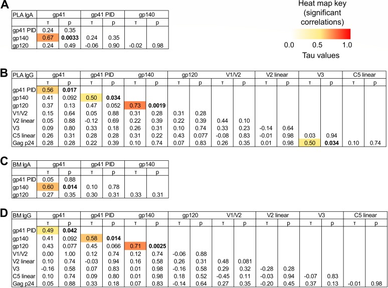
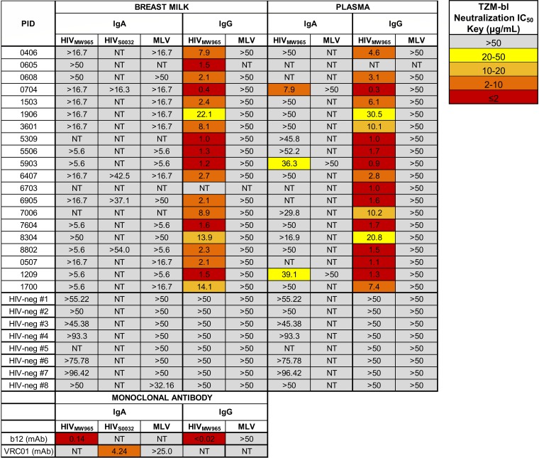
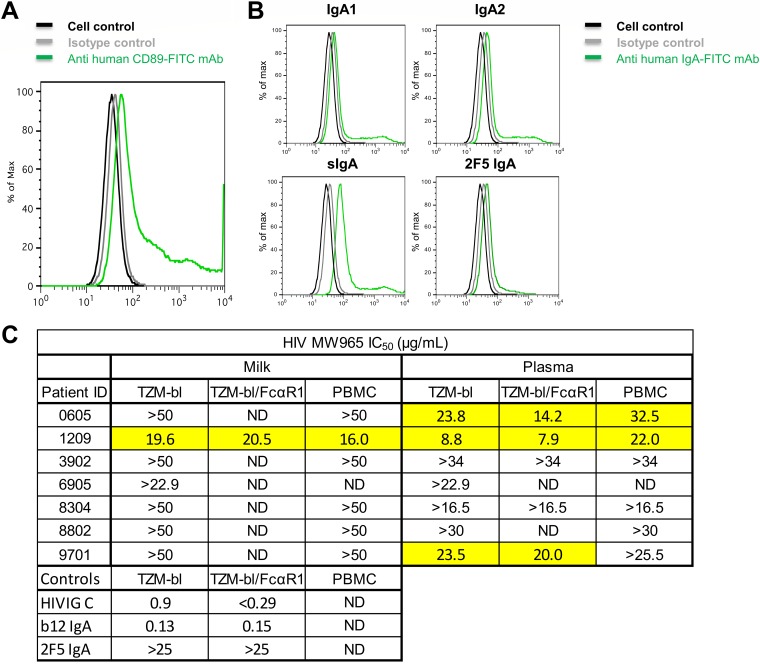
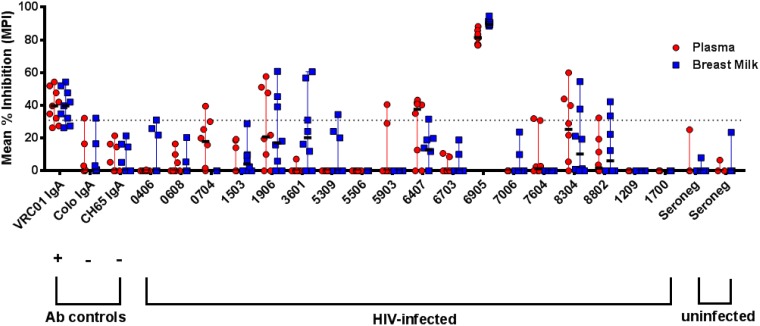
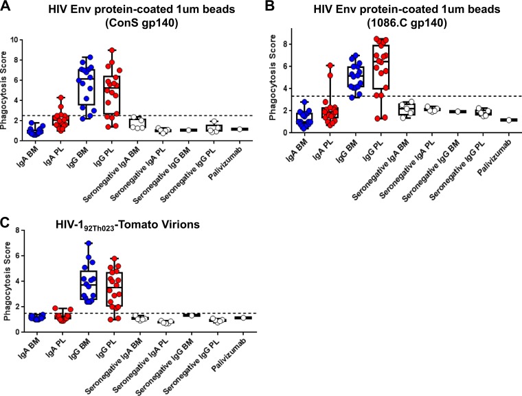
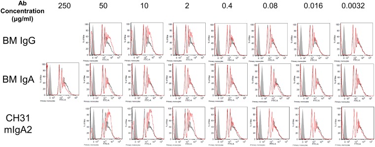
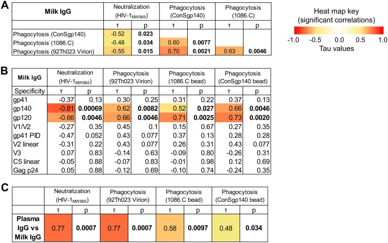
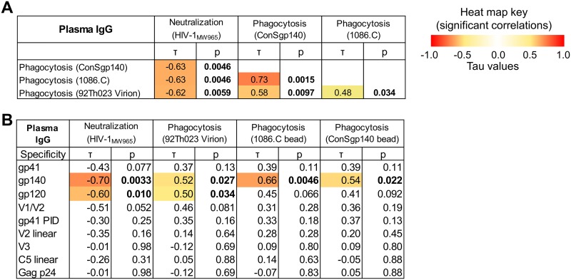
The humoral response to invading mucosal pathogens comprises multiple antibody isotypes derived from systemic and mucosal compartments. To understand the contribution of each antibody isotype/source to the mucosal humoral response, parallel investigation of the specificities and functions of antibodies within and across isotypes and compartments is required. The role of IgA against HIV-1 is complex, with studies supporting a protective role as well as a role for serum IgA in blocking effector functions. Thus, we explored the fine specificity and function of IgA in both plasma and mucosal secretions important to infant HIV-1 infection, i.e., breast milk. IgA and IgG were isolated from milk and plasma from 20 HIV-1-infected lactating Malawian women. HIV-1 binding specificities, neutralization potency, inhibition of virus-epithelial cell binding, and antibody-mediated phagocytosis were measured. Fine-specificity mapping showed IgA and IgG responses to multiple HIV-1 Env epitopes, including conformational V1/V2 and linear V2, V3, and constant region 5 (C5). Env IgA was heterogeneous between the milk and systemic compartments (Env IgA, τ = 0.00 to 0.63, P = 0.0046 to 1.00). Furthermore, IgA and IgG appeared compartmentalized as there was a lack of correlation between the specificities of Env-specific IgA and IgG (in milk, τ = -0.07 to 0.26, P = 0.35 to 0.83). IgA and IgG also differed in functions: while neutralization and phagocytosis were consistently mediated by milk and plasma IgG, they were rarely detected in IgA from both milk and plasma. Understanding the ontogeny of the divergent IgG and IgA antigen specificity repertoires and their effects on antibody function will inform vaccination approaches targeted toward mucosal pathogens.IMPORTANCE Antibodies within the mucosa are part of the first line of defense against mucosal pathogens. Evaluating mucosal antibody isotypes, specificities, and antiviral functions in relationship to the systemic antibody profile can provide insights into whether the antibody response is coordinated in response to mucosal pathogens. In a natural immunity cohort of HIV-infected lactating women, we mapped the fine specificity and function of IgA in breast milk and plasma and compared these with the autologous IgG responses. Antigen specificities and functions differed between IgG and IgA, with antiviral functions (neutralization and phagocytosis) predominantly mediated by the IgG fraction in both milk and plasma. Furthermore, the specificity of milk IgA differed from that of systemic IgA. Our data suggest that milk IgA and systemic IgA should be separately examined as potential correlates of risk. Preventive vaccines may need to employ different strategies to elicit functional antiviral immunity by both antibody isotypes in the mucosa.

Keywords: HIV-1; IgA; effector functions; mucosal immunity.


Figures

FIG 1
FIG 1
Env antigen specificities of milk IgG and IgA and plasma and milk IgAs do not correlate, while milk and plasma IgG Env antigen-specificities are strongly correlated. (A, C, D, F and H) For 16 HIV-1+ lactating women, milk and plasma samples were obtained, and IgA and IgG were purified from both sample types. For IgA (A) and sIgA (D), binding scores to gp41, gp41 PID, gp140, and gp120 antigens are shown (see Materials and Methods for antigens and calculation). For IgA, scores for binding to gp70 V1/V2, linear V2, linear V3, and linear C5 antigen subspecificities are also shown (C). For IgG, scores are shown for binding to Env gp41, gp41 PID, gp140, and gp120, as well as Gag p24 antigens (F), and to gp70 V1/V2, linear V2, gp70 V3, and linear C5 antigen subspecificities (H). Background-subtracted MFI values below 0 are shown with a value of 0.01. (B, E, G, I, J and K) Correlations between compartments for each antigen specificity or antigen specificity ratio. Kendall’s tau values and corresponding corrected P values (see Materials and Methods) are reported. Boldface P values indicate significant correlations at a P value of <0.05, with corresponding tau values color coded on a scale of 0 to 1.
FIG 2
FIG 2
Heterogeneity of the Env antigen specificities of breast milk (BM) and plasma (PLA) IgGa and IgAa between individuals. For each of 16 HIV-1+ individuals, the relationships between selected pairwise binding scores are displayed as percentages. N.D. indicates that either one of the pair of values was a missing value or exceeded the limit of quantitation (<100 MFI or >23,000 MFI).
FIG 3
FIG 3
Env antigen-specific IgA and IgG responses in milk and plasma seldom correlate with other Env antigen-specific responses in HIV-infected lactating women. To determine whether immune responses to the various HIV-1 antigens were associated with each other, correlations between binding scores for selected antigen specificities were tested for plasma IgA (A), plasma IgG (B), breast milk (BM) IgA (C), and breast milk IgG (D). Kendall’s tau values and corresponding corrected P values (see Materials and Methods) are reported. Boldface P values indicate significant correlations at a P value of <0.05, with corresponding tau values color coded on a scale of 0 to 1.
FIG 4
FIG 4
Milk and plasma IgG, but not IgA, mediate tier 1 HIVMW965 neutralization. To identify HIV-1 neutralization function in antibody fractions of milk and plasma, we determined the neutralization IC50 in TZM-bl cells for IgA and IgG antibodies isolated from milk and plasma samples of 20 participants. HIV-specific neutralization activities against the clade C tier 1 HIV variants MW965 (and also S0032 for selected samples) are shown, as well as nonspecific antiviral neutralization activities against murine leukemia virus (MLV). Eight HIV-negative controls are also shown, as well as positive-control recombinant monoclonal antibodies (b12 and VRC01, CD4 binding site broadly neutralizing antibodies) in IgA and IgG backbones. No milk IgA samples neutralized HIV-1MW965 or HIV-1S0032, whereas three plasma IgA samples had detectable neutralization.
FIG 5
FIG 5
IgA-mediated tier 1 HIV-1 neutralization is not enhanced in FcαRI-expressing cells or peripheral blood mononuclear cells. (A) To gain insight into the role of CD89 in HIV infectivity and neutralization, the cDNAs for the human FcαRI and the γ chain of the FcεR were transduced into TZM-bl cells using lentiviral constructs. Specific monoclonal antibodies and flow cytometry detected surface expression of FcαRI on this new TZM-bl cell line. (B) Flow cytometry diagrams show the capability of the TZM-bl/FcαRI cell line to bind to human monomeric IgA1 and monomeric IgA2, human colostrum secretory IgA, and monoclonal 2F5 IgA. (C) To determine whether neutralization activity is enhanced in the presence of Fc alpha receptor 1 (FcαRI), we examined the neutralization IC50 using TZM-bl cells transduced with FcαRI and also in human peripheral blood mononuclear cells (PBMCs). Milk and plasma IgAs were tested for six participants with a variety of neutralization phenotypes in TZM-bl cells. The positive controls HIVIG (clade C) and b12 IgA MAb and the negative-control 2F5 MAb (broadly neutralizing, but does not neutralize MW965) were also tested. ND, not done for the indicated sample. Neutralization was not enhanced in either FcαRI-transduced TZM-bl cells or in peripheral blood mononuclear cells.
FIG 6
FIG 6
Breast milk and plasma IgAs may inhibit C.1086 HIV-1 virion binding to epithelial cells. Inhibition of binding to the colonic epithelial cell line HT-29 was assessed using the tier 2 clade C virus HIV-1 C.1086, with 7 to 12 replicates performed over three independent experiments. VRC01 IgA was used as a positive control, and HIV-negative colostrum IgA and anti-influenza virus CH65 IgA were used as negative controls. A dotted line indicates the mean percent inhibition (MPI) cutoff of 31%, calculated as 2 standard deviations plus the MPI of anti-influenza virus hemagglutinin MAb CH65 IgA relative to that of the no-antibody condition.
FIG 7
FIG 7
Breast milk and plasma IgGs, but not IgAs, mediate phagocytosis of HIV-1 virions and Env-coated beads. To determine the phagocytosis function in antibody fractions of breast milk (BM) and plasma (PL), we tested IgA and IgG from milk and plasma of 16 HIV+ women for phagocytosis of beads coated with HIV-1 Env ConS gp140 (A), beads coated with HIV-1 Env 1086.C gp140 (B), and fully infectious fluorescent HIV-1 virions (HIV-192Th023-Tomato) (C). Antibodies from an additional five HIV-negative women were also tested as negative controls, as well as the anti-respiratory syncytial virus antibody palivizumab. The black dotted line indicates the positivity cutoff, determined using the mean +3 standard deviations of values of the negative controls used in the assay.
FIG 8
FIG 8
Lack of detectable milk IgA-mediated phagocytosis despite increasing IgA concentration. To determine if lack of milk IgA phagocytosis activity was due to lower HIV-1 specific activity in IgA than that in IgG, higher concentrations of IgA and IgG were purified from the milk of an HIV-positive lactating woman recruited in the United states. Breast milk (BM) IgG, breast milk IgA, and a control antibody, CH31 mIgA2, were tested for phagocytosis of HIV ConS gp140-coated beads at 5-fold dilutions as indicated, and flow cytometry diagrams indicative of the phagocytosis results are shown. The red traces indicate sample antibody setup while black traces indicate the no-antibody control setup, and gray fill indicates the no-target control setup. Antibody-mediated phagocytosis is indicated by a greater area under the curve for the red trace than that for the black trace. Milk IgG and CH31 mIgA2 showed antibody-mediated phagocytosis activity down to 2 μg/ml, while milk IgA showed no antibody-mediated phagocytosis activity even at 250 μg/ml.
FIG 9
FIG 9
Functional IgG activity in milk and plasma are correlated and linked to antibodies targeting gp140 and gp120. (A) To determine if the functional activity of anti-HIV-1 antibodies in milk were associated with each other, correlations were tested between neutralization (HIV-1MW965) and phagocytosis (ConS gp140 beads, 1086.C beads, and 92Th023 virions) for milk IgG. Kendall’s tau values and corresponding corrected P values (see Materials and Methods) are reported. Boldface P values indicate significant correlations at a P value of <0.05, with corresponding absolute tau values color coded on a scale of 0 to 1. (B) To determine candidate antigen specificities that could be targeted for functional activity by milk IgG antibodies, correlations were tested between antigen-specific binding and functional activity. (C) To identify whether functional activity was conserved across milk and plasma antibodies, correlations were tested between milk and plasma IgGs for each antibody function (neutralization and phagocytosis).
FIG 10
FIG 10
Correlations between antibody functions and epitope specificity in plasma IgG. (A) To determine if the functional activity of anti-HIV-1 antibodies in milk were associated with each other, correlations were tested between neutralization (HIVMW965) and phagocytosis (ConS gp140 beads, 1086.C beads, 92Th023 virions) for plasma IgG. Kendall’s tau values and corresponding corrected P values (see Materials and Methods) are reported. Boldface P values indicate significant correlations at a P value of <0.05, with corresponding absolute tau values color coded on a scale of 0 to 1. (B) To determine candidate antigen specificities that could be targeted for functional activity by plasma IgG antibodies, correlations were tested between antigen-specific binding and functional activity.
FIG 11
FIG 11
Virion phagocytosis is not dependent on spinoculation and requires specific Fc-FcR interaction. (A) To assess whether virion phagocytosis was dependent on the spinoculation procedure in the experimental setup, parallel conditions were set up with and without the spinoculation step (see Materials and Methods), as indicated in the legend on the figure. Spinoculation was performed at 1,200 × g for 1 h at 4°C. Antibodies were added at 12.5 μg/ml unless otherwise specified. Three pairs of antibody and virus were chosen based on their known properties for virion phagocytosis in the presence of spinoculation (top row). All three pairs retained virion phagocytosis signal even when spinoculation was omitted though overall specific and nonspecific virion phagocytosis was decreased relative to values for the experiments with spinoculation. This remained true despite decreasing the concentration of antibody (bottom left), virus (bottom-middle), or both antibody and virus (bottom right). (B and C) To further assess whether virion phagocytosis was dependent on Fc-FcR interactions, the CD4 binding site broadly neutralizing antibody CH31 was recombinantly expressed in human mIgA1, IgG1, IgG3, and IgG4 backbones and examined for virus capture of HIV-1BaL virions or phagocytosis of HIV-1BaL-Tomato virions. For virus capture, the median and range of three replicates from a single experiment are reported, and for phagocytosis, the median and range of data from four different human primary monocyte donors representing two independent experiments are reported. A positivity cutoff of 20% for infectious virus capture and 1.77 for virion phagocytosis was determined based on the mean +3 standard deviations of the values for historical negative controls. CH31 mIgA1, IgG1, and IgG3 mediated virion phagocytosis, as reported, while CH31 IgG4 did not, despite its ability to capture virions, showing that virion phagocytosis is dependent on Fc-FcR interactions.

Similar articles

Cited by

References

    1. Plotkin SA. 2013. Complex correlates of protection after vaccination. Clin Infect Dis 56:1458–1465. doi:10.1093/cid/cit048. - DOI - PubMed
    1. Azevedo MS, Yuan L, Iosef C, Chang KO, Kim Y, Nguyen TV, Saif LJ. 2004. Magnitude of serum and intestinal antibody responses induced by sequential replicating and nonreplicating rotavirus vaccines in gnotobiotic pigs and correlation with protection. Clin Diagn Lab Immunol 11:12–20. - PMC - PubMed
    1. Prejean J, Song R, Hernandez A, Ziebell R, Green T, Walker F, Lin LS, An Q, Mermin J, Lansky A, Hall HI. 2011. Estimated HIV incidence in the United States, 2006–2009. PLoS One 6:e17502. doi:10.1371/journal.pone.0017502. - DOI - PMC - PubMed
    1. Besredka A. 1927. Local immunization. Bailliere, Tindall Cox, London, UK.
    1. Kozlowski PA, Cu-Uvin S, Neutra MR, Flanigan TP. 1997. Comparison of the oral, rectal, and vaginal immunization routes for induction of antibodies in rectal and genital tract secretions of women. Infect Immun 65:1387–1394. - PMC - PubMed

Publication types

MeSH terms