. 2019 Apr 3;93(8):e01907-18.
doi: 10.1128/JVI.01907-18. Print 2019 Apr 15.

The Molecular Basis for Antigenic Drift of Human A/H2N2 Influenza Viruses

Affiliations

The Molecular Basis for Antigenic Drift of Human A/H2N2 Influenza Viruses

M Linster et al. J Virol. .

Abstract

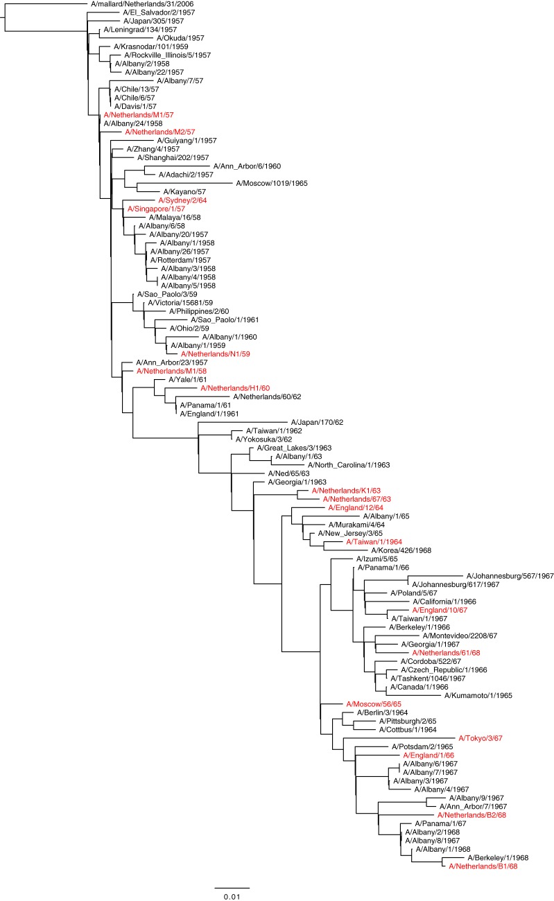
Influenza A/H2N2 viruses caused a pandemic in 1957 and continued to circulate in humans until 1968. The antigenic evolution of A/H2N2 viruses over time and the amino acid substitutions responsible for this antigenic evolution are not known. Here, the antigenic diversity of a representative set of human A/H2N2 viruses isolated between 1957 and 1968 was characterized. The antigenic change of influenza A/H2N2 viruses during the 12 years that this virus circulated was modest. Two amino acid substitutions, T128D and N139K, located in the head domain of the H2 hemagglutinin (HA) molecule, were identified as important determinants of antigenic change during A/H2N2 virus evolution. The rate of A/H2N2 virus antigenic evolution during the 12-year period after introduction in humans was half that of A/H3N2 viruses, despite similar rates of genetic change.IMPORTANCE While influenza A viruses of subtype H2N2 were at the origin of the Asian influenza pandemic, little is known about the antigenic changes that occurred during the twelve years of circulation in humans, the role of preexisting immunity, and the evolutionary rates of the virus. In this study, the antigenic map derived from hemagglutination inhibition (HI) titers of cell-cultured virus isolates and ferret postinfection sera displayed a directional evolution of viruses away from earlier isolates. Furthermore, individual mutations in close proximity to the receptor-binding site of the HA molecule determined the antigenic reactivity, confirming that individual amino acid substitutions in A/H2N2 viruses can confer major antigenic changes. This study adds to our understanding of virus evolution with respect to antigenic variability, rates of virus evolution, and potential escape mutants of A/H2N2.

Keywords: antigenic evolution; influenza virus A/H2N2; molecular drift; pandemic.


Figures

FIG 1
FIG 1
Maximum likelihood phylogenetic tree based on HA1 amino acid sequences of human A/H2N2 viruses. Virus isolates used for antigenic characterization are highlighted in red.
FIG 2
FIG 2
Antigenic map of human A/H2N2 influenza viruses as measured in HI assays with ferret postinfection antisera. Circles indicate the position of viruses, squares represent two ferret antisera raised against each of the viruses A/Japan/305/57, A/Singapore/1/57, A/Netherlands/K1/63, A/England/1/66, A/Tokyo/3/67, and A/Netherlands/B1/68. The underlying grid depicts the scale of antigenic difference between the viruses, with each square representing one antigenic unit or a 2-fold difference in HI titer. Years of isolation of the A/H2N2 virus isolates are indicated, ranging from 1957 (red) to 1968 (blue).
FIG 3
FIG 3
Summary of substitutions responsible for antigenic differences between NL/M1/57 and NL/B1/68. Antigenic maps showing the antigenic change caused by individual amino acid substitutions introduced into NL/M1/57 (A) or NL/B1/68 (B) and combinations of mutations introduced into NL/M1/57 (C) or NL/B1/68 (D). Viruses are shown as circles of different colors, with a diamond indicating the mutant virus with the largest antigenic distance to the corresponding wild-type strain. Sera are indicated as open squares. The underlying map of wild-type viruses from Fig. 2 is shown in gray, and its positioning is kept constant. The arrows indicate the antigenic distance of a double mutant that spans a long distance between the earliest and latest isolates of A/H2N2. Structure of an HA trimer (E) with individual monomers in shades of gray, the RBS in yellow, and mutations near the RBS with a measurable effect on antigenicity in orange. The two mutations with the biggest combined effect in panel C are colored in red (T128D and N139K).
FIG 4
FIG 4
Rates of genetic and antigenic evolution of A/H2N2 and A/H3N2 virus during 12 years of circulation in humans. Genetic (A) and antigenic (B) distances of the A/H2N2 (red squares) and A/H3N2 (blue circles) viruses from the first human virus isolates in 1957 (A/Netherlands/M1/1957) and 1968 (A/Bilthoven/16190/1968). Rates are derived from the slope of the best-fit regression line.

Similar articles

Cited by

References

    1. Blumenfeld HL, Kilbourne ED, Louria DB, Rogers DE. 1959. Studies on influenza in the pandemic of 1957–1958. I. An epidemiologic, clinical and serologic investigation of an intrahospital epidemic, with a note on vaccination efficacy. J Clin Invest 38:199–212. doi:10.1172/JCI103789. - DOI - PMC - PubMed
    1. Kilbourne ED. 2006. Influenza pandemics of the 20th century. Emerg Infect Dis 12:9–14. doi:10.3201/eid1201.051254. - DOI - PMC - PubMed
    1. Viboud C, Simonsen L, Fuentes R, Flores J, Miller MA, Chowell G. 2016. Global mortality impact of the 1957–1959 influenza pandemic. J Infect Dis 213:738–745. doi:10.1093/infdis/jiv534. - DOI - PMC - PubMed
    1. Cobos AJ, Nelson CG, Jehn M, Viboud C, Chowell G. 2016. Mortality and transmissibility patterns of the 1957 influenza pandemic in Maricopa County, Arizona. BMC Infect Dis 16:405. doi:10.1186/s12879-016-1716-7. - DOI - PMC - PubMed
    1. Lindstrom SE, Cox NJ, Klimov A. 2004. Genetic analysis of human H2N2 and early H3N2 influenza viruses, 1957–1972: evidence for genetic divergence and multiple reassortment events. Virology 328:101–119. doi:10.1016/j.virol.2004.06.009. - DOI - PubMed

Publication types

MeSH terms

Substances