. 2019 Jan 30;10(1):490.
doi: 10.1038/s41467-019-08353-4.

Template-directed RNA polymerization and enhanced ribozyme catalysis inside membraneless compartments formed by coacervates

Affiliations

Template-directed RNA polymerization and enhanced ribozyme catalysis inside membraneless compartments formed by coacervates

Raghav R Poudyal et al. Nat Commun. .

Abstract

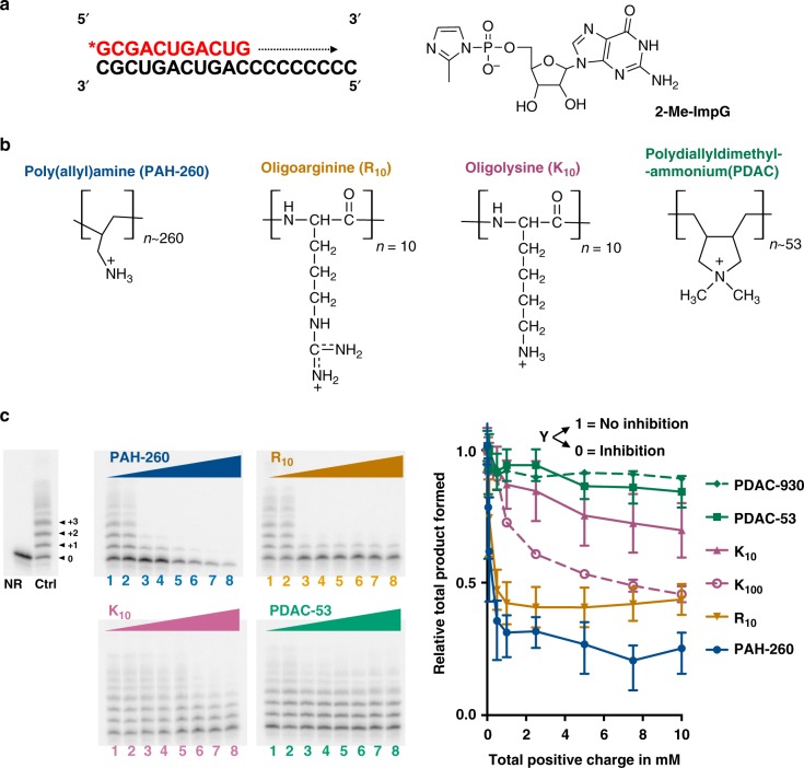
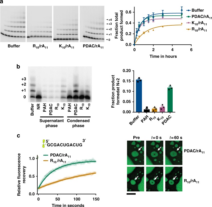
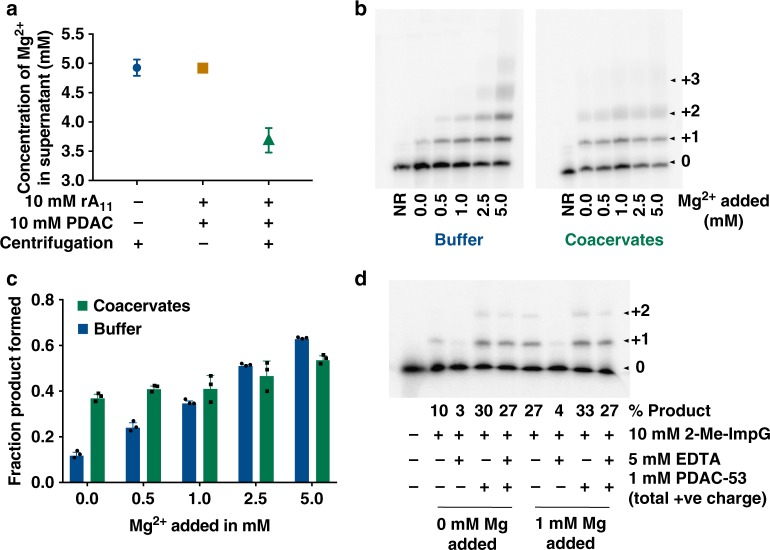
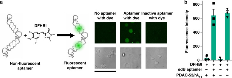
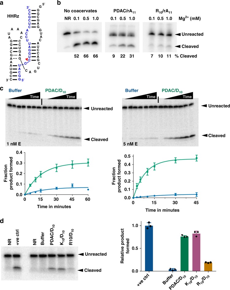
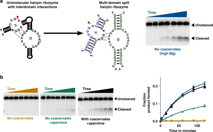
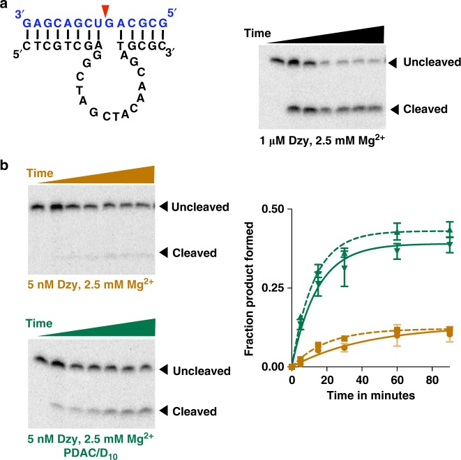
Membraneless compartments, such as complex coacervates, have been hypothesized as plausible prebiotic micro-compartments due to their ability to sequester RNA; however, their compatibility with essential RNA World chemistries is unclear. We show that such compartments can enhance key prebiotically-relevant RNA chemistries. We demonstrate that template-directed RNA polymerization is sensitive to polycation identity, with polydiallyldimethylammonium chloride (PDAC) outperforming poly(allylamine), poly(lysine), and poly(arginine) in polycation/RNA coacervates. Differences in RNA diffusion rates between PDAC/RNA and oligoarginine/RNA coacervates imply distinct biophysical environments. Template-directed RNA polymerization is relatively insensitive to Mg2+ concentration when performed in PDAC/RNA coacervates as compared to buffer, even enabling partial rescue of the reaction in the absence of magnesium. Finally, we show enhanced activities of multiple nucleic acid enzymes including two ribozymes and a deoxyribozyme, underscoring the generality of this approach, in which functional nucleic acids like aptamers and ribozymes, and in some cases key cosolutes localize within the coacervate microenvironments.


Conflict of interest statement

The authors declare no competing interests.

Figures

Fig. 1
Fig. 1
Effect of polyamines on template-directed polymerization. a Primer (red)-template (black) complex used for template-directed polymerization using guanosine 5′-(phosphor)-2-methylimidazolide (2-Me-ImpG). b Structures of polyamines used. c Representative denaturing polyacrylamide gel images show extension of 32P-labeled primer. Lane Ctrl indicates reaction without any cationic polymer and NR indicates no reaction in the absence of monomer, lanes 1–8 contain 0.050, 0.10, 0.50, 1.0, 2.5, 5.0, 7.5, and 10 mM total positive charge from PAH-260 (blue), R10 (orange), K10 (dark pink), and PDAC-53 (green). Reactions were carried out for 5 h and contained < 50 nM 5ʹ-end-labeled primer and 1 µM template in 25 mM Tris–HCl pH 8.0 and 5 mM MgCl2. Fraction converted to product was normalized to the yield in the absence of any cation ctrl. Error bars represent S.E.M. from three independent experiments. Uncropped gel images are shown in Supplementary Figure 14a
Fig. 2
Fig. 2
Coacervates of PDAC/rA11 RNA support template-directed polymerization. a Template-directed polymerization reactions in the presence of coacervates. Coacervates were formed by adding respective polycations (10 mM total positive charge) to solutions containing 10 mM 2-me-ImpG and 7.5 mM total negative charge from rA11 in 5 mM Mg and 25 mM Tris–HCl pH 8.0. Reactions were initiated by adding pre-annealed 5′-end-labeled RNA primer and unlabeled template, and incubated at room temperature and time points were taken at 0 min, 15 min, 30 min, 1 h, 1.5 h, 2 h, 3 h, and 5 h. Samples were separated by denaturing PAGE. Total product formed was calculated by quantifying all the visible bands in the given lane. Data were fit to first order exponential. Error bars represent S.E.M from three independent experiments. b Reactions were initiated as a except the Mg concentration was decreased to 1 mM to slow the reactions. Reactions were immediately centrifuged at 14,000×g after initiation. The bulk phase was then separated from the condensed phase and the two phases were allowed to react separately for 5 h. Quantifications of product yields are shown for condensed phases. Error bars represent S.E.M. from three independent experiments. c FRAP recovery curves for Cy3-labeled RNA primer in PDAC/rA11 and R10/rA11 coacervates at 10 mM charge-balanced condition. Shown as best fits to Eq. (2) (five independent trials). Representative images of PDAC/rA11 (top) and R10/rA11 (bottom). Scale bar is 5 µm. Uncropped gel images are shown in Supplementary Figure 14b, c
Fig. 3
Fig. 3
Template-directed polymerization at sub-optimal Mg concentrations. a Mg 2+ levels were measured by atomic absorption spectroscopy. Solutions with 5 mM MgCl2 were formed in 25 mM Tris–HCl pH 8.0 with indicated anions and cations, followed by centrifugation at 14,000×g for 2 min. A portion of the supernatant phase was removed and diluted in water prior to measurement. Error bars represent the range of values from two experiments. b Yields of template-directed polymerization at different amounts of added Mg in presence and absence of PDAC-53/rA11 coacervates. c Quantifications from b. Error bars represent S.E.M. from three independent experiments. d Template-directed polymerization reactions were assembled as previously described in the presence or absence of indicated molecules and ions. Uncropped gel images are shown in Supplementary Figure 15a, b
Fig. 4
Fig. 4
Active RNA aptamer folding inside PDAC/rA11 coacervates. a (Left) Model of stabilized dimeric broccoli (sdB) RNA drawn in NUPACK, and confocal microscopy images (right) of PDAC-53/rA11 coacervates containing sdB RNA complexed with DFHBI dye. No fluorescence is seen when coacervates contain only DFHBI dye or an inactive mutant of the aptamer. Coacervates contained 100 nM sdB RNA and 10 µM DFHBI in 25 mM Tris pH 8.0, 5 mM MgCl2 and 5 mM KCl. 488 nm laser was used for excitation and emission window was 500–550 nm. Scale bar is 20 µm. b Bulk fluorescence measurement of sdB RNA aptamer in the presence and absence of PDAC-53/rA11 coacervates. Error bars represent S.E.M. (n = 3) from three independent experiments
Fig. 5
Fig. 5
Enhanced ribozyme activity in coacervates. a Structure of the hammerhead ribozyme–substrate complex. The cleavage site is indicated with red arrow. b Gel images showing ribozyme cleavage at different Mg2+ concentrations in the coacervate phase after 1 h. Dilute and coacervate phases were separated immediately after the reaction initiation. Reactions contained 25 mM Tris·HCl pH 8.0, 5 mM NaCl and indicated Mg2+. c Representative gel images showing ribozyme cleavage in buffer or in PDAC-53/D10 coacervates at 1 nM and 5 nM ribozyme concentration; data were quantified from the gels above and fit to Eq. (1). Reactions in buffer (blue trace) and PDAC/D10 coacervates (green trace) were performed at room temperature in 25 mM Tris–HCl pH 8.0, 1 mM MgCl2, and 2.5 mM KCl. For 1 and 5 nM enzyme, observed rate constants are 0.05 ± 0.02 and 0.05 ± 0.02 min−1 for reactions in buffer and 0.06 ± 0.01 and 0.12 ± 0.01 for reactions in coacervates. d Representative gel image showing ribozyme cleavage after 1 h of reaction in 25 mM Tris–HCl pH 7.5, 1 mM MgCl2, and 2.5 mM KCl. NR contained no ribozyme, +ve ctrl contained 250 nM ribozyme, and all other lanes contained 2.5 nM enzyme. Coacervates contained 15 mM total positive charge from various polycations and 15 mM total negative charge from D10. Product yields were normalized to “+ve ctrl”. All error bars represent S.E.M. (n = 3) from three independent experiments. Uncropped gel images are shown in Supplementary Figure 16a, b
Fig. 6
Fig. 6
Concentration of cofactors in coacervates enhances of hairpin ribozyme catalysis. a Consensus secondary structure of the unimolecular hairpin ribozyme with conserved nucleotides (left), the site for cleavage is indicated with red arrowhead. Multi-domain split hairpin ribozyme where loop A consists of the substrate (blue) and the substrate-binding strand (black) and loop B domains separated (green). Gel image showing cleavage of the substrate is shown. Reaction were performed in buffer containing 25 mM Tris–HCl pH 7.5, 25 mM MgCl2 and 25 mM KCl. Reactions contained 1 nM substrate, 1 µM substrate-binding strand, and 1 µM of the loop B domain. b Substrate cleavage of loop A domain by the ribozyme at sub-optimal conditions. All reactions were performed in 25 mM Tris·HCl pH 7.5, 2.5 mM MgCl2, and 2.5 mM KCl with additional components as indicated. Reactions contained 1 nM substrate, 50 nM substrate-binding strand, and 25 nM of the loop B domain. Coacervates were formed by PDAC-53 and spermine at 5 mM total positive charge from each and 10 mM total negative charge from polyaspartic acid (D30). Time points were taken at 0, 2.5, 15, 30, 60, 90, and 120 min. Plots showing fraction product cleaved versus time is shown in right. Fractions product formed were fit to Eq. (1). Colors in the plot correspond to labels of gel images. All error bars represent S.E.M. (n = 3) from three independent experiments. Uncropped gel images are shown in Supplementary Figure 17a
Fig. 7
Fig. 7
Coacervate-mediated stimulation of a DNAzyme. a Structure of the 10–23 DNAzyme. The enzyme strand is shown in black and the substrate is shown in blue. The red arrow indicates the cleavage site. Gel image shows efficient cleavage of the substrate (0.25 pM) by the enzyme (1 µM) in 25 mM Tris–HCl pH 8.0 containing 2.5 mM MgCl2 and 2.5 mM KCl. Time points were taken at 0, 2.5, 5, 15, 30, 60, and 90 min. b Reactions contained 5 nM of the enzyme strand and 0.25 pM substrate strand in 25 mM Tris–HCl pH 8.0 containing 2.5 mM MgCl2 and 2.5 mM KCl. Coacervates were formed by adding PDAC-53 and D10 at 10 mM total charge from each. Time points were taken at 0, 5, 15, 30, 60, 90, and 120 min. Fraction product formed were calculated from gels shown in b and Supplementary Figure 13 and data were fit to Eq. (1). Green solid and dashed lines indicate experiments performed in 2.5 mM Mg2+ or 5 mM Mg2+, respectively. All error bars represent S.E.M. (n = 3) from three independent experiments. Uncropped gel images are shown in Supplementary Figure 17b

References

    1. Joyce GF. The antiquity of RNA-based evolution. Nature. 2002;418:212–221. doi: 10.1038/418214a. - DOI - PubMed
    1. Benner SA, Ellington AD. Modern metabolism as a palimpsest of the RNA world. Proc. Natl Acad. Sci. USA. 1989;86:7054–7058. doi: 10.1073/pnas.86.18.7054. - DOI - PMC - PubMed
    1. Lorsch JR, Szostak JW. In vitro evolution of new ribozymes with polynucleotide kinase activity. Nature. 1994;371:31–36. doi: 10.1038/371031a0. - DOI - PubMed
    1. Poudyal RR, et al. Selective inactivation of functional RNAs by ribozyme-catalyzed covalent modification. ACS Synth. Biol. 2017;6:528–534. doi: 10.1021/acssynbio.6b00222. - DOI - PubMed
    1. Tuerk C, MacDougal S, Gold L. RNA pseudoknots that inhibit human immunodeficiency virus type 1 reverse transcriptase. Proc. Natl Acad. Sci. USA. 1992;89:6988–6992. doi: 10.1073/pnas.89.15.6988. - DOI - PMC - PubMed

Publication types