. 2019 Jan 30;9(1):988.
doi: 10.1038/s41598-018-37399-5.

Progesterone Treatment Attenuates Glycolytic Metabolism and Induces Senescence in Glioblastoma

Affiliations

Progesterone Treatment Attenuates Glycolytic Metabolism and Induces Senescence in Glioblastoma

Fahim Atif et al. Sci Rep. .

Abstract

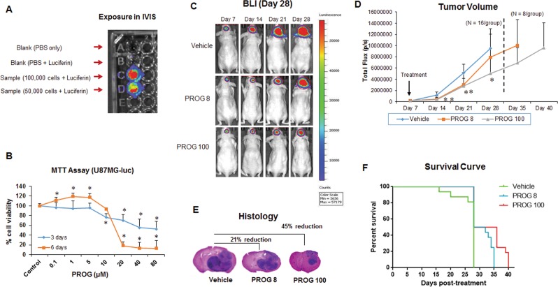
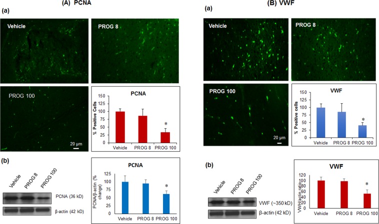
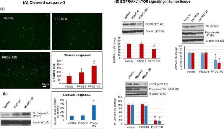
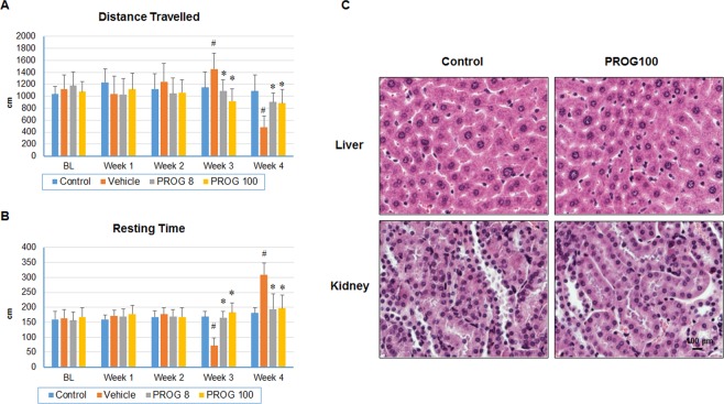
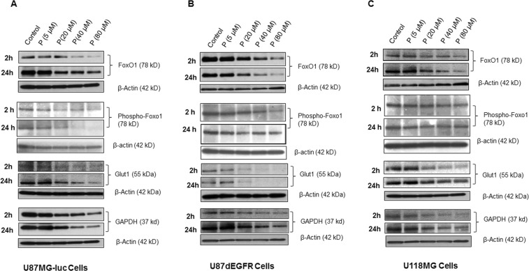
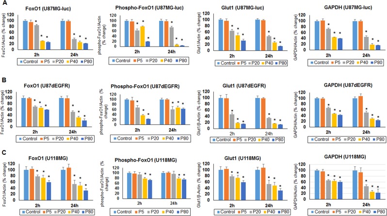
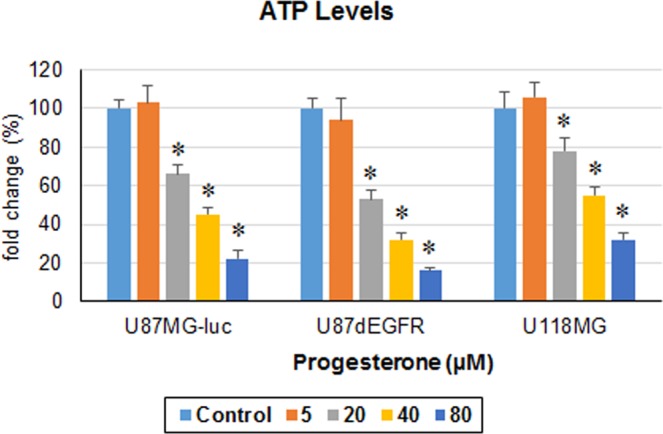
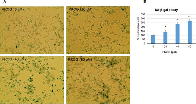
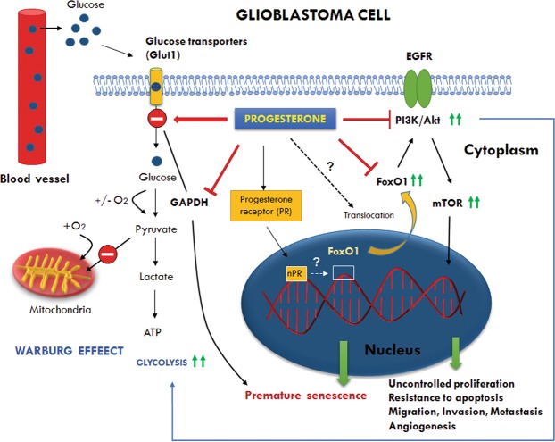
We examined the effect of progesterone treatments on glycolytic metabolism and senescence as possible mechanisms in controlling the growth of glioblastoma multiforme (GBM). In an orthotopic mouse model, after tumor establishment, athymic nude mice received treatment with progesterone or vehicle for 40 days. Compared to controls, high-dose progesterone administration produced a significant reduction in tumor size (~47%) and an increased survival rate (~43%) without any demonstrable toxicity to peripheral organs (liver, kidney). This was accompanied by a significant improvement in spontaneous locomotor activity and reduced anxiety-like behavior. In a follow-up in vitro study of U87MG-luc, U87dEGFR and U118MG tumor cells, we observed that high-dose progesterone inhibited expression of Glut1, which facilitated glucose transport into the cytoplasm; glyceraldehyde 3-phosphate dehydrogenase (GAPDH; a glycolysis enzyme); ATP levels; and cytoplasmic FoxO1 and Phospho-FoxO1, both of which control glycolytic metabolism through upstream PI3K/Akt/mTOR signaling in GBM. In addition, progesterone administration attenuated EGFR/PI3K/Akt/mTOR signaling, which is highly activated in grade IV GBM. High-dose progesterone also induced senescence in GBM as evidenced by changes in cell morphology and β-galactocidase accumulation. In conclusion, progesterone inhibits the modulators of glycolytic metabolism and induces premature senescence in GBM cells and this can help to reduce/slow tumor progression.


Conflict of interest statement

A US patent (#US 8,435,972 B2) was issued to FA and DGS on May 7, 2013 for the use of progesterone and compositions related thereto.

Figures

Figure 1
Figure 1
Effect of progesterone on tumor growth and survival rate. (A) Luciferase assay; (B) MTT reduction assay in U87MG-luc cells (n = 6 each); (C) representative images of BLI; (D) tumor volume (E) brain-tumor histology (H&E); and (F) survival rate of tumor-bearing mice in different groups (n = 16 each). Values are expressed as mean ± SD in different groups. *P < 0.01: Significant difference compared to control/vehicle group.
Figure 2
Figure 2
In vivo effect of progesterone on markers of (A) proliferation and (B) angiogenesis in tumor tissue. Representative photomicrographs of IHC and cell counting (a) and representative western blot bands with densitometric analysis (b) from different groups. Values are expressed as mean ± SD in different groups (n = 8 each). Significant difference: *P < 0.01 compared to control. For both PCNA and VWF proteins, bands were cropped from different parts of the same gel (Supplementary Figure S1).
Figure 3
Figure 3
In vivo effect of progesterone on markers of (A) apoptosis and (B) PI3K/Akt/mTOR signaling in tumor tissue. Representative photomicrographs of IHC and cell counting (a) and representative western blot bands with densitometric analysis (b) from different groups. Values are expressed as mean ± SD in different groups (n = 8 each). Significant difference: *P < 0.01 compared to control. Bands were cropped from different parts of the same gel, or from different gels (Supplementary Figure S2).
Figure 4
Figure 4
Progesterone improves spontaneous locomotor activity deficits without any organ toxicity in tumor-bearing mice. (A) Distance travelled; (B) resting time in different groups (n = 16); (C) high-dose progesterone toxicity in liver and kidney. Values are expressed as means ± SD. *P < 0.01: Significant difference compared to control group.
Figure 5
Figure 5
Effect of high-dose progesterone on markers of glycolytic metabolism in (A) U87MG-luc, (B) U87dEGFR, and (C) U118MG cells in vitro. Representative blots from different groups. Values are expressed as mean ± SD in different groups (n = 8 each). Significant difference: *P < 0.01 compared to control. Bands were cropped from different parts of the same gel, or from different gels (Supplementary Figure S3).
Figure 6
Figure 6
Effect of high-dose progesterone on the markers of glycolytic metabolism in (A) U87MG-luc, (B) U87dEGFR, and (C) U118MG cells in vitro. Densitometric analysis of blots (from Fig. 5) from different groups. Values are expressed as mean ± SD in different groups (n = 8 each). Significant difference: *P < 0.01 compared to control.
Figure 7
Figure 7
Progesterone inhibits ATP levels in GBM cells in vitro. ATP levels in different GBM cell lines after 24 h exposure to progesterone. Values are expressed as mean ± SD in different groups (n = 8 each). Significant difference: *P < 0.01 compared to control.
Figure 8
Figure 8
Progesterone induces premature senescence in GBM cells in vitro. Representative photomicrographs (A) and cell counting of SA-β-gal positive cells (B) in different groups. Values are expressed as mean ± SD in different groups (n = 8 each). Significant difference: *P < 0.01 compared to control.
Figure 9
Figure 9
Schematic representation of the modulatory effects of progesterone on glycolytic metabolism, PI3K/Akt/mTOR signaling, and FoxO1 transcription factor in GBM. The figure shows the preference for glycolysis over mitochondrial oxidation despite sufficient levels of oxygen (the Warburg effect) which supports high anabolic activity of cancer cells for uncontrolled proliferation, migration, invasion and metastasis. Glut1 (Glucose transporter isoform 1) facilitates glucose transport into the cytoplasm and its upregulation is observed in different cancers including GBM. GAPDH (Glyceraldehyde 3-phosphate dehydrogenase) is a glycolysis enzyme whose overexpression is positively correlated with tumor progression in many cancers. Depletion of GAPDH is reported to induce senescence in tumor cells. FoxO1 (Forkhead box family O1) is a transcription factor which is critical for the regulation of cell cycle exit and arrest at G1, and induction of apoptosis. FoxO1 deregulation/inactivation leads to uncontrolled proliferation, and resistance to apoptosis. FoxO1 controls glycolytic metabolism through upstream PI3K/Akt/mTOR and downstream c-Myc activation. Our data showed that progesterone modulates glycolytic metabolism and induces premature senescence in GBM cells by inhibiting Glut1, GAPDH and cytoplasmic FoxO1 activity. Dotted arrows and question mark represent the modulatory effect of progesterone by an as yet unknown mechanism. How progesterone modulates nuclear FoxO1 activity or its translocation to the cytoplasm is still not known and needs to be defined.

References

    1. American Brain Tumor Association. American Brain Tumor Association Brain Tumor Statistics. (Chicago, IL, 2014). http://abta.pub30.convio.net/about-us/news/brain-tumor-statistics/.
    1. Adamson C, et al. Glioblastoma multiforme: a review of where we have been and where we are going. Expert Opin Investig Drugs. 2009;18:1061–1083. doi: 10.1517/13543780903052764. - DOI - PubMed
    1. Sun T, et al. Sexually dimorphic RB inactivation underlies mesenchymal glioblastoma prevalence in males. J Clin Invest. 2014;124:4123–4133. doi: 10.1172/JCI71048. - DOI - PMC - PubMed
    1. Central Brain Tumor Registry of the United States. CBTRUS Staistical Report: Primary Brain and Central Nervous System Tumors Diagnosed in the United States in 2004–2006. (CBTRUS, Hinsdale, Illinois, 2010).
    1. Verhaak RG, et al. Integrated genomic analysis identifies clinically relevant subtypes of glioblastoma characterized by abnormalities in PDGFRA, IDH1, EGFR, and NF1. Cancer Cell. 2010;17:98–110. doi: 10.1016/j.ccr.2009.12.020. - DOI - PMC - PubMed

MeSH terms