Identification of Key Signaling Pathways Orchestrating Substrate Topography Directed Osteogenic Differentiation Through High-Throughput siRNA Screening
- PMID: 30700820
- PMCID: PMC6353928
- DOI: 10.1038/s41598-018-37554-y
Identification of Key Signaling Pathways Orchestrating Substrate Topography Directed Osteogenic Differentiation Through High-Throughput siRNA Screening
Abstract
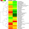
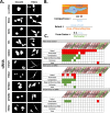
Fibrous scaffolds are used for bone tissue engineering purposes with great success across a variety of polymers with different physical and chemical properties. It is now evident that the correct degree of curvature promotes increased cytoskeletal tension on osteoprogenitors leading to osteogenic differentiation. However, the mechanotransductive pathways involved in this phenomenon are not fully understood. To achieve a reproducible and specific cellular response, an increased mechanistic understanding of the molecular mechanisms driving the fibrous scaffold mediated bone regeneration must be understood. High throughput siRNA mediated screening technology has been utilized for dissecting molecular targets that are important in certain cellular phenotypes. In this study, we used siRNA mediated gene silencing to understand the osteogenic differentiation observed on fibrous scaffolds. A high-throughput siRNA screen was conducted using a library collection of 863 genes including important human kinase and phosphatase targets on pre-osteoblast SaOS-2 cells. The cells were grown on electrospun poly(methyl methacrylate) (PMMA) scaffolds with a diameter of 0.938 ± 0.304 µm and a flat surface control. The osteogenic transcription factor RUNX2 was quantified with an in-cell western (ICW) assay for the primary screen and significant targets were selected via two sample t-test. After selecting the significant targets, a secondary screen was performed to identify osteoinductive markers that also effect cell shape on fibrous topography. Finally, we report the most physiologically relevant molecular signaling mechanisms that are involved in growth factor free, fibrous topography mediated osteoinduction. We identified GTPases, membrane channel proteins, and microtubule associated targets that promote an osteoinductive cell shape on fibrous scaffolds.
Conflict of interest statement
The authors declare no competing interests.
Figures


Similar articles
-
Dual drug-delivering polycaprolactone-collagen scaffold to induce early osteogenic differentiation and coupled angiogenesis.Biomed Mater. 2020 May 19;15(4):045008. doi: 10.1088/1748-605X/ab7978. Biomed Mater. 2020. PMID: 32427577
-
Electrospun scaffolds for multiple tissues regeneration in vivo through topography dependent induction of lineage specific differentiation.Biomaterials. 2015 Mar;44:173-85. doi: 10.1016/j.biomaterials.2014.12.027. Epub 2015 Jan 12. Biomaterials. 2015. PMID: 25617136
-
Synergistic interaction of platelet derived growth factor (PDGF) with the surface of PLLA/Col/HA and PLLA/HA scaffolds produces rapid osteogenic differentiation.Colloids Surf B Biointerfaces. 2016 Mar 1;139:68-78. doi: 10.1016/j.colsurfb.2015.11.053. Epub 2015 Nov 28. Colloids Surf B Biointerfaces. 2016. PMID: 26700235
-
Substrate curvature sensing through Myosin IIa upregulates early osteogenesis.Integr Biol (Camb). 2013 Nov;5(11):1407-16. doi: 10.1039/c3ib40068a. Epub 2013 Oct 9. Integr Biol (Camb). 2013. PMID: 24104522
-
Metformin induces osteoblastic differentiation of human induced pluripotent stem cell-derived mesenchymal stem cells.J Tissue Eng Regen Med. 2018 Feb;12(2):437-446. doi: 10.1002/term.2470. Epub 2017 Aug 11. J Tissue Eng Regen Med. 2018. PMID: 28494141 Free PMC article.
Cited by
-
Driving Cells with Light-Controlled Topographies.Adv Sci (Weinh). 2019 May 20;6(14):1801826. doi: 10.1002/advs.201801826. eCollection 2019 Jul 17. Adv Sci (Weinh). 2019. PMID: 31380197 Free PMC article.
-
Gingival Response to Dental Implant: Comparison Study on the Effects of New Nanopored Laser-Treated vs. Traditional Healing Abutments.Int J Mol Sci. 2020 Aug 22;21(17):6056. doi: 10.3390/ijms21176056. Int J Mol Sci. 2020. PMID: 32842709 Free PMC article. Clinical Trial.
-
Human mesenchymal stem cell morphology, migration, and differentiation on micro and nano-textured titanium.Bioact Mater. 2019 Sep 19;4:249-255. doi: 10.1016/j.bioactmat.2019.08.001. eCollection 2019 Dec. Bioact Mater. 2019. PMID: 31667441 Free PMC article.
-
Improving mechanical and antibacterial properties of PMMA via polyblend electrospinning with silk fibroin and polyethyleneimine towards dental applications.Bioact Mater. 2020 Apr 15;5(3):510-515. doi: 10.1016/j.bioactmat.2020.04.005. eCollection 2020 Sep. Bioact Mater. 2020. PMID: 32322761 Free PMC article.
References
MeSH terms
Substances
LinkOut - more resources
Full Text Sources

