Review
. 2019 Jan 25:41:2.
doi: 10.1186/s41021-019-0119-6. eCollection 2019.

Mechanism and regulation of DNA damage recognition in nucleotide excision repair

Affiliations
Review

Mechanism and regulation of DNA damage recognition in nucleotide excision repair

Masayuki Kusakabe et al. Genes Environ. .

Abstract

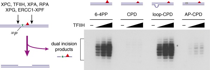
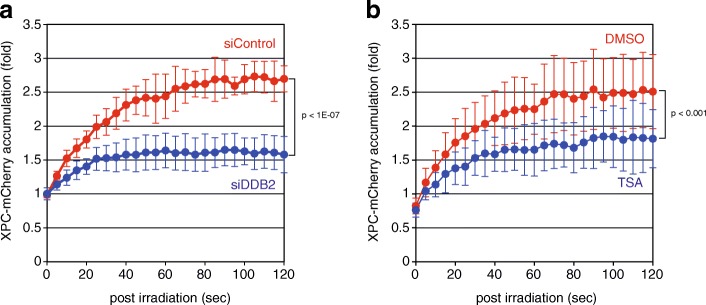
Nucleotide excision repair (NER) is a versatile DNA repair pathway, which can remove an extremely broad range of base lesions from the genome. In mammalian global genomic NER, the XPC protein complex initiates the repair reaction by recognizing sites of DNA damage, and this depends on detection of disrupted/destabilized base pairs within the DNA duplex. A model has been proposed that XPC first interacts with unpaired bases and then the XPD ATPase/helicase in concert with XPA verifies the presence of a relevant lesion by scanning a DNA strand in 5'-3' direction. Such multi-step strategy for damage recognition would contribute to achieve both versatility and accuracy of the NER system at substantially high levels. In addition, recognition of ultraviolet light (UV)-induced DNA photolesions is facilitated by the UV-damaged DNA-binding protein complex (UV-DDB), which not only promotes recruitment of XPC to the damage sites, but also may contribute to remodeling of chromatin structures such that the DNA lesions gain access to XPC and the following repair proteins. Even in the absence of UV-DDB, however, certain types of histone modifications and/or chromatin remodeling could occur, which eventually enable XPC to find sites with DNA lesions. Exploration of novel factors involved in regulation of the DNA damage recognition process is now ongoing.

Keywords: Chromatin; DNA damage recognition; Nucleotide excision repair; TFIIH; UV-DDB; XPA; XPC.


Conflict of interest statement

Not applicable.Not applicable.The authors declare that they have no competing interests.Springer Nature remains neutral with regard to jurisdictional claims in published maps and institutional affiliations.

Figures

Fig. 1
Fig. 1
A model for DNA damage recognition and verification in mammalian GG-NER. The XPC protein complex recognizes and binds to DNA sites with disrupted/destabilized base pair(s) regardless of the presence (left) or absence (right) of relevant lesions. After loading of the TFIIH complex, the XPD helicase subunit in TFIIH scans a DNA strand in 5′-3′ direction in conjunction with XPA, and the presence of a lesion is finally verified by blockage of this translocation. Even though a lesion is not present exactly at the XPC-bound site, this scanning mechanism may provide NER with a chance to find some nearby lesions
Fig. 2
Fig. 2
Stimulation of in vitro dual incision reactions with CPD substrates by the presence of mismatched bases or an AP site. Internally 32P-labeled DNA substrates containing a site-specific CPD (~ 200 bp in length) were incubated with six purified recombinant NER factors, and excised oligonucleotides containing both the 32P label and CPD were detected by denaturing polyacrylamide gel electrophoresis and the following autoradiography as described previously [12, 50]
Fig. 3
Fig. 3
Kinetic analyses of XPC recruitment to DNA damage sites in living cells. DNA damage was induced with the 780-nm femtosecond laser and three-photon absorption within subnuclear regions of human osteosarcoma U2OS cells stably expressing mCherry-fused XPC. Cells were pre-treated either with siRNA targeting DDB2 (a) or with a histone deacetylase inhibitor, trichostatin A (TSA) (b). Time-lapse images were acquired every 5 s with the Olympus FV-3000 confocal laser scanning microscope, and relative fluorescence intensities of the irradiated areas were quantified. Mean values and standard deviations were calculated from 10 (a) and 17 (b) samples, respectively

References

    1. Gillet LCJ, Schärer OD. Molecular mechanisms of mammalian global genome nucleotide excision repair. Chem Rev. 2006;106:253–276. doi: 10.1021/cr040483f. - DOI - PubMed
    1. Friedberg EC, Walker GC, Siede W, Wood RD, Schultz RA, Ellenberger T. DNA repair and mutagenesis, second edition. Washington, DC: ASM Press; 2006.
    1. Sugasawa K. Molecular mechanisms of DNA damage recognition for mammalian nucleotide excision repair. DNA Repair (Amst) 2016;44:110–117. doi: 10.1016/j.dnarep.2016.05.015. - DOI - PubMed
    1. Riedl T, Hanaoka F, Egly JM. The comings and goings of nucleotide excision repair factors on damaged DNA. EMBO J. 2003;22:5293–5303. doi: 10.1093/emboj/cdg489. - DOI - PMC - PubMed
    1. Sugasawa K, Ng JMY, Masutani C, Iwai S, van der Spek PJ, Eker APM, et al. Xeroderma pigmentosum group C protein complex is the initiator of global genome nucleotide excision repair. Mol Cell. 1998;2:223–232. doi: 10.1016/S1097-2765(00)80132-X. - DOI - PubMed