Review
. 2019 Mar;13(1):91-102.
doi: 10.1007/s12105-018-0981-8. Epub 2019 Mar 7.

Ulcerated Lesions of the Oral Mucosa: Clinical and Histologic Review

Affiliations
Review

Ulcerated Lesions of the Oral Mucosa: Clinical and Histologic Review

Sarah G Fitzpatrick et al. Head Neck Pathol. 2019 Mar.

Abstract

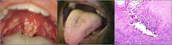
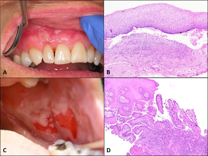
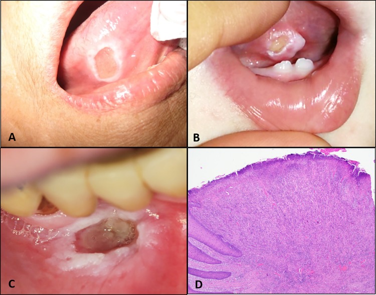
Ulcerated lesions of the oral cavity have many underlying etiologic factors, most commonly infection, immune related, traumatic, or neoplastic. A detailed patient history is critical in assessing ulcerative oral lesions and should include a complete medical and medication history; whether an inciting or triggering trauma, condition, or medication can be identified; the length of time the lesion has been present; the frequency of episodes in recurrent cases; the presence or absence of pain; and the growth of the lesion over time. For multiple or recurrent lesions the presence or history of ulcers on the skin, genital areas, or eyes should be evaluated along with any accompanying systemic symptoms such as fever, arthritis, or other signs of underlying systemic disease. Biopsy may be indicated in many ulcerative lesions of the oral cavity although some are more suitable for clinical diagnosis. Neoplastic ulcerated lesions are notorious in the oral cavity for their ability to mimic benign ulcerative lesions, highlighting the essential nature of biopsy to establish a diagnosis in cases that are not clinically identifiable or do not respond as expected to treatment. Adjunctive tests may be required for final diagnosis of some ulcerated lesions especially autoimmune lesions. Laboratory tests or evaluation to rule out systemic disease may be also required for recurrent or severe ulcerations especially when accompanied by other symptoms. This discussion will describe the clinical and histopathologic characteristics of a variety of ulcerated lesions found in the oral cavity.

Keywords: Aphthous stomatitis; Herpetic gingivostomatitis; Oral erythema multiforme; Oral lichen planus; Oral lymphoma; Oral medication related ulcer; Oral squamous cell carcinoma; Oral ulcer; Oral vesiculobullous lesions; Traumatic oral ulcer.


Conflict of interest statement

Conflict of interest

All authors declare no conflicts of interests.

Ethics Approval

This article does not contain any studies with human participants or animal performed by any of the authors.

Informed Consent

Not applicable for this article.

Figures

Fig. 1
Fig. 1
Bacterial ulceration. a Syphilitic ulceration of the soft palate. b Hematoxylin and eosin (H&E) medium power magnification demonstrating ulceration with perivascular and diffuse inflammation consisting of plasma cells and neutrophils. cTreponema pallidum immunohistochemical staining exhibiting abundant organisms (high power). Clinical and histologic photos courtesy of Dr. Brenda Nelson
Fig. 2
Fig. 2
Viral ulceration. a Primary hepetic infection involving the soft palate and oropharynx in a young adult (photo courtesy Dr. Donald Cohen). b Multiple coalescing ulcerations of the tongue and lips of secondary HSV related ulceration in an HIV positive patient (photo courtesy University of Florida Oral Pathology Biopsy Service Archives). c HSV related epithelial changes with prominent Tzanck cell formation (H&E medium power magnification)
Fig. 3
Fig. 3
Fungal ulceration. a Deep ulceration of the tongue secondary to histoplasmosis infection (photo courtesy Dr. Indraneel Bhattacharyya). b Fungal organisms consistent with histoplasmosis within inflamed tissue with numerous macrophages (H&E high power magnification)
Fig. 4
Fig. 4
Immune related ulcerations. a Aphthous stomatitis (photo courtesy Dr. Indraneel Bhattacharyya). b Erythema multiforme (photo courtesy Dr. Ashley Clark). c Ulcerated oral lichen planus (photo courtesy Dr. Mary Hartigan and Dr. Alan Fetner). d Histologic appearance of oral lichen planus demonstrating band-like lymphocytic infiltration in the superficial lamina propria and degeneration of the basal cell layer (H&E low power magnification)
Fig. 5
Fig. 5
Vesiculobullous immune related ulcerations. a Mucous membrane pemphigoid affecting the gingiva (photo courtesy Dr. Angela Wilson). b MMP exhibiting subepithelial separation and chronic inflammation (H&E low power magnification). c Pemphigus vulgaris affecting the buccal mucosa (photo courtesy Dr. Christopher Lee). d PV exhibiting intraepithelial separation (H&E low power magnification)
Fig. 6
Fig. 6
Acute traumatic ulcerations. a Chemical burn due to topical aspirin placement over implant sites (photo courtesy Dr. Shawn Lottier). b Post-anesthetic ulceration reaction (photo courtesy University of Florida Oral Pathology Biopsy Service Archives)
Fig. 7
Fig. 7
Chronic traumatic ulcerations. a Traumatic ulcerative granuloma with stromal eosinophilia (TUGSE) on lateral tongue (photo courtesy Dr. Prashant Pandya). b Riga-Fede disease on ventral tongue adjacent to natal teeth in an infant (photo courtesy Dr. Naveen Allin). c Factitious traumatic injury to the labial vestibule secondary to picking habit (photo courtesy Dr. Donald Cohen). d Deep TUGSE ulceration extending into skeletal muscle (H&E low power magnification)
Fig. 8
Fig. 8
Neoplastic ulcerations. a Squamous cell carcinoma of the soft palate (photo courtesy Dr. Hardeep Chehal). b Invasive squamous cell carcinoma (right) and adjacent ulceration (left) (H&E low power magnification × 2). c T-cell lymphoma of the upper lip (photo courtesy Dr. Donald Cohen). d Atypical lymphocytic proliferation in T-cell lymphoma (H&E medium power magnification). e EBVMCU of the maxillary hard palate (photo courtesy Dr. Leah Strange). f EBVMCU demonstrating ulceration overlying atypical lymphocytic proliferation with Reed Sternberg like CD30 + cells (H&E medium power magnification)

References

    1. Leão JC, Gueiros LA, Porter SR. Oral manifestations of syphilis. Clinics. 2006;61:161–166. - PubMed
    1. Alam F, Argiriadou AS, Hodgson TA, et al. Primary syphilis remains a cause of oral ulceration. Br Dent J. 2000;189:352–354. - PubMed
    1. Little JW. Syphilis: an update. Oral Surg Oral Med Oral Pathol Oral Radiol Endod. 2005;100:3–9. - PubMed
    1. Stewart RJ, Tsang CA, Pratt RH, et al. Tuberculosis —United States, 2017. MMWR Morb Mortal Wkly Rep. 2018;67:317–323. - PMC - PubMed
    1. Krawiecka E, Szponar E. Tuberculosis of the oral cavity: an uncommon but still a live issue. Adv Dermatol Allergol. 2015;32(4):302–306. - PMC - PubMed

LinkOut - more resources