. 2019 Sep;32(6):749-761.
doi: 10.1002/ca.23343. Epub 2019 Feb 19.

What is a tongue tie? Defining the anatomy of the in-situ lingual frenulum

Affiliations

What is a tongue tie? Defining the anatomy of the in-situ lingual frenulum

Nikki Mills et al. Clin Anat. 2019 Sep.

Abstract

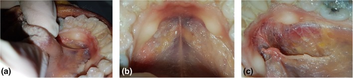
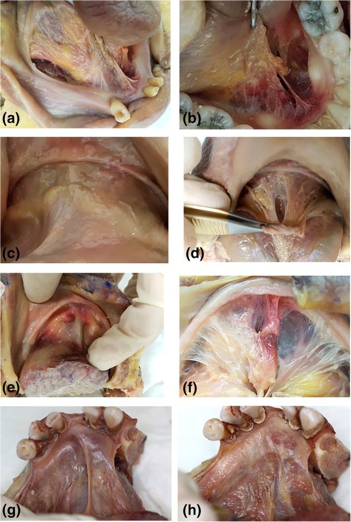
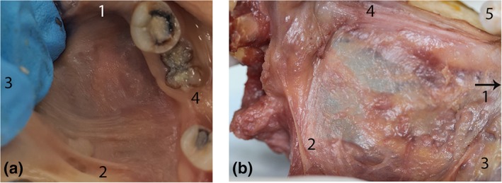
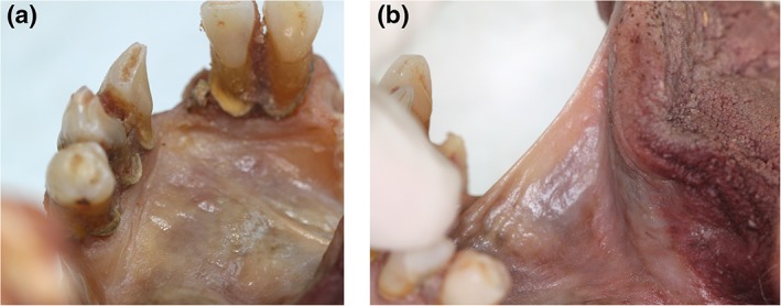
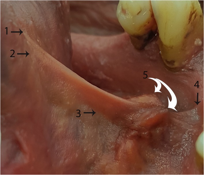
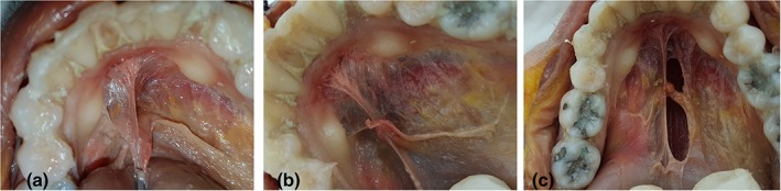
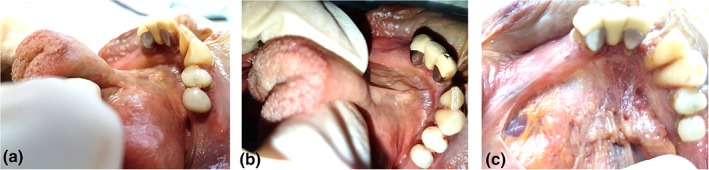
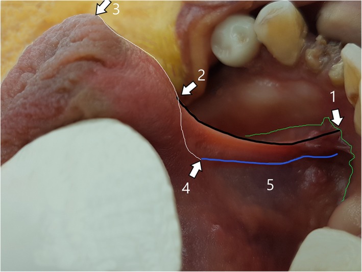
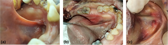
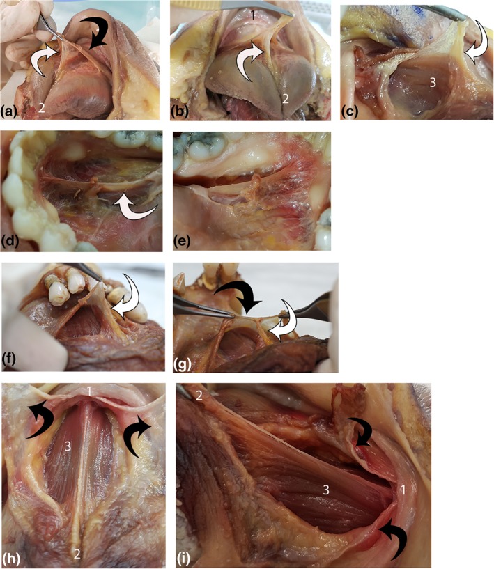
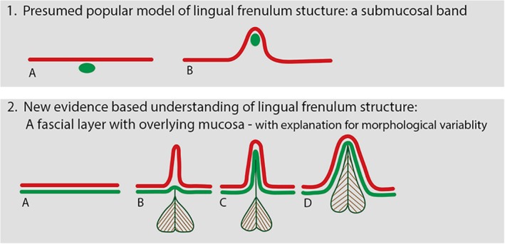
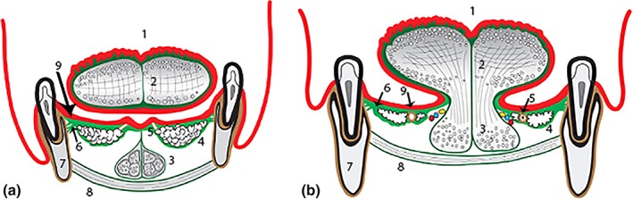
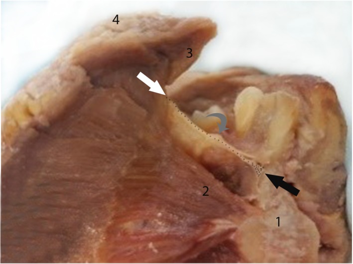
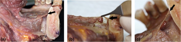
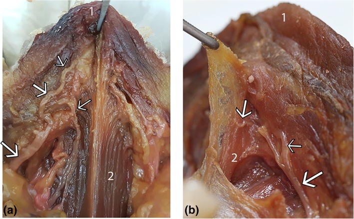
Surgical release of the lingual frenulum (frenotomy) has become an increasingly common procedure, performed from birth through to adulthood. Surprisingly, detailed anatomy of the in-situ lingual frenulum has never been described, and no anatomical basis has been proposed for the individual variability in frenulum morphology. The lingual frenulum is frequently referred to as a "cord" or "submucosal band" of connective tissue, yet there is no evidence to support this anatomical construct. This paper aims to describe the anatomy of the in-situ lingual frenulum and its relationship to floor of mouth structures. Fresh tissue microdissection of the lingual frenulum and floor of mouth was performed on nine adult cadavers with photo-documentation and description of findings. The lingual frenulum is a dynamic structure, formed by a midline fold in a layer of fascia that inserts around the inner arc of the mandible, forming a diaphragm-like structure across the floor of mouth. This fascia is located immediately beneath the oral mucosa, fusing centrally with the connective tissue on the tongue's ventral surface. The sublingual glands and submandibular ducts are enveloped by the fascial layer and anterior genioglossus fibers are suspended beneath it. Lingual nerve branches are located superficially on the ventral surface of the tongue, immediately deep to the fascia. The lingual frenulum is not a discrete midline structure. It is formed by dynamic elevation of a midline fold in the floor of mouth fascia. With this study, the clinical concept of ankyloglossia and its surgical management warrant revision. Clin. Anat. 32:749-761, 2019. © 2019 The Authors. Clinical Anatomy published by Wiley Periodicals, Inc. on behalf of American Association of Clinical Anatomists.

Keywords: ankyloglossia; congenital; fascia; floor of mouth; frenotomy; lingual frenulum; lingual nerve; oral cavity; tongue tie.


Figures

Figure 1
Figure 1
Floor of mouth fascia—mandibular attachment. (a, b) Mucosa intact, tongue elevated, and retracted to create tension along frenulum. (c) Mucosa removed, exposing floor of mouth fascia, and attachment of the fascia around the inner surface of the mandible. [Color figure can be viewed at http://wileyonlinelibrary.com]
Figure 2
Figure 2
Variations in thickness and height of midline mandibular attachment of floor of mouth fascia. All images: tongue elevation and retraction to place fascia under tension. Specimen 1: (a) mucosa removed, thin/transparent fascia, elevated midline mandibular attachment. Specimen 2: (b) mucosa removed, thin/transparent fascia, elevated midline mandibular attachment. Specimen 3: (c) mucosa intact (d) mucosa removed: thick/opaque fascia, no elevation of midline mandibular attachment. Specimen 4: (e) mucosa intact (f) mucosa removed: thin/transparent fascia, slight elevation of midline mandibular attachment. Specimen 5: (g) mucosa intact, (h) mucosa removed: very thick/opaque fascia, slight elevation of midline mandibular attachment. [Color figure can be viewed at http://wileyonlinelibrary.com]
Figure 3
Figure 3
Continuity of floor of mouth fascia around lateral sides of the tongue. (a) Mucosa intact: lateral floor of mouth. Tongue retracted medially to create tension in fascia. (b) Posterior‐most aspect of floor of mouth fascia (left side, posterior tongue medialized to display space between tongue and mandible, mucosa removed). Here, the fascia is a thin, transparent layer, with high distensibility, continuous posteriorly as a submucosal layer extending into the piriform fossa. (1) Anterior (2) Posterior (3) Medial (tongue) (4) Lateral (mandible) (5) Molar tooth. [Color figure can be viewed at http://wileyonlinelibrary.com]
Figure 4
Figure 4
Tongue elevation creating tension in floor of mouth fascia. Both images of same specimen. (a) Tongue in “neutral” position: no tension in floor of mouth fascia. (b) Tongue elevated: tension drawing up the floor of mouth fascia (with overlying mucosa) to form a midline fold (recognizable as the lingual frenulum). [Color figure can be viewed at http://wileyonlinelibrary.com]
Figure 5
Figure 5
“Surface anatomy” of the lingual frenulum—Example 1 Tongue elevated to create tension in the floor of mouth fascia, raising the fold of the frenulum. (1) Highest point of midline mucosal attachment to ventral tongue. (2) Highest point of midline floor of mouth fascia attachment to ventral tongue. (3) Genioglossus—drawn into base of lingual frenulum (suspended from floor of mouth fascia). (4) Highest point of midline fascial attachment on the inner surface of mandible. (5) (White arrows): Submandibular duct openings—suspended from floor of mouth fascia. [Color figure can be viewed at http://wileyonlinelibrary.com]
Figure 6
Figure 6
Height of midline fascial attachment to mandible—elevated. (a, b) Floor of mouth with mucosa removed. Fascial attachment to mandible elevated in midline, creating “eiffel tower” appearance. (c) Window created in fascia (to right of midline) to show underlying sublingual space and genioglossus. [Color figure can be viewed at http://wileyonlinelibrary.com]
Figure 7
Figure 7
Height of midline fascial attachment to mandible—not elevated. (a and b): Mucosa in situ. (c) Mucosa removed: height of midline fascial attachment to mandible not elevated relative to height of fascial attachment on either side of midline. [Color figure can be viewed at http://wileyonlinelibrary.com]
Figure 8
Figure 8
“Surface anatomy” of the lingual frenulum—Example 2. (1) midline mandibular attachment of floor of mouth fascia (higher than its attachment either side of midline). (2) Midline ventral tongue attachment of floor of mouth fascia (same height as mucosal attachment). (3) Tip of tongue (junction of ventral and dorsal tongue surfaces). (4) Location of genioglossus merging into body of tongue. (5) Superior edge of genioglossus fibers, drawn up into base of lingual frenulum with tongue elevation. Green line: height of attachment of floor of mouth fascia to mandible (with overlying mucosa closely applied) nb: the attachment is higher in midline, creating an “eiffel tower” appearance. Black line: (between points 1 and 2): fold of the lingual frenulum—opaque, with the fascial layer elevated up to the top of the fold. White line: (between points 3 and 4): midline ventral tongue surface. Distance between points 2 and 3: the “free length” of the tongue. Blue line: superior‐most aspect of genioglossus fibers, being drawn up into frenulum. [Color figure can be viewed at http://wileyonlinelibrary.com]
Figure 9
Figure 9
Variable morphology of lingual frenulum under tension. (a) Transparent: mucosal fold elevating just above fascial fold, genioglossus at base of frenulum. (b) Opaque: mucosa and fascia drawn up to top of fold, genioglossus drawn up into mid‐frenulum. (c) Thick/bulky: genioglossus drawn up into frenulum together with mucosa and fascia, less defined fold. [Color figure can be viewed at http://wileyonlinelibrary.com]
Figure 10
Figure 10
Suspension of genioglossus from floor of mouth fascia. Each line shows multiple views of a single specimen (four specimens total). (1) mandible, (2) ventral tongue tip, and (3) genioglossus. White arrow: connective tissue suspending genioglossus. Black arrow: floor of mouth fascia. (a, b): Suspension of genioglossus from floor of mouth fascia. (c) Connective tissue suspending genioglossus continuous with genioglossus epimysium. (d, e) Left and right lateral view of connective tissue suspending genioglossus. (f, g) Midline suspension of genioglossus from floor of mouth fascia. (h, i) Floor of mouth fascia divided (midline sagittal incision) and retracted, exposing genioglossus (under tension with tongue elevated and retracted). Suspending connective tissue and epimysium removed. [Color figure can be viewed at http://wileyonlinelibrary.com]
Figure 11
Figure 11
Anatomically based understanding of lingual frenulum structure. Diagram illustrating coronal section of floor of mouth: (1) Current “presumed” understanding of lingual frenulum structure: a submucosal band: (a): tongue relaxed, (b): tongue elevated, raising lingual frenulum. Red line: oral mucosa green oval: coronal section of connective tissue “band.” (2) Our newly proposed anatomically based understanding of lingual frenulum structure: red line: oral mucosa green line: floor of mouth fascia, with genioglossus suspended from fascia. (a): Tongue relaxed, floor of mouth fascia immediately beneath mucosa. (bd) Variations in frenulum morphology with tongue elevated to raise frenulum. (b) “Transparent” frenulum—mucosal fold elevates above fascia to form fold, with fascia remaining low/at base of fold. (c) “Opaque” frenulum—mucosal and fascia elevate together to form fold. (d) “Thick” frenulum”—mucosa and fascia elevate together, with genioglossus also drawn into fold. [Color figure can be viewed at http://wileyonlinelibrary.com]
Figure 12
Figure 12
Diagram of floor of mouth fascia (coronal section). (a) anterior floor of mouth (under blade of tongue). (b) postero‐lateral floor of mouth (under lateral sides of tongue). (1) Tongue—dorsal surface, (2) Tongue—intrinsic muscles (median septum and superficial connective tissue—dark green), (3) Anterior fibers of genioglossus (in diagram a: suspended from floor of mouth fascia, in diagram; b: merging into body of tongue), (4) Sublingual glands (enveloped by and suspended from floor of mouth fascia), (5) Submandibular duct (in diagram a: entering papilla at mucosal surface, in diagram b: embedded in fascia with sublingual glands), (6) Floor of mouth fascia—spans floor of mouth (bright green)—insertion into mandible immediately beneath oral mucosa), (7) Mandible, (8) Mylohyoid, and (9) Oral mucosa (red layer). [Color figure can be viewed at http://wileyonlinelibrary.com]
Figure 13
Figure 13
Hemi‐mandible: mid‐sagittal section through tongue and floor of mouth. Floor of mouth fascia (dotted line) with insertion anteriorly onto mandible (black arrow) and posteriorly onto ventral tongue (white arrow). “Window” through to sublingual space beneath fascia, showing submandibular duct (gray arrow) and sublingual glands suspended from fascia (visible as irregular tan‐colored tissue on under surface of fascia, between submandibular duct and mandibular insertion). (1) Mandible (2) Genioglossus (3) Ventral tongue surface (4) Dorsal tongue surface. [Color figure can be viewed at http://wileyonlinelibrary.com]
Figure 14
Figure 14
Section through mandible in parasagittal plane: showing floor of mouth fascia and sublingual space. All images are of same specimen: demonstrating suspension of sublingual glands from inferior surface of floor of mouth fascia. (a) Lateral view of specimen: showing ventral tongue surface and mandible, with floor of mouth fascia spanning between and sublingual space visible beneath fascia. (b) Specimen tilted to show inferior surface of fascia, with suspended sublingual glands. (c) Close‐up lateral view, showing mandibular attachment of fascia and suspended sublingual glands. Black arrow: indicating location of floor of mouth fascia attachment to mandible. (1) Mandible, (2) Sublingual glands (3) Ventral tongue surface. [Color figure can be viewed at http://wileyonlinelibrary.com]
Figure 15
Figure 15
Lingual nerve branches: location on ventral tongue. Two specimens (a and b): floor of mouth fascia removed, nerve branches shown to be located immediately beneath the fascia on the surface of the muscle, with branches passing toward the tongue tip and onto the connective tissue suspending genioglossus. (1) Ventral tongue tip (2) Genioglossus. White arrows: lingual nerve branches. [Color figure can be viewed at http://wileyonlinelibrary.com]

References

    1. Baker A. 2015. Surgical treatment of ankyloglossia. Oper Tech Otolaryngol Head Neck Surg 26:28–32.
    1. Barot VJ, Vishnoi SL, Chandran S, Bakutra GV. 2014. Laser: The torch of freedom for ankyloglossia. Indian J Plastic Surg 47:418–422. - PMC - PubMed
    1. Chinnadurai S, Francis D, Epstein R, Morad A, Kohanim S, McPheeters M. 2015. Treatment of ankyloglossia for reasons other than breastfeeding: A systematic review. Pediatrics 135:1467–1474. - PMC - PubMed
    1. Chu MW, Bloom DC. 2009. Posterior ankyloglossia: A case report. Int J Pediatr Otorhinolaryngol 73:881–883. - PubMed
    1. Coryllos E, Watson Genna, C , Salloum A. 2004. Congenital tongue tie and its impact on breastfeeding. American Academy of Pediatrics (Summer): Breastfeeding: Best for Mother and Baby:1–6.

LinkOut - more resources