. 2019 Apr;23(4):2558-2567.
doi: 10.1111/jcmm.14146. Epub 2019 Jan 31.

FBXL10 regulates cardiac dysfunction in diabetic cardiomyopathy via the PKC β2 pathway

Affiliations

FBXL10 regulates cardiac dysfunction in diabetic cardiomyopathy via the PKC β2 pathway

Leilei Yin et al. J Cell Mol Med. 2019 Apr.

Abstract

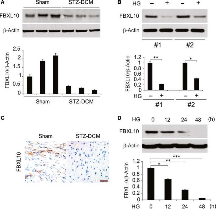
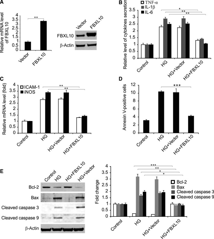
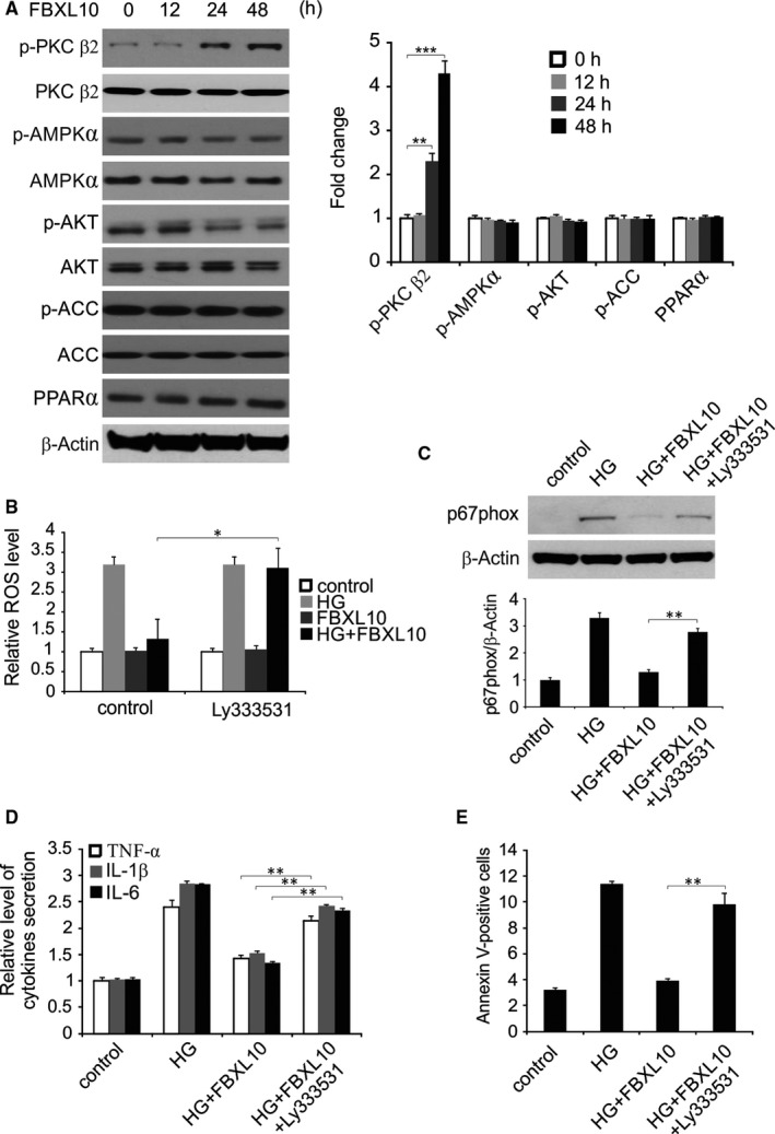
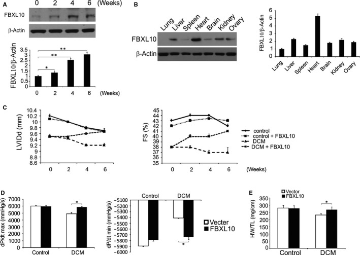
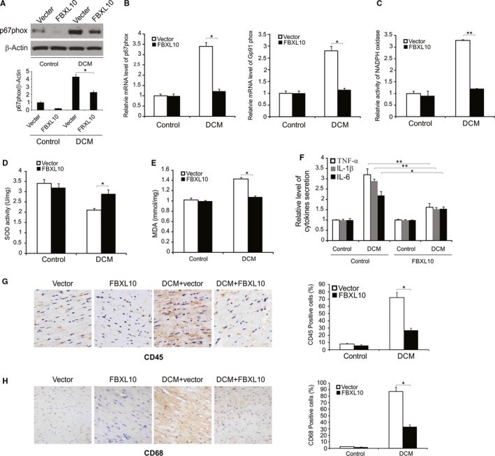
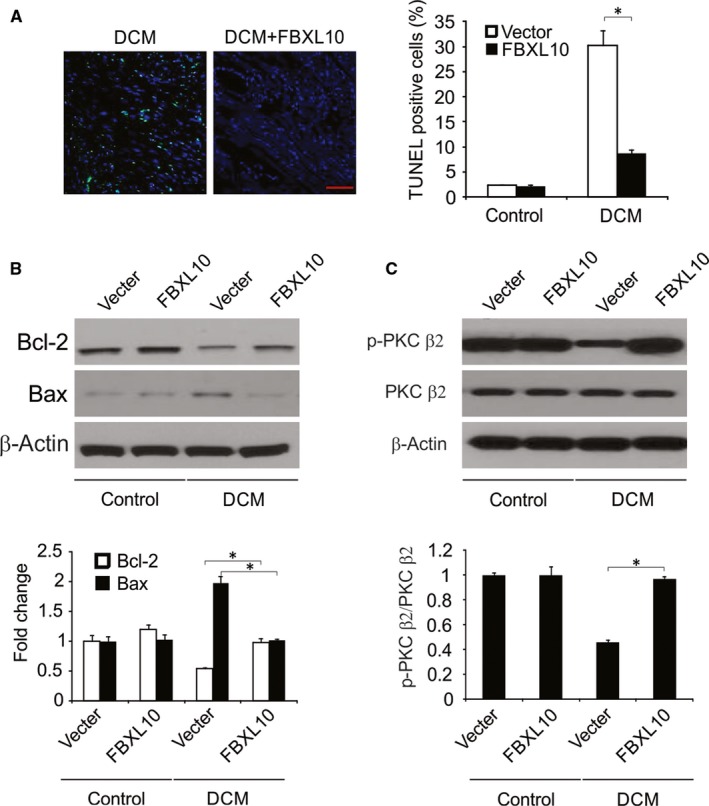
Diabetic cardiomyopathy (DCM) is a condition associated with significant structural changes including cardiac tissue necrosis, localized fibrosis, and cardiomyocyte hypertrophy. This study sought to assess whether and how FBXL10 can attenuate DCM using a rat streptozotocin (STZ)-induced DCM model system. In the current study, we found that FBXL10 expression was significantly decreased in diabetic rat hearts. FBXL10 protected cells from high glucose (HG)-induced inflammation, oxidative stress, and apoptosis in vitro. In addition, FBXL10 significantly activated PKC β2 signaling pathway in H9c2 cells and rat model. The cardiomyocyte-specific overexpression of FBXL10 at 12 weeks after the initial STZ administration attenuated oxidative stress and inflammation, thereby reducing cardiomyocyte death and preserving cardiac function in these animals. Moreover, FBXL10 protected against DCM via activation of the PKC β2 pathway. In conclusion, FBXL has the therapeutic potential for the treatment of DCM.

Keywords: Apoptosis; Diabetic cardiomyopathy; FBXL10; Inflammation; PKC β2; oxidative stress.


Conflict of interest statement

The authors confirm that there are no conflicts of interest.

Figures

Figure 1
Figure 1
FBXL10 was downregulated in the heart in STZ‐induced diabetic mice, in HG‐stimulated cardiomyocytes and H9c2 cells. (A) The expression of FBXL10 in STZ‐induced diabetic hearts were analyzed by Western blotting and normalized to β‐actin. Data represent the mean ± SD of three independent experiments. (B) The protein level of FBXL10 in cardiomyocytes was analyzed by Western blotting and normalized to β‐actin. Data represent the mean ± SD of three independent experiments. **, < 0.01; *, < 0.05. (C) Immunohistochemistry of FBXL10 in STZ‐induced diabetic hearts. Scale bar, 50 μm (n = 6). (D) H9c2 cells were treated with high glucose (HG, 33 mmol/L glucose), FBXL10 level was analyzed by Western blotting and normalized to β‐actin. Data represent the mean ± SD of three independent experiments. **< 0.01; *< 0.05
Figure 2
Figure 2
FBXL10 reduced HG‐induced inflammatory response and apoptosis. (A) H9c2 cells were transfected with FBXL10, FBXL10 expression was analyzed by Western blotting and real‐time RT‐PCR. Data represent the mean ± SD of three independent experiments. **< 0.01. (B) H9c2 cells with or without FBXL10 transfection were stimulated by HG. The secretion of TNF‐α, IL‐1β and IL‐6 was determined by ELISA. Data represent the mean ± SD of three independent experiments. **< 0.01; *< 0.05. (C) H9c2 cells with or without FBXL10 transfection were stimulated by HG. The expression of ICAM‐1 and iNOS was assessed by real time RT‐PCR. Data represent the mean ± SD of three independent experiments. **< 0.01. (D) H9c2 cells with or without FBXL10 transfection were stimulated by HG. Cell apoptosis was assessed by flow cytometry. Data represent the mean ± SD of three independent experiments. ***< 0.001. (E) H9c2 cells with or without FBXL10 transfection were stimulated by HG. Indicated protein level was detected by Western blotting and normalized to β‐actin. Data represent the mean ± SD of three independent experiments. ***< 0.001; **< 0.01; *< 0.05
Figure 3
Figure 3
FBXL10 suppressed HG‐induced production of ROS, inflammation and apoptosis in H9c2 cells via PKC β2. (A) H9c2 cells were transfected with FBXL10. Indicated protein level was detected by Western blotting at indicated time point and normalized to β‐actin. Data represent the mean ± SD of three independent experiments. ***< 0.001; **< 0.01. (B) H9c2 cells transfected with FBXL10 were treated with HG with or without Ly333531. ROS was analyzed by DCFH‐DA. Data represent the mean ± SD of three independent experiments. *< 0.05. (C) H9c2 cells transfected with FBXL10 were treated with HG with or without Ly333531. Protein level of P67phox were analyzed by Western blotting and normalized to β‐actin. Data represent the mean ± SD of three independent experiments. **< 0.01. (D) H9c2 cells transfected with FBXL10 were treated with HG with or without Ly333531. The secretion of TNF‐α, IL‐1β and IL‐6 was determined by ELISA. Data represent the mean ± SD of three independent experiments. **< 0.01. (E) H9c2 cells transfected with FBXL10 were treated with HG with or without Ly333531. Apoptosis was analyzed by Flow cytometry. Data represent the mean ± SD of three independent experiments. **< 0.01
Figure 4
Figure 4
FBXL10 improved diabetes‐induced cardiac dysfunction in vivo. (A) FBXL10 overexpression in rat hearts after infection with an AAV was analyzed by Western blotting and normalized to β‐actin. Data represent the mean ± SD of three independent experiments. **< 0.01; *< 0.05. (B) The expression of FBXL10 in heart and extracardiac organs after transfection was analyzed by Western blotting and normalized to β‐actin. (C) Alteration of LVIDd and FS after overexpression of FBXL10 (n = 6). (D) Effect of on haemodynamic measurements (n = 6). (E) The ratio of heart weight (HW) to tibia length (TL) (n = 6). *< 0.05
Figure 5
Figure 5
FBXL10 attenuated diabetes‐induced oxidative injury and inflammation in vivo. (A) The expression of p67phox in diabetic rat hearts was analyzed by Western blotting and normalized to β‐actin. Data represent the mean ± SD of three independent experiments. *< 0.05. (B) The mRNA level of P67phox and Gp91phox in diabetic hearts was analyzed by Real‐time PCR. Data represent the mean ± SD of three independent experiments. *< 0.05. (C) NADPH oxidase activity in diabetic hearts by FBXL10 overexpression. Data represent the mean ± SD of three independent experiments. **< 0.01. (D) Total SOD activity in diabetic hearts after FBXL10 overexpression. Data represent the mean ± SD of three independent experiments. *< 0.05. (E) Lipid peroxidation in diabetic hearts. Data represent the mean ± SD of three independent experiments. *< 0.05. (F) mRNA level of myocardial TNF‐α, IL‐1β and IL‐6 in rats with diabetes. Data represent the mean ± SD of three independent experiments. **< 0.01; *< 0.05. (G) CD45 expression in the rat heart with diabetes was analyzed by immunohistochemistry. Data represent the mean ± SD of three independent experiments. *< 0.05. (H) CD68 expression in the rat heart with diabetes was analyzed by immunohistochemistry. Data represent the mean ± SD of three independent experiments. *< 0.05
Figure 6
Figure 6
FBXL10 attenuated diabetes‐induced apoptosis in vivo. (A) TUNEL staining in diabetic hearts (n = 6). Scale bar, 20 μm. (B) The expression of Bcl‐2 and Bax in diabetic hearts was analyzed by Western blotting and normalized to β‐actin. Data represent the mean ± SD of three independent experiments. *< 0.05. (C) The expression of phosphor‐ and totle‐PKC β2 in the diabetic hearts was analyzed by Western blotting. The level of p‐PKC β2 was normalized to PKC β2. Data represent the mean ± SD of three independent experiments. *< 0.05

References

    1. Miki T, Yuda S, Kouzu H, Miura T. Diabetic cardiomyopathy: pathophysiology and clinical features. Heart Fail Rev. 2013;18:149‐166. - PMC - PubMed
    1. Tarquini R, Pala L, Brancati S, et al. Clinical approach to diabetic cardiomyopathy: a review of human studies. Curr Med Chem. 2018;25:1510‐1524. - PubMed
    1. Fang ZY, Prins JB, Marwick TH. Diabetic cardiomyopathy: evidence, mechanisms, and therapeutic implications. Endocr Rev. 2004;25:543‐567. - PubMed
    1. Toedebusch R, Belenchia A, Pulakat L. Diabetic cardiomyopathy: impact of biological sex on disease development and molecular signatures. Front Physiol. 2018;9:453. - PMC - PubMed
    1. Kannel WB, McGee DL. Diabetes and cardiovascular disease. the Framingham study. JAMA. 1979;241:2035‐2038. - PubMed

MeSH terms