. 2019 Mar;8(3):1169-1174.
doi: 10.1002/cam4.1969. Epub 2019 Jan 30.

Disruption of erythroid nuclear opening and histone release in myelodysplastic syndromes

Affiliations

Disruption of erythroid nuclear opening and histone release in myelodysplastic syndromes

Baobing Zhao et al. Cancer Med. 2019 Mar.

Abstract

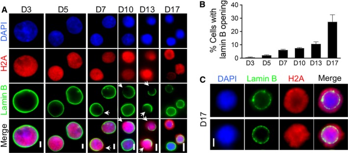
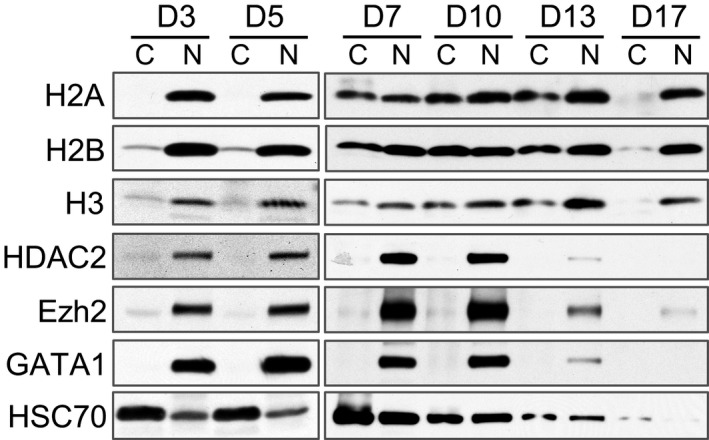
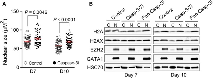
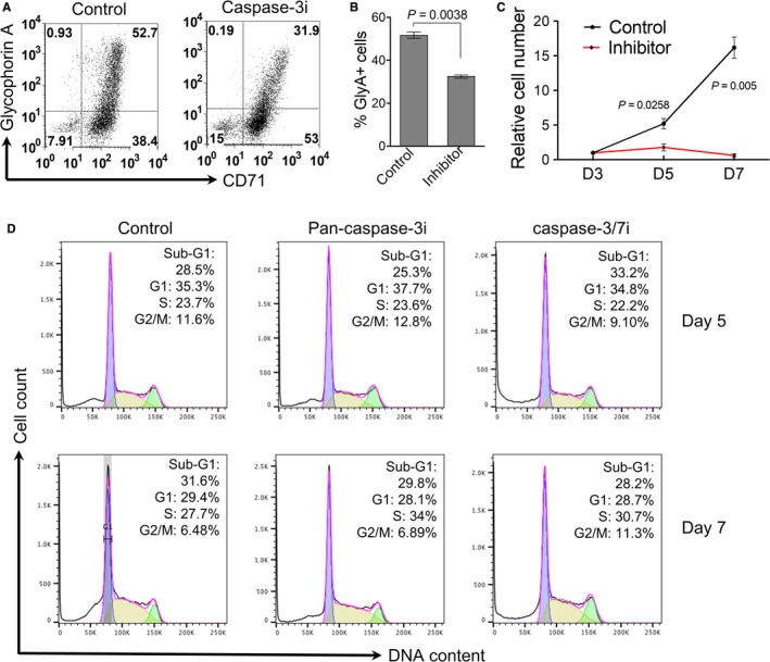
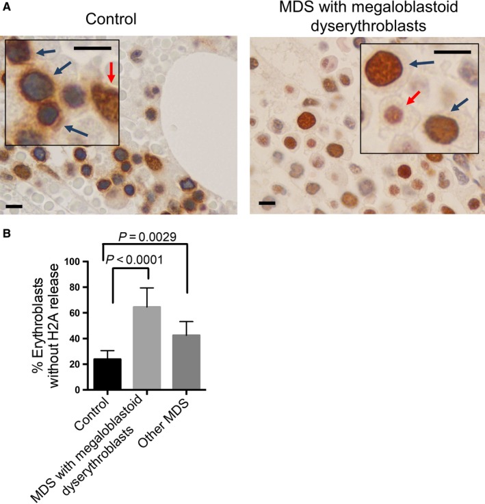
Mammalian terminal erythropoiesis involves several characteristic phenomena including chromatin condensation and enucleation. One of the newly identified features of terminal erythropoiesis in mouse is a dynamic nuclear opening and histone release process, which is required for chromatin condensation. However, it is unclear whether the same feature is present in human. Here, we use an in vitro human CD34-positive hematopoietic stem and progenitor cell culture system and reveal that nuclear openings and histone release are also identified during human terminal erythropoiesis. In contrast to mouse in which each erythroblast contains a single opening, multiple nuclear openings are present in human erythroblast, particularly during the late-stage differentiation. The nuclear opening and histone release process is mediated by caspase-3. Inhibition of caspase-3 blocks nuclear opening, histone release, chromatin condensation, and terminal differentiation. We confirm the finding of histone cytosolic release in paraffin-embedded human bone marrow in vivo. Importantly, we find that patients with myelodysplastic syndrome (MDS) exhibit significant defects in histone release in the dysplastic erythroblasts. Our results reveal developmentally conserved nuclear envelop and histone dynamic changes in human terminal erythropoiesis and indicate that disruption of the histone release process plays a critical role in the pathogenesis of dyserythropoiesis in MDS.

Keywords: chromatin condensation; enucleation; erythropoiesis; myelodysplastic syndromes.


Conflict of interest statement

None declared.

Figures

Figure 1
Figure 1
Caspase‐3‐mediated nuclear opening formation with histone release during human terminal erythroid differentiation. A, Immunofluorescent stains of lamin B and H2A in differentiating primary human erythroblasts at indicated days in culture. Arrows indicate lamin B openings. Scale bars: 5 µm. B, Quantitative analysis of the percentage of cells with lamin B opening in A. C, Immunofluorescent stains of lamin B and H2A in culture human erythroblasts on day 17. Scale bars: 2 µm
Figure 2
Figure 2
Histones are partially released into the cytoplasm during human terminal erythroid differentiation. Western blot analysis of indicated proteins from fractionated cytoplasmic (C) and nuclear (N) lysates of differentiating primary human erythroblasts on indicated days in culture with Epo. An equal number of cells were loaded in each lane
Figure 3
Figure 3
Caspase‐3 activation is required for nuclear condensation and histone release in human terminal erythropoiesis. A, The effect of caspase‐3 inhibitor on nuclear condensation. CD34+ cultured in Epo‐containing medium were treated with caspase‐3 inhibitor (Caspase‐3i) on day 3 in culture and collected on day 7 or day 10. Nuclear size was measured by calculating the DAPI‐positive area using the ImageJ software. Each dot represents a single cell. Data were obtained from three independent experiments. B, Western blot analysis of indicated proteins from fractionated cytoplasmic (C) and nuclear (N) lysates of indicated cells. CD34+ cells were treated with caspase‐3/7 inhibitor (casp‐3/7i) or pan‐caspase‐3 inhibitor (pan‐casp‐3i) on day 3 in culture and collected on day 7 or day 10. An equal number of cells were loaded in each lane
Figure 4
Figure 4
Caspase‐3 inhibition leads to the blockage of human erythroid cells differentiation and proliferation. A, CD34+ cells cultured in Epo‐containing medium were treated with caspase‐3 inhibitor on day 3 in culture and collected on day 7. Flow cytometric analysis of transferrin receptor (CD71) and glycophorin A were performed. B, Quantitative analysis of the percentage of glycophorin A positive cells (GlyA+) in A. Data were obtained from three independent experiments. C, CD34+ cells cultured in Epo‐containing medium were treated with caspase‐3 inhibitor on day 3, and the total cell number was counted on indicated days in culture. Data were shown as fold changes (day 3 as 1) and obtained from three independent experiments. D, Flow cytometric analysis of cell cycle distribution. Cells were treated as in C and analyzed by flow cytometry using propidium iodide (PI)‐staining. The percentages of populations of different cell cycle phases are shown in the upper‐right corner. The data are representative of three independent experiments
Figure 5
Figure 5
Myelodysplastic syndrome (MDS) patients exhibit reduced nuclear opening formation and histone release in erythroblasts. A, Representative immunohistochemical stains of H2A in normal control individuals and patients with myelodysplastic syndromes carrying megaloblastoid erythroblasts. Blue arrows indicate erythroblasts. Red arrows indicate other hematopoietic cells. Scale bars: 8 μm. The unstained cells are red blood cells due to hemorrhage during biopsy. B, Quantitative analysis of the percentage of erythroblasts without H2A release. Control: N = 6, MDS with megaloblastoid erythroblasts: N = 10, Other MDS: N = 7. 500 erythroblasts were counted in each case

References

    1. Ji P, Murata‐Hori M, Lodish HF. Formation of mammalian erythrocytes: chromatin condensation and enucleation. Trends Cell Biol. 2011;21(7):409‐415. - PMC - PubMed
    1. Hattangadi SM, Wong P, Zhang L, Flygare J, Lodish HF. From stem cell to red cell: regulation of erythropoiesis at multiple levels by multiple proteins, RNAs, and chromatin modifications. Blood. 2011;118(24):6258‐6268. - PMC - PubMed
    1. An X, Schulz VP, Mohandas N, Gallagher PG. Human and murine erythropoiesis. Curr Opin Hematol. 2015;22(3):206‐211. - PMC - PubMed
    1. Ji P. New insights into the mechanisms of mammalian erythroid chromatin condensation and enucleation. Int Rev Cell Mol Biol. 2015;316:159‐182. - PubMed
    1. Ji P, Jayapal SR, Lodish HF. Enucleation of cultured mouse fetal erythroblasts requires Rac GTPases and mDia2. Nat Cell Biol. 2008;10(3):314‐321. - PubMed

Publication types