Review
. 2019 Mar;100(3):350-366.
doi: 10.1099/jgv.0.001193. Epub 2019 Jan 31.

Vehicles of intercellular communication: exosomes and HIV-1

Affiliations
Review

Vehicles of intercellular communication: exosomes and HIV-1

Jennifer L Welch et al. J Gen Virol. 2019 Mar.

Abstract

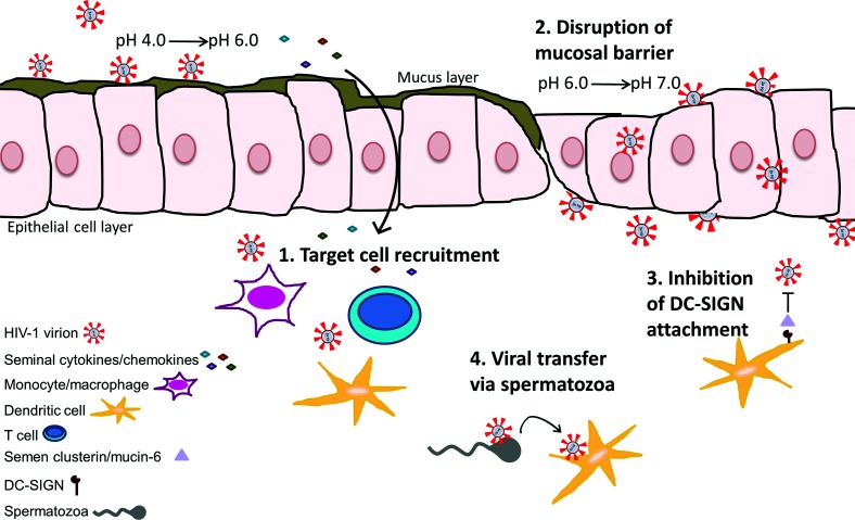
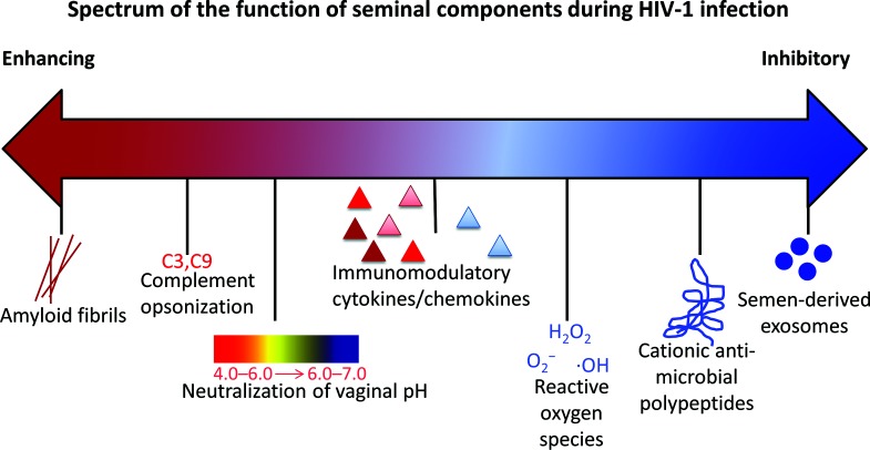
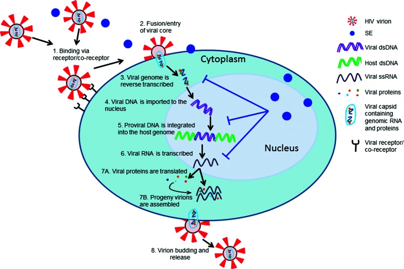
The terms extracellular vesicles, microvesicles, oncosomes, or exosomes are often used interchangeably as descriptors of particles that are released from cells and comprise a lipid membrane that encapsulates nucleic acids and proteins. Although these entities are defined based on a specific size range and/or mechanism of release, the terminology is often ambiguous. Nevertheless, these vesicles are increasingly recognized as important modulators of intercellular communication. The generic characterization of extracellular vesicles could also be used as a descriptor of enveloped viruses, highlighting the fact that extracellular vesicles and enveloped viruses are similar in both composition and function. Their high degree of similarity makes differentiating between vesicles and enveloped viruses in biological specimens particularly difficult. Because viral particles and extracellular vesicles are produced simultaneously in infected cells, it is necessary to separate these populations to understand their independent functions. We summarize current understanding of the similarities and differences of extracellular vesicles, which henceforth we will refer to as exosomes, and the enveloped retrovirus, HIV-1. Here, we focus on the presence of these particles in semen, as these are of particular importance during HIV-1 sexual transmission. While there is overlap in the terminology and physical qualities between HIV-1 virions and exosomes, these two types of intercellular vehicles may differ depending on the bio-fluid source. Recent data have demonstrated that exosomes from human semen serve as regulators of HIV-1 infection that may contribute to the remarkably low risk of infection per sexual exposure.

Keywords: HIV; exosome; semen; vesicle; virus.


Conflict of interest statement

The authors declare that there are no conflicts of interest.

Figures

Fig. 1.
Fig. 1.
Schematic representation of the effects of semen on the mucosal microenvironment during HIV-1 transmission. (1) Seminal-associated cytokines/chemokines may traverse the mucosal layer and recruit cells susceptible to HIV-1 infection, such as CD4+ T cells, dendritic cells and monocytes or macrophages. (2) The alkaline properties of semen raise the acidic vaginal pH from a range of 4.0–6.0 to one of 6.0–7.0, disrupting the protective mucosal layer and allowing more efficient viral dissemination through the epithelial cell layer. (3) Clusterin and mucin-6 molecules in semen prevent HIV-1 attachment to DC-SIGN on dendritic cells. (4) Spermatozoa may act as carriers of HIV-1 virions to susceptible cells. See text for references.
Fig. 2.
Fig. 2.
Spectrum of the function of seminal components during HIV-1 infection. HIV-enhancing components (red) and HIV-inhibitory components (blue) co-exist in seminal fluid. See text for references.
Fig. 3.
Fig. 3.
Electron micrograph of semen exosomes obtained by negative staining. Shown is a heterogeneous population of vesicles consisting of a range of sizes with singular or double membranes of differing densities (translucent light vs translucent dark particles). The white arrows highlight a few exosome particles.
Fig. 4.
Fig. 4.
Semen exosomes inhibit HIV-1 lifecycle steps. Schematic of the HIV-1 lifecycle. (1) HIV-1 virions bind to cell receptor/co-receptor. (2) Fusion and entry inserts the viral core in the cell cytoplasm. (3) The viral genome is reverse transcribed from single-stranded RNA to double-stranded DNA. (4) Viral double-stranded DNA is imported into the cell nucleus. (5) Viral double-stranded DNA is integrated into host double-stranded DNA. (6) Viral RNA is transcribed from integrated DNA. Concurrently, (7A) viral proteins are translated from viral RNA and (7B) progeny virions are assembled with viral proteins and viral RNA. (8) New HIV-1 virions bud from the cell plasma membrane. Semen exosomes inhibit HIV-1 at the steps of reverse transcription, proviral integration and viral transcription. See text for references.

Similar articles

Cited by

References

    1. Raposo G, Stoorvogel W. Extracellular vesicles: exosomes, microvesicles, and friends. J Cell Biol. 2013;200:373–383. doi: 10.1083/jcb.201211138. - DOI - PMC - PubMed
    1. Li M, Zeringer E, Barta T, Schageman J, Cheng A, et al. Analysis of the RNA content of the exosomes derived from blood serum and urine and its potential as biomarkers. Philos Trans R Soc Lond B Biol Sci. 2014;369:20130502. doi: 10.1098/rstb.2013.0502. - DOI - PMC - PubMed
    1. Abels ER, Breakefield XO. Introduction to Extracellular Vesicles: Biogenesis, RNA Cargo Selection, Content, Release, and Uptake. Cell Mol Neurobiol. 2016;36:301–312. doi: 10.1007/s10571-016-0366-z. - DOI - PMC - PubMed
    1. Cantin R, Diou J, Bélanger D, Tremblay AM, Gilbert C. Discrimination between exosomes and HIV-1: purification of both vesicles from cell-free supernatants. J Immunol Methods. 2008;338:21–30. doi: 10.1016/j.jim.2008.07.007. - DOI - PubMed
    1. UNAIDS Fact sheet- Latest statistics on the status of the AIDS epidemic. 2018.

Publication types

LinkOut - more resources