

Sclerosing pneumocytoma mixed with a typical carcinoid tumor: A case report and review of literature
- PMID: 30702609
- PMCID: PMC6380861
- DOI: 10.1097/MD.0000000000014315
Sclerosing pneumocytoma mixed with a typical carcinoid tumor: A case report and review of literature
Abstract
Rationale: Sclerosing pneumocytoma accompanied with other type of tumor in one patient is very rare. Here, we report a case of a sclerosing pneumocytoma mixed with a typical carcinoid tumor in a same neoplasm.
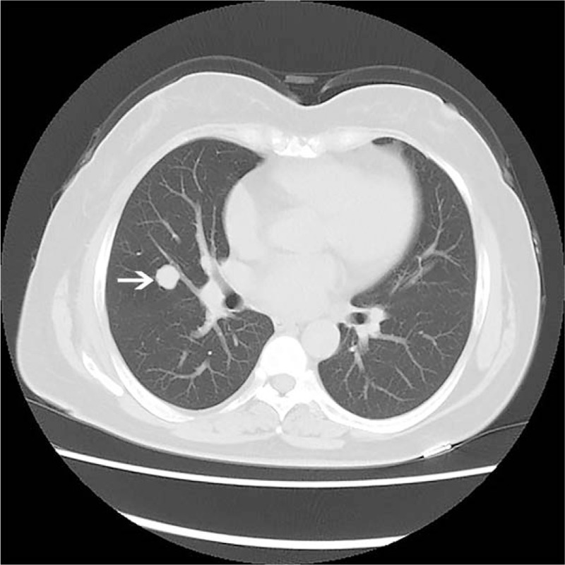
Patient concerns: A 55-year-old woman incidentally detected a space-occupying lesion of right lung in routine health examination. The patient was asymptomatic and there were no positive findings in routine laboratory examination, physical examination, and pulmonary function test. Computed tomography revealed a solitary round mass in the middle lobe of the right lung.
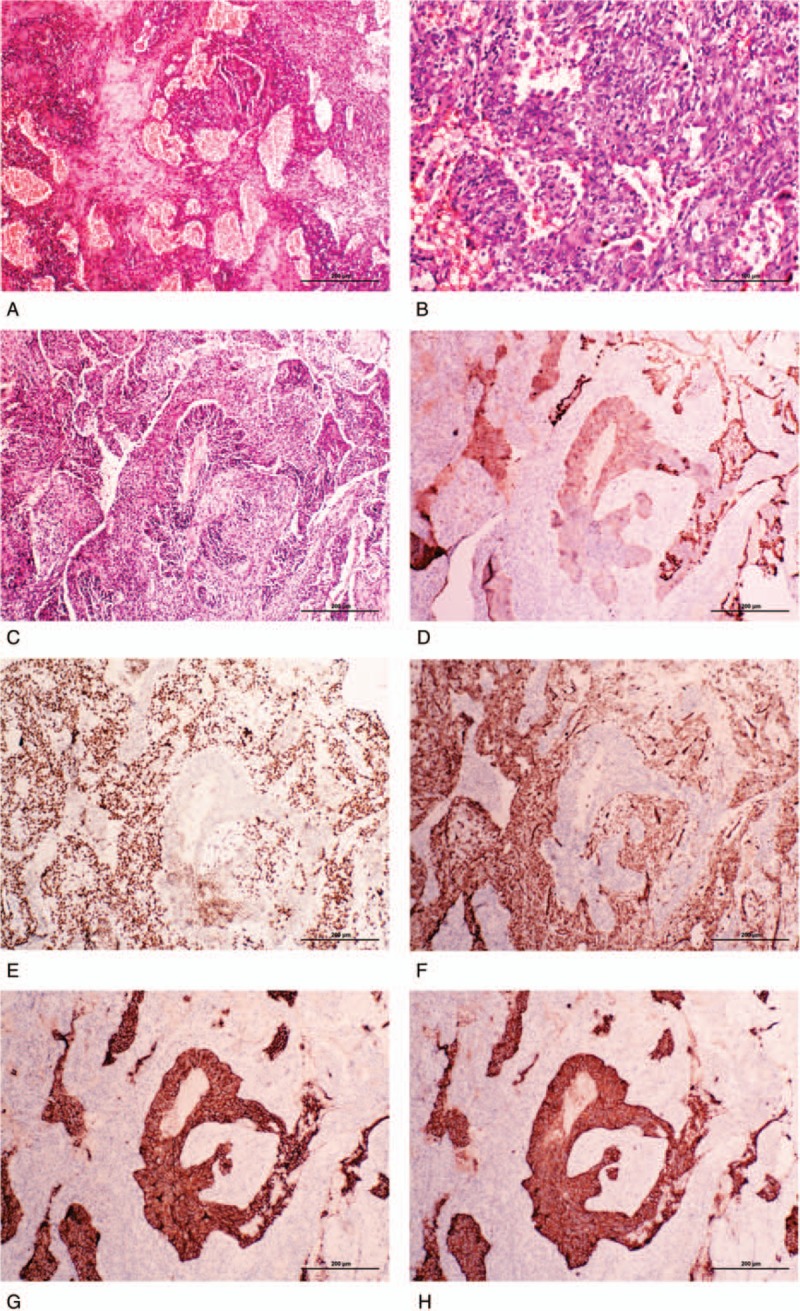
Diagnosis: The lesion was diagnosed as a sclerosing pneumocytoma accompanied with a typical carcinoid tumor of the right lung.
Intervention: The patient underwent thoracoscopic lobectomy in our hospital.
Outcomes: The postoperative course was uneventful.
Lessons: This case is rare and noteworthy for a lesion containing two different types of neoplasms, which may cause diagnostic difficulties.
Conflict of interest statement
The authors have no conflicts of interest to disclose.
Figures
References
-
- Liebow AA, Hubbell DS. Sclerosing hemangioma (histiocytoma, xanthoma) of the lung. Cancer 1956;9:53–75. - PubMed
-
- Devouassoux-Shisheboran M, Hayashi T, Linnoila RI, et al. A clinicopathologic study of 100 cases of pulmonary sclerosing hemangioma with immunohistochemical studies: TTF-1 is expressed in both round and surface cells, suggesting an origin from primitive respiratory epithelium. Am J Surg Pathol 2000;24:906–16. - PubMed
-
- Keylock JB, Galvin JR, Franks TJ. Sclerosing hemangioma of the lung. Arch Pathol Lab Med 2009;133:820–5. - PubMed
-
- Lin XY, Fan CF, Dong XJ, et al. Expression and significance of stem cell markers in pulmonary sclerosing haemangioma. Histopathology 2012;61:178–85. - PubMed
Publication types
MeSH terms
LinkOut - more resources
Full Text Sources
Medical
