

Parathyroid adenoma presenting with spontaneous cervical and anterior mediastinal hemorrhage: A case report
- PMID: 30702621
- PMCID: PMC6380857
- DOI: 10.1097/MD.0000000000014347
Parathyroid adenoma presenting with spontaneous cervical and anterior mediastinal hemorrhage: A case report
Abstract
Rationale: Spontaneous anterior cervical or mediastinal hemorrhage is a rare presentation of parathyroid adenoma.
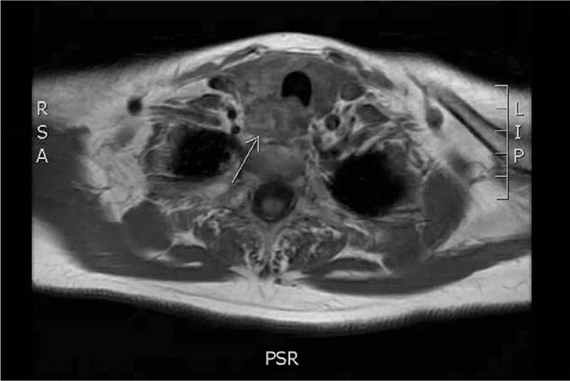
Patient concerns: A 69-year-old woman presented with neck hematoma and dysphagia and was found to have a soft tissue mass adjacent to her thyroid gland as seen on MRI and neck ultrasound.
Diagnosis: Laboratory testing demonstrated elevated calcium and parathyroid hormone supporting diagnosis of parathyroid adenoma.
Interventions: She underwent right inferior parathyroidectomy and en bloc right hemithyroidectomy due to significant fibrosis.
Outcomes: Pathology confirmed hypercellular parathyroid and normal thyroid tissue. Postoperatively, patient's calcium and parathyroid hormone levels had normalized.
Lessons: In conclusion, imaging may not always be specific in identifying the source of neck hematoma and so laboratory studies should be done to rule out parathyroid adenoma as the underlying etiology.
Conflict of interest statement
The authors have no conflicts of interest to declare.
Figures


References
-
- Hammett-Stabler CA, Maygarden SJ. Reisner HM. Pathology of the endocrine system. McGraw-Hill, Pathology: A Modern Case Study. New York, NY: 2014.
-
- Fitzgerald PA. Papadakis MA. Endocrine disorders. McGraw-Hill, Current Medical Diagnosis & Treatment. New York, NY: 2018.
-
- Capps RD. Multiple parathyroid tumors with massive mediastinal and subcutaneous hemorrhage. Am J Med Sci 1934;188:801–4.
Publication types
MeSH terms
LinkOut - more resources
Full Text Sources
Research Materials
