Review
. 2019 Mar 15:484:34-41.
doi: 10.1016/j.mce.2019.01.019. Epub 2019 Jan 28.

The structural basis of the arrestin binding to GPCRs

Affiliations
Review

The structural basis of the arrestin binding to GPCRs

Vsevolod V Gurevich et al. Mol Cell Endocrinol. .

Abstract

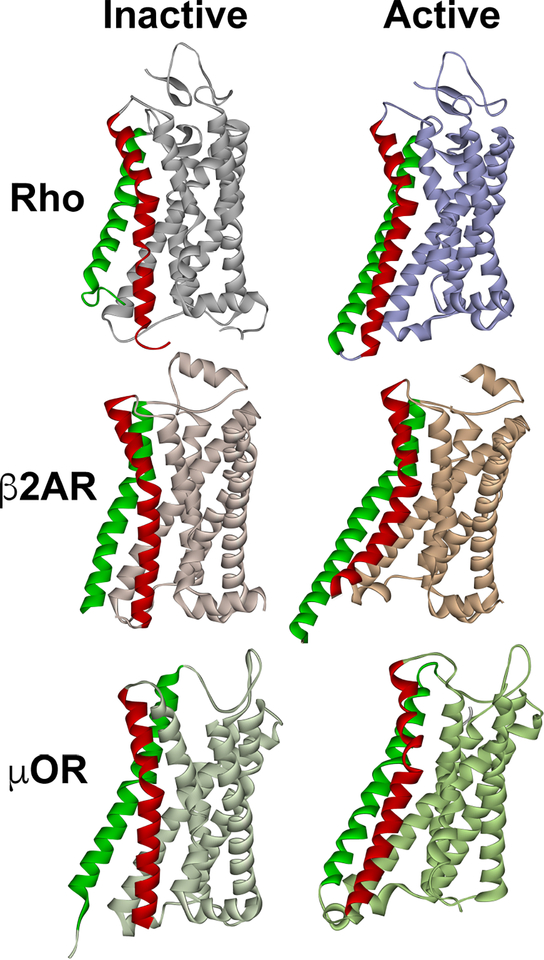
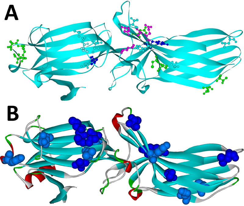
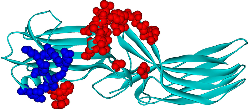
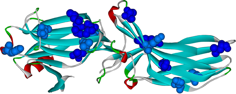
G protein-coupled receptors (GPCRs) are the largest family of signaling proteins targeted by more clinically used drugs than any other protein family. GPCR signaling via G proteins is quenched (desensitized) by the phosphorylation of the active receptor by specific GPCR kinases (GRKs) followed by tight binding of arrestins to active phosphorylated receptors. Thus, arrestins engage two types of receptor elements: those that contain GRK-added phosphates and those that change conformation upon activation. GRKs attach phosphates to serines and threonines in the GPCR C-terminus or any one of the cytoplasmic loops. In addition to these phosphates, arrestins engage the cavity that appears between trans-membrane helices upon receptor activation and several other non-phosphorylated elements. The residues that bind GPCRs are localized on the concave side of both arrestin domains. Arrestins undergo a global conformational change upon receptor binding (become activated). Arrestins serve as important hubs of cellular signaling, emanating from activated GPCRs and receptor-independent.

Keywords: Arrestin; GPCR; Phosphates; Protein engineering; Receptor specificity; Signaling.


Figures

Fig. 1.
Fig. 1.. The structure of inactive and active GPCRs.
The most pronounced activation-induced conformational change in all GPCRs is the outward movement of TM5 (green) and TM6 (red). In case of TM6, activation-induced shift is 14 A in Gs-coupled β2AR (Rasmussen et al., 2011), but smaller in Gi-coupled receptors, 10 A in both rhodopsin (Kang et al., 2018; Kang et al., 2015) and μ-opioid receptor (Koehl et al., 2018). The structures of inactive (dark) rhodopsin (Rho) (PDB ID: 1GZM (Li et al., 2004)), active rhodopsin (from complex with arrestin-1; PDB ID: 4ZWJ (Kang et al., 2015)), inactive β2AR (PDB ID: 2RH1 (Rasmussen et al., 2007)), active β2AR (from complex with Gs; PDB ID: 3SN6 (Rasmussen et al., 2011)), inactive μ-opioid receptor (μOR) (PDB ID: 4DKL (Manglik et al., 2012)), and active μOR (from complex with Gi; PDB ID: 6DDE (Koehl et al., 2018)) are shown. The common feature of active GPCR structures is the cavity between helices that opens on the cytoplasmic side. In case of Gi-coupled GPCRs (Rho and μOR) the outward movement of the helices is smaller, so that this cavity is narrower than in case of Gs-coupled β2AR. The dimensions of this cavity might underlie GPCR selectivity for particular G proteins, whereas non-visual arrestins likely accommodate GPCRs that couple to all types of G proteins.
Fig. 2.
Fig. 2.. Arrestin elements implicated in receptor binding.
A. The residues in positions identified as important for the receptor preference of different arrestin proteins (the structure of arrestin-2 is shown; PDB ID: 1G4M (Han et al., 2001); the residues indicated are those of arrestin-2) are shown as ball-and stick models colored as follows: light blue, those identified by reduced mobility of the spin label in receptor-bound arrestins using EPR (Val70, Leu71, Leu73, Val167, Leu191, Ser234, Thr246, Tyr249) (Hanson et al., 2007; Hanson et al., 2006); green, those identified by site-directed mutagenesis and receptor binding in vitro and in cells (Leu48, Glu50, Arg51, Asp240, Cys251, Pro252, Asp259, Thr261) (Gimenez et al., 2014; Gimenez et al., 2012; Prokop et al., 2017; Vishnivetskiy et al., 2011); dark blue, those identified by both of these methods (Leu68, Tyr238); magenta, those identified by direct contact with the receptor in the crystal structure of the arrestin-1 complex with rhodopsin (Lys138, Asn245, Ala247, Gln248) (Kang et al., 2015), including those confirmed by in-cell assays (Lys138, Gln248) (Prokop et al., 2017). Receptor-binding residues identified by several independent methods are localized on the concave sides of the two arrestin domains, with the highest concentration in the central crest of the receptor-binding side of arrestins. B. The residues that change mobility upon receptor binding, according to SDSL-EPR study (Vishnivetskiy et al., 2011) are shown as CPK models (dark blue – strong immobilization; light blue – less pronounced immobilization). The same crystal structure of arrestin-2 (PDB ID: 1G4M) was used. Both panels were generated using Accelrys DS ViewerPro 6.0 (Dassault Systemes, BIOVIA Corp, San Diego, CA).
Fig. 3.
Fig. 3.. Arrestin-1 residues contacting rhodopsin in co-crystal.
Mouse arrestin-1 residues that directly interact with rhodopsin are shown as CPK models (based on rhodopsin-bound mouse arrestin-1 structure from complex A, PDB ID: 4ZWJ (Kang et al., 2015)). Those that bind receptor-attached phosphates or negative charges in the rhodopsin C-terminus are colored dark blue (Zhou et al., 2017)(mouse arrestin-1 residues Lys15, Lys16, Arg19, Lys111, Lys167, Lys168, Arg172), those that interact with unphosphorylated parts of the rhodopsin molecule are colored red (Kang et al., 2015; Zhuo et al., 2014) (Val12, Ile13, Phe14, Gln70, Glu71, Ile73, Asp74, Met76, Gly77, Leu78, Arg82, Asp83, Leu84, Lys142, Leu250, Tyr251, Ser252, Asp254, Tyr255, Arg292, Thr320).
Fig. 4.
Fig. 4.. Elements of arrestin-1 that move upon rhodopsin binding.
Bovine arrestin-1 structure in it basal conformation (PDB ID: 1CF1 (Hirsch et al., 1999)) is shown, with the elements that move upon rhodopsin binding (Kim et al., 2012) colored dark red. These include the finger loop (residues 67–78 (Hanson et al., 2006)), the 139-loop (Kim et al., 2012) (a.k.a. the middle loop in arrestin-2 (Shukla et al., 2013)), as well as loops at the distal tips of the N-domain (residues 155–168) and C-domain (residues 336–344).
Fig. 5.
Fig. 5.. Activation of arrestin-3 and switch regions.
Conformational changes upon arrestin-3 activation are shown: the release of the arrestin C-terminus (magenta; magenta arrow shows the direction of the movement; aSwIV) anchored to the body of the molecule via the three-element interaction in the basal state (PDB ID: 3P2D (Zhan et al., 2011a)), the movement of the finger loop and helix formation in its tip (red; red arrow shows the direction of the movement), the twist of the two domains relative to each other (N-domain, gray; C-domain, teal). Movements of several loops, likely creating effector docking sites, are also shown: middle loop (dark blue; dark blue arrow indicates the direction of movement); the “leg” of the α-helix I (green, with green arrowhead in a circle; aSwI), inter-domain hinge (yellow with yellow arrowhead in a circle; aSwIIa), register-shifted β-strand XI in the C-domain (dark brown; aSwIIb), and the lariat loop (orange with orange arrow; in the active structure (PDB ID: 5TV1 (Chen et al., 2017)) it is not resolved; aSwIII). All arrestin subtypes have switches II, III, and IV, whereas only arrestin-1 has polyproline motifs in the switch I. Note that only the interactions of clathrin and AP2 with the aSwIV were experimentally confirmed so far (Kim and Benovic, 2002).

References

    1. Ahmed MR, Zhan X, Song X, Kook S, Gurevich VV, and Gurevich EV (2011). Ubiquitin ligase parkin promotes Mdm2-arrestin interaction but inhibits arrestin ubiquitination. Biochemistry, 3749–3763. - PMC - PubMed
    1. Altenbach C, Kusnetzow AK, Ernst OP, Hofmann KP, and Hubbell WL (2008). High-resolution distance mapping in rhodopsin reveals the pattern of helix movement due to activation. Proc Natl Acad Sci U S A 105, 7439–7444. - PMC - PubMed
    1. Azevedo AW, Doan T, Moaven H, Sokal I, Baameur F, Vishnivetskiy SA, Homan KT, Tesmer JJ, Gurevich VV, Chen J, et al. (2015). C-terminal threonines and serines play distinct roles in the desensitization of rhodopsin, a G protein-coupled receptor. Elife 4, doi: 10.7554/eLife.05981. - DOI - PMC - PubMed
    1. Baillie GS, Adams DR, Bhari N, Houslay TM, Vadrevu S, Meng D, Li X, Dunlop A, Milligan G, Bolger GB, et al. (2007). Mapping binding sites for the PDE4D5 cAMP-specific phosphodiesterase to the N- and C-domains of beta-arrestin using spot-immobilized peptide arrays. Biochem J 404, 71–80. - PMC - PubMed
    1. Benovic JL, DeBlasi A, Stone WC, Caron MG, and Lefkowitz RJ (1989). Beta-adrenergic receptor kinase: primary structure delineates a multigene family. Science 246, 235–240. - PubMed

Publication types

Substances

LinkOut - more resources