Review
. 2019 Jun:56:87-96.
doi: 10.1016/j.sbi.2018.12.007. Epub 2019 Jan 28.

Polypeptide GalNAc-Ts: from redundancy to specificity

Affiliations
Review

Polypeptide GalNAc-Ts: from redundancy to specificity

Matilde de Las Rivas et al. Curr Opin Struct Biol. 2019 Jun.

Abstract

Mucin-type O-glycosylation is a post-translational modification (PTM) that is predicted to occur in more than the 80% of the proteins that pass through the Golgi apparatus. This PTM is initiated by a family of polypeptide GalNAc-transferases (GalNAc-Ts) that modify Ser and Thr residues of proteins through the addition of a GalNAc moiety. These enzymes are type II membrane proteins that consist of a Golgi luminal catalytic domain connected by a flexible linker to a ricin type lectin domain. Together, both domains account for the different glycosylation preferences observed among isoenzymes. Although it is well accepted that most of the family members share some degree of redundancy toward their protein and glycoprotein substrates, it has been recently found that several GalNAc-Ts also possess activity toward specific targets. Despite the high similarity between isoenzymes, structural differences have recently been reported that are key to understanding the molecular basis of both their redundancy and specificity. The present review focuses on the molecular aspects of the protein substrate recognition and the different glycosylation preferences of these enzymes, which in turn will serve as a roadmap to the rational design of specific modulators of mucin-type O-glycosylation.


Figures

Figure 1.
Figure 1.. Summary of the known peptide and glycopeptide specificities of the GalNAc-T family.
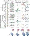
Phylogenetic tree of the GalNAc-Ts showing their 1) random peptide derived peptide substrate motifs as Sequence Logos [7••,23••,32••,46] 2) neighboring prior glycosylation preferences (1-3 residues) due to catalytic domain interactions [4••], and 3) their long range prior glycosylation preferences (~6-~17 residues) due to lectin domain binding [23••,40••]. Note that ‘-ND-’ stands for not determined while ‘---’ indicates no or weak activity. Also note that GalNAc-T8, -T9, -T18 and -T19 exhibit nearly undetectable activities against most substrates [47] and have not been well characterized. The models at the bottom show the different substrate binding modes that lead to the indicated specificities. The catalytic and lectin domains are shown as oval-shaped figures in blue and red, respectively. (Glyco)peptides are indicated in purple while the yellow squares denote the position of prior GalNAc moieties in the glycopeptides. Arrows indicate the position of GalNAc transfer to the acceptor site.
Figure 2.
Figure 2.. Cartoon and surface representation of GalNAc-Ts structures.
Catalytic and lectin domains are shown in purple and salmon respectively, while flexible loop and linker are displayed in yellow. (a) Extended (left panel) and compact (right panel) forms of monomeric HsGalNAc-T2. (b) Cartoon and surface representation of MmGalNAc-T1, HsGalNAc-T4, DmPGANT9A and HsGalNAc-T10. (c) Surface representation of the HsGalNAc-T2-UDP-MUC5AC-13 complex. The overall structure is shown in salmon, monoglycopeptide MUC5AC-13 and the flexible loop are depicted in cyan and yellow, respectively. The flexible loop of the enzyme is shown in its closed and open conformations.
Figure 3.
Figure 3.. Interactions between GalNAc-Ts and their substrates.
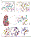
(a) Close-up view of the catalytic domain of HsGalNAc-T2 with two different peptides, EA2 (cyan sticks; left panel) and glycopeptide MUC5AC-13 (GlyThrThrProSerProValProThrThrSerThrThr*SerAlaPro) (yellow sticks; right panel), which are similarly recognized by HsGalNAc-T2 through a hydrophobic patch. UDP is depicted as sticks with magenta carbon atoms and Mn2+ is shown as a purple sphere. (b) On the left panel, surface representation of the HsGalNAc-T4-UDP-Diglycopeptide 6 (GlyAlaThr*3GlyAlaGlyAlaGlyAlaGlyThr*11Thr12ProGlyProGly) complex. Peptide backbone is depicted in cyan with the two GalNAc groups as blue and red sticks; the enzyme flexible loop is shown in its closed conformation in yellow. On the right panel, close-up view of the main interactions between HsGalNAc-T4 catalytic domain glycopeptide binding-site and the GalNAc group on T*11 of diglycopeptide 6. The GalNAc-T4 residues forming the peptide-binding site are depicted in salmon and yellow and the glycopeptide is depicted in cyan, with the GalNAc groups shown as blue and red sticks. Mn2+ and water molecules are depicted as green and red spheres, respectively, and hydrogen bonds appears as dotted yellow lines. Please note that we only show water-mediated interactions in which only the water molecule act as a bridge between the residues. (c) Main interactions between GalNAc-T2, -T4 and -T10 isoenzymes lectin domain (shown as salmon, purple and slate, respectively) and the GalNAc moiety (shown as sticks with orange carbon atoms).
Figure 4.
Figure 4.. Superposition of HsGalNAc-T2 and HsGalNAc-T4.
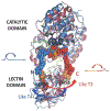
Superimposed cartoon representations of HsGalNAc-T2-UDP-MUC5AC-13 glycopeptide (GlyThrThrProSerProValProThrThrSerThrThr*SerAlaPro) complex depicted in red and HsGalNAc-T4-UDP-diglycopeptide 6 (GlyAlaThr*3GlyAlaGlyAlaGlyAlaGlyThr*11Thr12ProGlyProGly) complex depicted in blue. The MUC5AC-13, diglycopeptide 6 and GalNAc moieties are shown in red, blue and orange atoms, respectively. The arrows indicate the direction of the long-range glycosylation preference of each enzyme, based on the orientation of their respective lectin domains with respect their catalytic domains. Note that the critical Asp residues of the lectin domain GalNAc-binding sites are indicated for clarification purposes.

References

    1. Bennett EP, Mandel U, Clausen H, Gerken TA, Fritz TA, Tabak LA: Control of mucin-type O-glycosylation: A classification of the polypeptide GalNAc-transferase gene family. Glycobiology (2011) 22(6):736–756. - PMC - PubMed
    2. This work provides an overview of the GalNAc-Ts family members, as well as a classification of its isoenzymes according to their glycosylation preferences.

    1. Hollingsworth MA, Swanson BJ: Mucins in cancer: Protection and control of the cell surface. Nature Reviews Cancer (2004) 4(1):45–60. - PubMed
    1. Kufe DW: Mucins in cancer: Function, prognosis and therapy. Nature Reviews Cancer (2009) 9(12):874–885. - PMC - PubMed
    1. Revoredo L, Wang S, Bennett EP, Clausen H, Moremen KW, Jarvis DL, Ten Hagen KG, Tabak LA, Gerken TA: Mucin-type O-glycosylation is controlled by short-and long-range glycopeptide substrate recognition that varies among members of the polypeptide GalNAc transferase family. Glycobiology (2015) 26(4):360–376. - PMC - PubMed
    2. This work digs into the glycosylation preferences of the different GalNAc-Ts isoenzymes and demonstrate that catalytic and lectin domains have unique functions and work in concert to direct glycosylation. The authors propose a more exhaustive re-classification of the family members.

    1. Pratt MR, Hang HC, Ten Hagen KG, Rarick J, Gerken TA, Tabak LA, Bertozzi CR: Deconvoluting the functions of polypeptide N-α-acetylgalactosaminyltransferase family members by glycopeptide substrate profiling. Chemistry & biology (2004) 11(7):1009–1016. - PubMed

Publication types

MeSH terms

Substances

LinkOut - more resources