

Clinical role, safety and diagnostic accuracy of percutaneous transthoracic needle biopsy in the evaluation of pulmonary consolidation
- PMID: 30704502
- PMCID: PMC6357395
- DOI: 10.1186/s12931-019-0982-5
Clinical role, safety and diagnostic accuracy of percutaneous transthoracic needle biopsy in the evaluation of pulmonary consolidation
Abstract
Background: To determine the clinical role, safety, and diagnostic accuracy of percutaneous transthoracic needle biopsy in the evaluation of pulmonary consolidation.
Methods: A retrospective review of all computed tomography (CT)-guided percutaneous transthoracic needle biopsies (PTNB) at a tertiary care hospital over a 4-year period was performed to identify all cases of PTNB performed for pulmonary consolidation. For each case, CT Chest images were reviewed by two thoracic radiologists. Histopathologic and microbiologic results were obtained and clinical follow-up was performed.
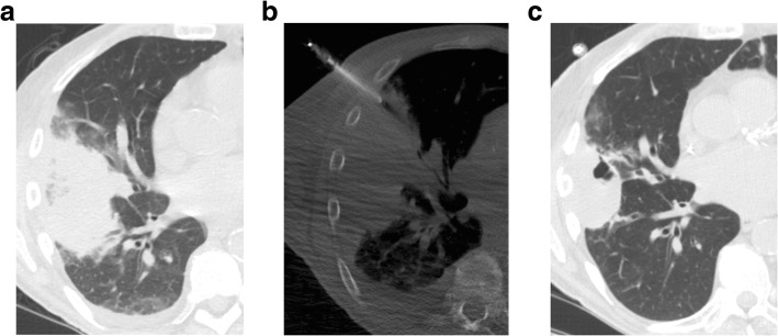
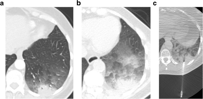
Results: Thirty of 1090 (M:F 17:30, mean age 67 years) patients underwent PTNB for pulmonary consolidation (2.8% of all biopsies). A final diagnosis was confirmed in 29 patients through surgical resection, microbiology, or clinicoradiologic follow-up for at least 18 months after biopsy. PTNB had an overall diagnostic accuracy of 83%. A final diagnosis of malignancy was made in 20/29 patients, of which 19 were correctly diagnosed by PTNB, resulting in a sensitivity of 95% and specificity of 100% for malignancy. In all cases of primary lung cancer, adequate tissue for molecular testing was obtained. A benign final diagnosis was made in 9 patients, infection in 5 cases and non-infectious benign etiology in 4 cases. PTNB correctly diagnosed all cases of infection. Minor complications occurred in 13% (4/30) of patients.
Conclusions: Pulmonary consolidation can be safely evaluated with CT-guided percutaneous needle biopsy. Diagnostic yield is high, especially for malignancy. PTNB of pulmonary consolidation should be considered following non-diagnostic bronchoscopy.
Keywords: Consolidation; Hemoptysis; Percutaneous transthoracic needle biopsy; Pneumothorax.
Conflict of interest statement
Ethics approval and consent to participate
The study was a retrospective single-center study and performed with IRB approval from the Partners Human Research Committee (Project number 2014P001409). Due to the retrospective design, the institutional ethical review board waived the need for informed consent.
Consent for publication
Not applicable
Competing interests
The authors declare that they have no competing interests.
Publisher’s Note
Springer Nature remains neutral with regard to jurisdictional claims in published maps and institutional affiliations.
Figures
References
MeSH terms
LinkOut - more resources
Full Text Sources
Medical
