. 2019 Jan 31;38(1):44.
doi: 10.1186/s13046-019-1047-9.

CYT997(Lexibulin) induces apoptosis and autophagy through the activation of mutually reinforced ER stress and ROS in osteosarcoma

Affiliations

CYT997(Lexibulin) induces apoptosis and autophagy through the activation of mutually reinforced ER stress and ROS in osteosarcoma

Zongyi Wang et al. J Exp Clin Cancer Res. .

Abstract

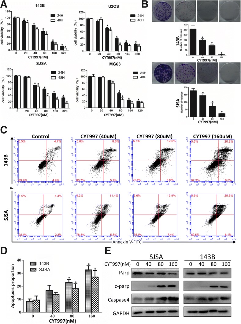
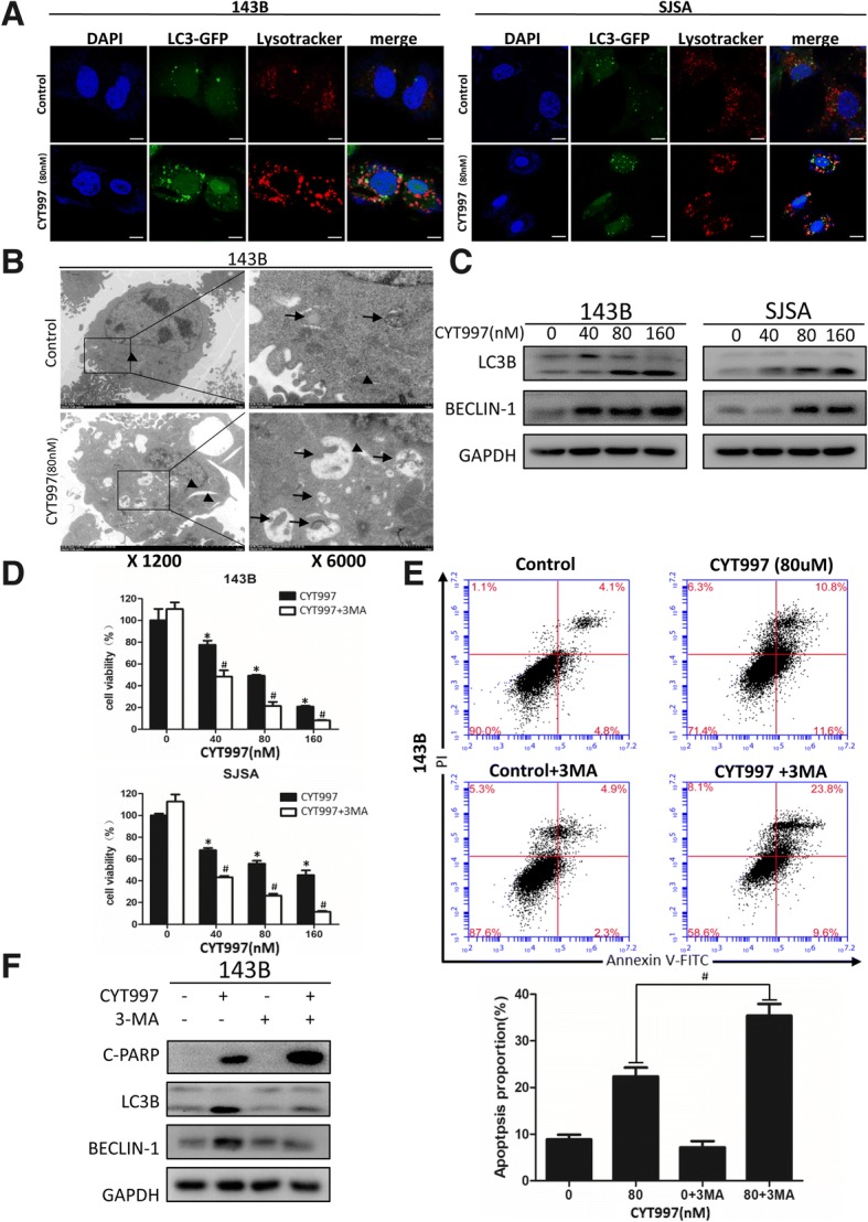
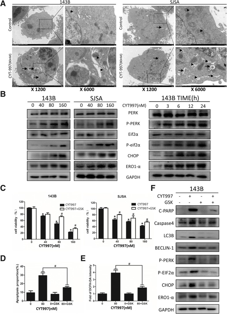
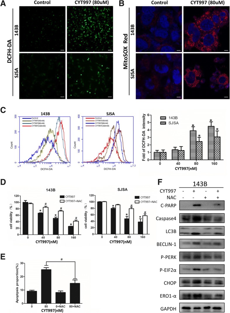
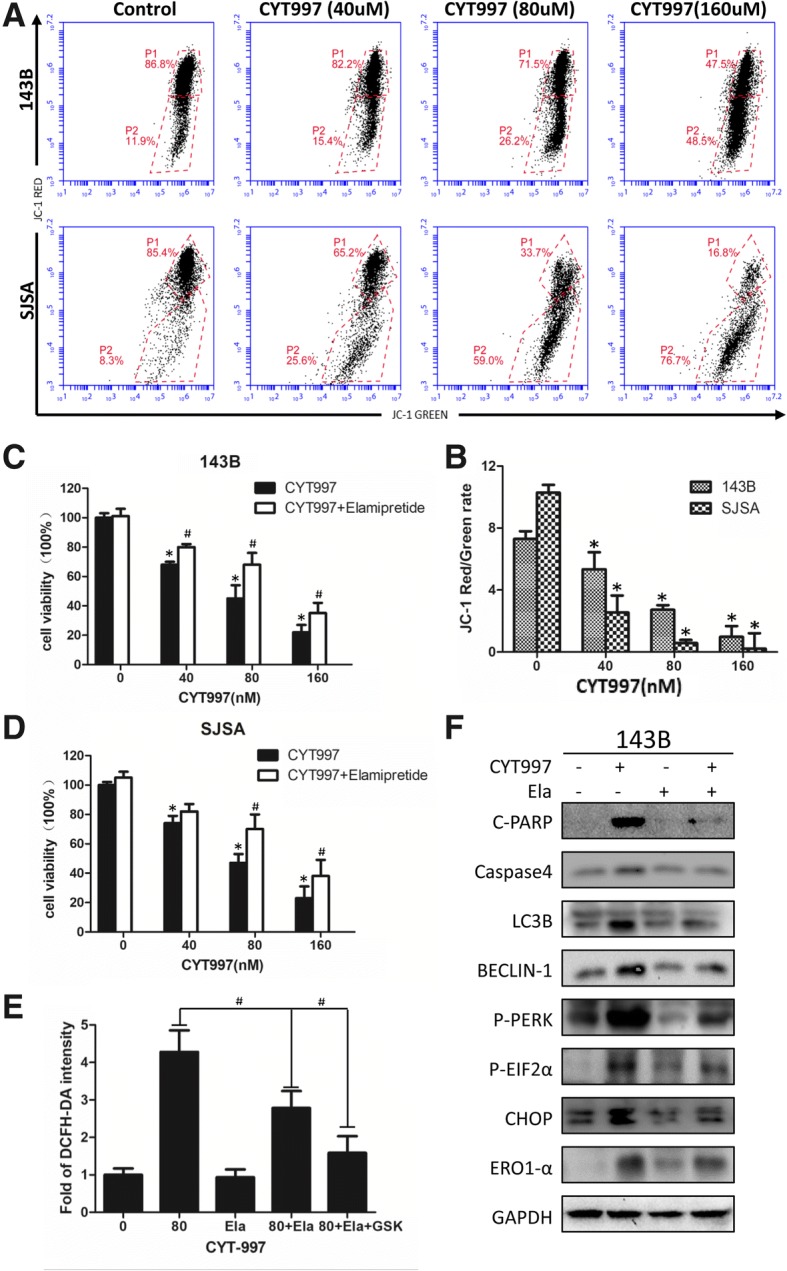
Background: Osteosarcoma (OS) is a common malignant cancer in children and adolescents and has a cure rate that has not improved in the last two decades. CYT997 (lexibulin) is a novel potent microtubule-targeting agent with various anticancer activities, such as proliferation inhibition, vascular disruption, and cell cycle arrest and apoptosis induction, in multiple cancers. However, the direct cytotoxic mechanisms of CYT997 have not yet been fully characterized.

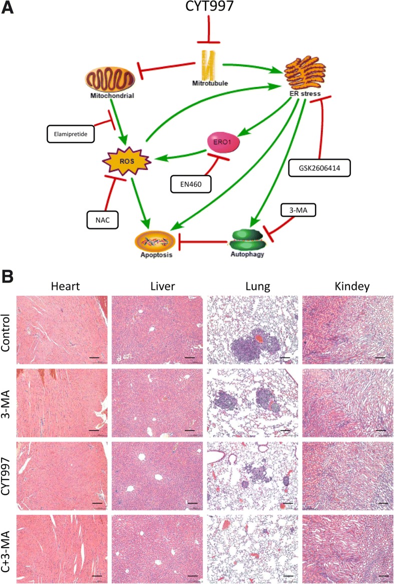
Methods: We evaluated apoptosis and autophagy in human osteosarcomas after treatment with CYT997 and investigated the underlying mechanisms. To explore relationships, we used the reactive oxygen species (ROS) scavenger N-acetyl cysteine (NAC), PERK inhibitor GSK2606414, ERO1 inhibitor EN460 and mitochondrial targeted protection peptide elamipretide. BALB/c-nu mice were inoculated with 143B tumor cells to investigate the in vivo effect of CYT997.

Results: We explored the efficacy and mechanism of CYT997 in osteosarcoma (OS) in vitro and in vivo and demonstrated that CYT997 potently suppresses cell viability and induces apoptosis and autophagy. CYT997 triggered production of ROS and exerted lethal effects via endoplasmic reticulum (ER) stress in OS cells. NAC attenuated these effects. The PERK inhibitor GSK2606414, which can block the ER stress pathway, reduced ROS production and enhanced cell viability. Moreover, activation of ERO1 in the ER stress pathway was responsible for inducing ROS production. ROS produced by the mitochondrial pathway also aggravate ER stress. Protection of mitochondria can reduce apoptosis and autophagy. Finally, CYT997 prominently reduced tumor growth in vivo.

Conclusions: This study suggests that CYT997 induces apoptosis and autophagy in OS cells by triggering mutually enhanced ER stress and ROS and may thus be a promising agent against OS.

Keywords: CYT997; ER stress; ERO1A; Osteosarcoma; ROS.


Conflict of interest statement

Ethics approval and consent to participate

All procedures in this study were performed in accordance with the ethical standards of the Animal Care and Use Committee of Shanghai General Hospital and Shanghai Jiaotong University.

Consent for publication

Not applicable.

Competing interests

The authors declare that they have no competing interests.

Publisher’s Note

Springer Nature remains neutral with regard to jurisdictional claims in published maps and institutional affiliations.

Figures

Fig. 1
Fig. 1
CYT997 inhibited cell proliferation and induced apoptosis in human osteosarcoma cells. a. 143B, SJSA, U2OS, and MG63 osteosarcoma cell lines were treated with CYT997 (0, 20, 40, 80, 160 and 320 μM) for 24 and 48 h. Cell viability was measured by CCK-8 assays. b. 143B and SJSA cells treated with CYT997 (0, 40, 80, 160 μM). Colony formation was evaluated by colony formation assays. c-d 143B and SJSA cells were treated with CYT997 for 24 h and analyzed using PI/Annexin V-FITC flow cytometry. Histograms indicate the proportion of apoptotic cells from three separate experiments. e Cells were treated with various concentrations of CYT997 for 24 h, and apoptosis-related proteins such as cleaved PARP, and caspase-4 were analyzed by western blotting. *P < 0.05, significantly different compared with the control group
Fig. 2
Fig. 2
CYT997 induced autophagy in OS cells, and inhibition of autophagy increased CYT997-induced apoptosis. a Osteosarcoma cell lines 143B and SJSA were transiently transfected with GFP-LC3-encoding plasmids for 24 h, treated with or without CYT997 (80 nM) for 24 h and stained with LysoTracker Red DND-99 (50 nM). Green color represents the formation of autophagosomes, and red color shows cellular acidic compartments, indicative of lysosomes and autolysosomes. Colocalization of autophagosomes and lysosomes was examined by confocal microscopy. Scale bars = 20 μm. b CYT997 induced accumulation of autophagosomes in osteosarcoma cells, as shown in the electron micrographs. Arrows indicate autophagosomes, and arrowheads indicate ER. c Osteosarcoma cells were treated with CYT997 (80 nM) for 24 h. Autophagy-related proteins, LC3B and beclin-1, were analyzed by western blotting. d 143B and SJSA cells were preincubated with 3-methyladenine (3-MA) (5 mM) for 2 h and then treated with CYT997 (80 nM) for 24 h, followed by cell proliferation detection using CCK-8 assays. e Osteosarcoma cells were preincubated with 3-MA (5 mM) and then treated with CYT997(80 nM) for 24 h and analyzed using PI/Annexin V-FITC flow cytometry. Histograms indicate the proportion of apoptotic cells from three separate experiments. f Cells were treated with 80 nM CYT997 and 3-MA for 24 h, and the levels of c-PARP, LC3B and Beclin-1 were assessed by western blotting. *P < 0.05, significantly different compared with the control group. # P < 0.05, significantly different compared with the CYT997 treatment group
Fig. 3
Fig. 3
CYT997 induced endoplasmic reticulum (ER) stress in osteosarcoma cell lines, and inhibition of the ER stress pathway decreased CYT997-induced apoptosis, autophagy and reactive oxygen species (ROS) production. a Osteosarcoma cell lines 143B and SJSA incubated with CYT997 (80 μM) for 24 h were visualized by electron microscopy; arrowheads point to the ER, and arrows indicate autophagosomes. b ER stress-related proteins including PERK, p-PERK, eif2α, p-eif2α, CHOP, and ERO1 were detected by western blotting after incubation of the cells with various concentrations of CYT997 for 24 h or treatment with CYT997 (80 nM) for different durations. c 143B and SJSA cells were cotreated with GSK2606414 (2 μM) and different concentrations of CYT997 for 24 h, followed by cell proliferation detection using CCK-8 assays. d 143B cells treated with CYT997 (80 nM) and/or GSK2606414 (2 μM) were analyzed using PI/Annexin V-FITC flow cytometry. Histograms indicate the proportion of apoptotic cells from three separate experiments. e 143B cells treated with CYT997 (80 nM) and/or GSK2606414 (2 μM) for 24 h, stained with 10 μM 2′,7′-dichlorodihydrofluorescein diacetate (DCFH-DA) at 37 °C in the dark for 30 min. Histograms indicate the fold change in DCFH-DA intensity relative to the control group from three separate experiments. f 143B cells were treated with CYT997 (80 nM) and/or GSK2606414 (2 μM) for 24 h, and apoptosis, autophagy and ER stress-related proteins including c-PARP, caspase-4, LC3B, Beclin-1, p-PERK, p-eif2α, CHOP and ERO1 were analyzed by western blotting. *P < 0.05, significantly different compared with the control group. # P < 0.05, significantly different compared with the CYT997 treatment group
Fig. 4
Fig. 4
CYT997 induced intracellular reactive oxygen species (ROS) generation, and ROS scavenging decreased CYT997-induced apoptosis, autophagy and endoplasmic reticulum (ER) stress (a-b). Osteosarcoma cells 143B and SJSA were treated with or without CYT997 (80 nM) for 24 h. Cells were stained with 10 μM 2′,7′-dichlorodihydrofluorescein diacetate (DCFH-DA) or 1 μM MitoSOX Red dye at 37 °C in the dark for 30 min and then observed by fluorescence microscopy and confocal microscopy. Scale bars = 50 and 5 μm. c Fluorescence intensity of DCFH-DA was detected using flow cytometry. Histograms indicate the fold change in DCFH-DA intensity relative to the control group from three separate experiments. d 143B and SJSA cells were cotreated with NAC (5 mM) and different concentrations of CYT997 for 24 h, followed by the CCK-8 assay for cell proliferation. e 143B cells treated with CYT997 (80 nM) and/or NAC (5 mM) analyzed using PI/Annexin V-FITC flow cytometry. Histograms indicate the proportion of apoptotic cells from three separate experiments. f 143B cells were treated with CYT997 (80 nM) and/or N-acetyl-L-cysteine (NAC) (5 mM) for 24 h, and apoptosis, autophagy and ER stress-related proteins including c-PARP, caspase-4, LC3B, Beclin-1, p-PERK, p-eif2α, CHOP and ERO1 were analyzed by western blotting. *P < 0.05, significantly different compared with the control group. # P < 0.05, significantly different compared with the CYT997 treatment group
Fig. 5
Fig. 5
ERO1 regulates reactive oxygen species (ROS) production, and inhibition or overexpression of ERO1 can respectively decrease or increase ROS levels. a 143B and SJSA cells were cotreated with EN460 (8 μM) and different concentrations of CYT997 for 24 h, followed by cell proliferation detection using CCK-8 assays. b 143B cells treated with CYT997 (80 nM) and/or EN460 (8 μM) 24 h were analyzed using PI/Annexin V-FITC flow cytometry. Histograms indicate the proportion of apoptotic cells from three separate experiments. c 143B cells were treated with CYT997 (80 nM) and/or EN460 (8 μM) for 24 h and stained with 10 μM 2′,7′-dichlorodihydrofluorescein diacetate (DCFH-DA) at 37 °C in the dark for 30 min. Histograms indicate the fold change in DCFH-DA intensity relative to the control group from three separate experiments. d 143B cells were treated with CYT997 (80 nM) and/or EN460 (8 μM) for 24 h, and apoptosis, autophagy and ER stress-related proteins including c-PARP, caspase-4, LC3B, Beclin-1, CHOP and ERO1 were analyzed by western blotting. e 143B cells were transfected with ERO1-encoding vectors and then treated with or without GSK2606414 for 24 h. Protein levels of ERO1 were detected by western blotting. f 143B cells transfected with ERO1-encoding vectors were treated with different concentrations of CYT997 and 5 nM of NAC for 24 h, followed by cell proliferation detection using CCK-8 assays. g 143B cells were transfected with ERO1-encoding vectors and treated with or without GSK2606414 (2 μM) for 24 h. H2O2 levels were then measured using a hydrogen peroxide assay kit. Histograms indicate the fold change in H2O2 concentration relative to the control group from three separate experiments. *P < 0.05, significantly different compared with the control group. # P < 0.05, significantly different compared with the CYT997 treatment group
Fig. 6
Fig. 6
ROS produced by the mitochondrial pathway also aggravate ER stress. a, b) JC-1 staining by flow cytometry was used to measure the mitochondrial membrane potential (MMP) after CYT997 treatment. Histograms indicate the red/green ratio of JC-1 fluorescence. c, d 143B and SJSA cells were cotreated with Elamipretide (10 nM) and different concentrations of CYT997 for 24 h, followed by cell proliferation detection using CCK-8 assays. e 143B cells were treated with CYT997 (80 nM), Elamipretide(10 nM) and/or GSK2606414 (2 μM) for 24 h and stained with 10 μM 2′,7′-dichlorodihydrofluorescein diacetate (DCFH-DA) at 37 °C in the dark for 30 min. Histograms indicate the fold change in DCFH-DA intensity relative to the control group from three separate experiments. f 143B cells were treated with CYT997 (80 nM) and/or Elamipretide (10 nM) for 24 h, and apoptosis, autophagy and ER stress-related proteins including c-PARP, caspase-4, LC3B, Beclin-1, p-PERK, p-eif2α, CHOP and ERO1 were analyzed by western blotting. *P < 0.05, significantly different compared with the control group. # P < 0.05, significantly different compared with the CYT997 treatment group
Fig. 7
Fig. 7
CYT997 suppressed the growth of human osteosarcoma xenografts in vivo, and 3-MA could enhance the in vivo anti-tumor effect. a BALB/c-nu mice bearing 143B human orthotopic osteosarcoma xenografts were divided into four groups: [1] mice treated with PBS only, [2] mice treated with 3-MA only, [3] mice treated with CYT997 only and [4] mice treated with CYT997 plus 3-MA. CYT997 plus 3-MA showed more significant anti-tumor effects than did CYT997-only treatment. b Tumor weights of the tumors in the four groups were recorded. c The weight of mice in the two groups were recorded. d Levels of c-PARP, LC3B, p-eif2α, CHOP, and ERO1 in tumor xenograft tissues were measured by western blotting. e Immunohistochemical staining of tumor specimens. Scale bars = 50 μm *P < 0.05, significantly different compared with the control group
Fig. 8
Fig. 8
a Proposed mechanism of lexibulin-induced apoptosis and autophagy in osteosarcoma cells. b H&E staining of important organs. Scale bars = 50 μm

Similar articles

Cited by

References

    1. Raymond AK, Jaffe N. Osteosarcoma multidisciplinary approach to the management from the pathologist's perspective. Cancer Treat Res. 2009;152:63–84. doi: 10.1007/978-1-4419-0284-9_4. - DOI - PubMed
    1. Luetke A, Meyers PA, Lewis I, Juergens H. Osteosarcoma treatment - where do we stand? A state of the art review. Cancer Treat Rev. 2014;40:523–532. doi: 10.1016/j.ctrv.2013.11.006. - DOI - PubMed
    1. Burns CJ, Fantino E, Phillips ID, Su S, Harte MF, Bukczynska PE, et al. CYT997: a novel orally active tubulin polymerization inhibitor with potent cytotoxic and vascular disrupting activity in vitro and in vivo. Mol Cancer Ther. 2009;8:3036–3045. doi: 10.1158/1535-7163.MCT-09-0076. - DOI - PubMed
    1. Burns CJ, Harte MF, Bu X, Fantino E, Joffe M, Sikanyika H, et al. Discovery of CYT997: a structurally novel orally active microtubule targeting agent. Bioorg Med Chem Lett. 2009;19:4639–4642. doi: 10.1016/j.bmcl.2009.06.079. - DOI - PubMed
    1. Lickliter JD, Francesconi AB, Smith G, Burge M, Coulthard A, Rose S, et al. Phase I trial of CYT997, a novel cytotoxic and vascular-disrupting agent. Br J Cancer. 2010;103:597–606. doi: 10.1038/sj.bjc.6605841. - DOI - PMC - PubMed

MeSH terms