. 2019 Jan 31;10(1):47.
doi: 10.1186/s13287-019-1152-x.

Microvesicles from human adipose stem cells promote wound healing by optimizing cellular functions via AKT and ERK signaling pathways

Affiliations

Microvesicles from human adipose stem cells promote wound healing by optimizing cellular functions via AKT and ERK signaling pathways

Sen Ren et al. Stem Cell Res Ther. .

Abstract

Background: Human adipose stem cells (ASCs) have emerged as a promising treatment paradigm for skin wounds. Recent works demonstrate that the therapeutic effect of stem cells is partially mediated by extracellular vesicles, which comprise exosomes and microvesicles. In this study, we investigate the regenerative effects of isolated microvesicles from ASCs and evaluate the mechanisms how ASC microvesicles promote wound healing.

Methods: Adipose stem cell-derived microvesicles (ASC-MVs) were isolated by differential ultracentrifugation, stained by PKH26, and characterized by electron microscopy and dynamic light scattering (DLS). We examined ASC-MV effects on proliferation, migration, and angiogenesis of keratinocytes, fibroblasts, and endothelial cells both in vitro and in vivo. Next, we explored the underlying mechanisms by gene expression analysis and the activation levels of AKT and ERK signaling pathways in all three kinds of cells after ASC-MV stimulation. We then assessed the effect of ASC-MVs on collagen deposition, neovascularization, and re-epithelialization in an in vivo skin injury model.

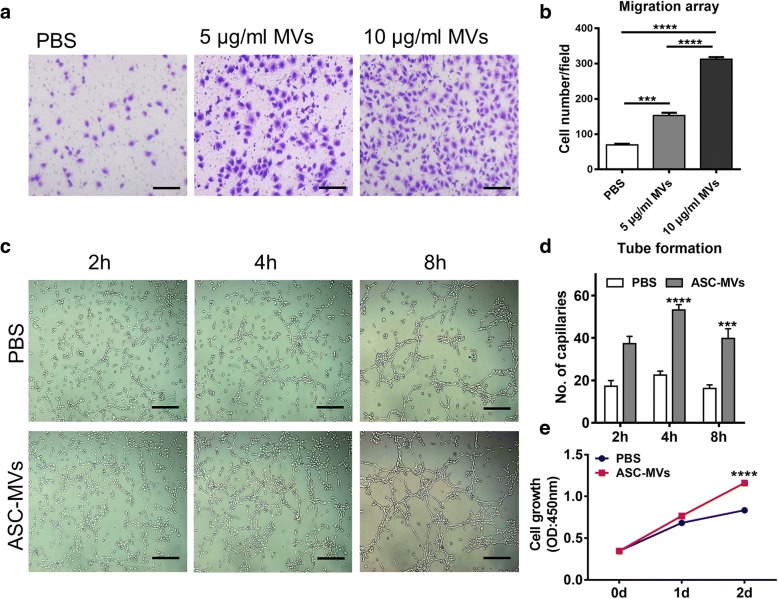
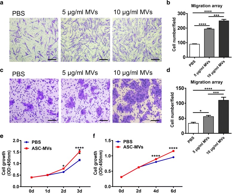
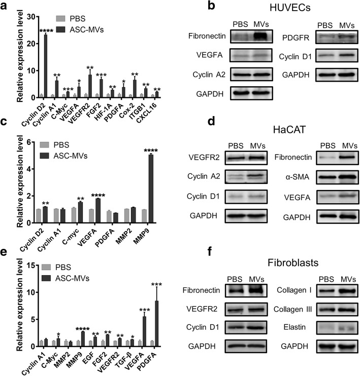
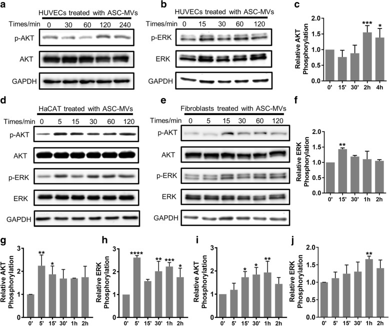
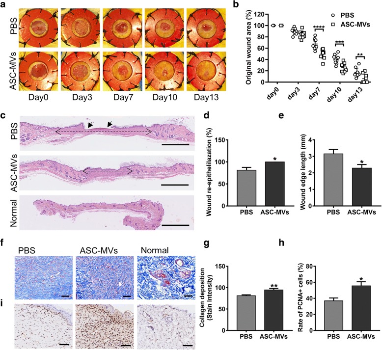
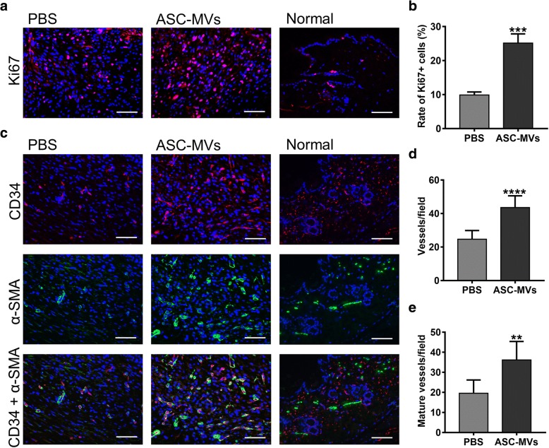
Results: ASC-MVs could be readily internalized by human umbilical vein endothelial cells (HUVECs), HaCAT, and fibroblasts and significantly promoted the proliferation, migration, and angiogenesis of these cells both in vitro and in vivo. The gene expression of proliferative markers (cyclin D1, cyclin D2, cyclin A1, cyclin A2) and growth factors (VEGFA, PDGFA, EGF, FGF2) was significantly upregulated after ASC-MV treatment. Importantly, ASC-MVs stimulated the activation of AKT and ERK signaling pathways in those cells. The local injection of ASC-MVs at wound sites significantly increased the re-epithelialization, collagen deposition, and neovascularization and led to accelerated wound closure.

Conclusions: Our data suggest that ASC-MVs can stimulate HUVEC, HaCAT, and fibroblast functions. ASC-MV therapy significantly accelerates wound healing, and the benefits of ASC-MVs may due to the involvement of AKT and ERK signaling pathways. This illustrates the therapeutic potential of ASC-MVs which may become a novel treatment paradigm for cutaneous wound healing.

Keywords: Adipose stem cells (ASCs); Extracellular vesicles; Microvesicles (MVs); Wound healing.


Conflict of interest statement

Ethics approval and consent to participate

All animal experiments were conducted according to the guidelines and standards of the Experimental Animal Center of Tongji Medical College, Huazhong University of Science and Technology.

The use of human adipose tissue and foreskin was approved by the Ethics Committee of Tongji Medical College, Huazhong University of Science and Technology (No. 2018-S288). We obtained the written informed consent from all the patients participated in this study.

Consent for publication

Not applicable.

Competing interests

The authors declare that they have no competing interests.

Publisher’s Note

Springer Nature remains neutral with regard to jurisdictional claims in published maps and institutional affiliations.

Figures

Fig. 1
Fig. 1
Isolation, characterization, and internalization of ASC-MVs. a Microvesicles isolated from ASC culture media were evidenced by electron microscopy. b Measurement of ASC-MV population by dynamic light scattering (DLS) demonstrated a single-peaked pattern (90–900 nm in diameter), which indicates isolated MVs were free of contamination. c Confocal images of HaCAT, HUVECs, and human foreskin fibroblasts incubated with either PBS or 20 μg PKH26-labeled MVs for 24 h
Fig. 2
Fig. 2
ASC-MVs induce HUVECs migration, proliferation, and angiogenesis. a Images of migrated HUVECs taken at 24 h after treatments of PBS, 5 μg/ml ASC-MVs, and 10 μg/ml ASC-MVs, respectively. Scar bar, 100 μm. b Qualification of migrated cells given different treatments (n = 3). c Enhanced tube formation in HUVECs treated with 20 μg/ml ASC-MVs compared with the controls at different time point. Scar bar, 50 μm. d Qualification of closed tubular structures shown in c (n = 3). e Increased HUVEC proliferation at day 2 after 20 μg/ml ASC-MV treatment compared with the controls as evidenced by CCK8 assay (n = 5). **p < 0.01, ***p < 0.001, ****p < 0.0001
Fig. 3
Fig. 3
Modulation of HaCAT and fibroblast function by ASC-MV administration. a Images of migrated fibroblasts taken at 24 h after treatments of PBS, 5 μg/ml ASC-MVs, and 10 μg/ml ASC-MVs, respectively. Scar bar, 100 μm. b Qualification of data shown in a (n = 3). c Images of migrated HaCAT given the above treatment. Scar bar, 100 μm. d Qualification of data shown in c (n = 3). e Increased HaCAT proliferation at days 2 and 3 after 20 μg/ml ASC-MV treatment compared with the controls as evidenced by CCK8 assay (n = 5). f Increased fibroblast proliferation at days 4 and 6 after 20 μg/ml ASC-MV treatment (n = 6). *p < 0.05, **p < 0.01, ***p < 0.001, ****p < 0.0001
Fig. 4
Fig. 4
Effect of ASC-MV treatment on gene expression in all three kinds of cells. qRT-PCR analysis of a cluster of gene expression in cells treated with either PBS or 20 μg/ml ASC-MVs. a Upregulated genes in HUVECs. c Upregulated genes in HaCAT. e Upregulated genes in fibroblasts. Western blot analysis of gene expression in cells given above treatments. b Upregulated genes in HUVECs. d HaCAT. f Fibroblasts. GAPDH served as an internal control. N = 3. *p < 0.05, **p < 0.01, ***p < 0.001, ****p < 0.0001
Fig. 5
Fig. 5
Effect of ASC-MVs on the activation level of AKT and ERK signaling pathways. Western blot analysis of the phosphorylation level of AKT and ERK1/2 in cells treated with 20 μg/ml ASC-MVs for the indicated lengths of time. a The ratio of p-AKT/AKT in HUVECs was examined and included on the blots at different time points. b The ratio of p-ERK/ERK in HUVECs was determined. d The ratio of p-ERK/ERK and p-AKT/AKT in HaCAT was determined. e The ratio of p-ERK/ERK and p-AKT/AKT in fibroblasts was determined. Qualification of the ratio of p-ERK/ERK and p-AKT/AKT at different time points. c, f Qualified data shown in a and b. g, h Qualified data shown in d. i, j Qualified data shown in e. N = 3. *p < 0.05, **p < 0.01, ***p < 0.001, ****p < 0.0001
Fig. 6
Fig. 6
Effect of ASC-MVs on wound healing in mice. a Gross view of excisional wounds in mice treated with either 50 μg ASC-MVs or an equal volume of PBS at different time points (n = 9). b Measurement of wound areas shown in a. c H&E staining analysis of wound sections following different treatments at day 13 post-wounding. The single-headed arrows indicate the un-epithelialized areas. The double-headed arrows indicate the edges of the granulation. Scar bar, 1 mm. d Qualification of wound re-epithelialization shown in c. e Qualification of granulation tissue formation shown in c. f Evaluation of collagen deposition by Masson staining at day 13 post-wounding. g Qualification of the stain intensity of blue collagen shown in f. h Qualification of the ratio of PCNA+ cells in wound beds. i Immunohistochemical staining for PCNA expression in wound sections. Scar bar, 100 μm. *p < 0.05, **p < 0.01, ***p < 0.001, ****p < 0.0001
Fig. 7
Fig. 7
ASC-MVs enhanced neovascularization and cellular proliferation in vivo as evidenced by immunofluorescence analysis. a Ki-67 (red color) immunofluorescence staining of wound sections treated with either 50 μg ASC-MVs or an equal volume of PBS at day 13 post-wounding. b Qualification of the ratio of Ki67+ cells in wound beds. c Immunofluorescent triple staining of wound sections given the above treatments at day 13 post-wounding. Smooth muscle cells (a-SMA), endothelial cells (CD34), and cell nuclei (DAPI) were stained with green, red, and blue colors. Newly formed vessels express CD34. Mature vessels express both CD34 and a-SMA. d Enumeration of newly formed vessels stained with red color. e Enumeration of mature vessels co-stained with red and green colors. Scar bar, 100 μm. N = 7. **p < 0.01, ***p < 0.001, ****p < 0.0001

References

    1. Singer AJ, Clark RA. Cutaneous wound healing. N Engl J Med. 1999;341:738–746. doi: 10.1056/NEJM199909023411006. - DOI - PubMed
    1. Falanga V. Wound healing and its impairment in the diabetic foot. Lancet. 2005;366:1736–1743. doi: 10.1016/S0140-6736(05)67700-8. - DOI - PubMed
    1. Gurtner GC, Werner S, Barrandon Y, Longaker MT. Wound repair and regeneration. Nature. 2008;453:314–321. doi: 10.1038/nature07039. - DOI - PubMed
    1. Heublein H, Bader A, Giri S. Preclinical and clinical evidence for stem cell therapies as treatment for diabetic wounds. Drug Discov Today. 2015;20:703–717. doi: 10.1016/j.drudis.2015.01.005. - DOI - PubMed
    1. Mizuno H, Tobita M, Uysal AC. Concise review: adipose-derived stem cells as a novel tool for future regenerative medicine. Stem Cells. 2012;30:804–810. doi: 10.1002/stem.1076. - DOI - PubMed

Publication types

Substances