. 2019 Mar 29;294(13):4911-4923.
doi: 10.1074/jbc.RA118.007135. Epub 2019 Jan 31.

Engineering a murine cell line for the stable propagation of hamster prions

Affiliations

Engineering a murine cell line for the stable propagation of hamster prions

Matthew E C Bourkas et al. J Biol Chem. .

Abstract

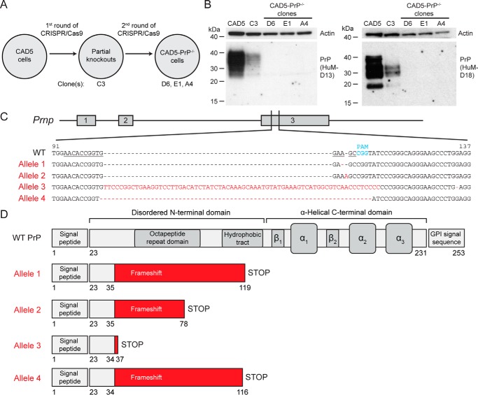
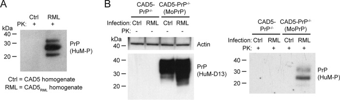
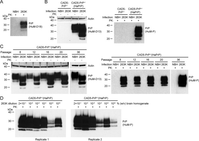
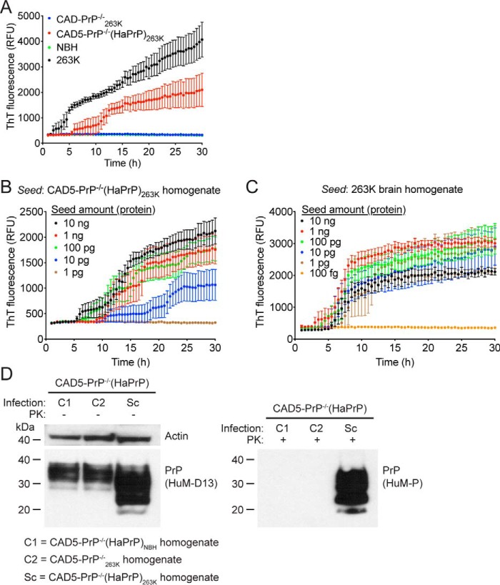
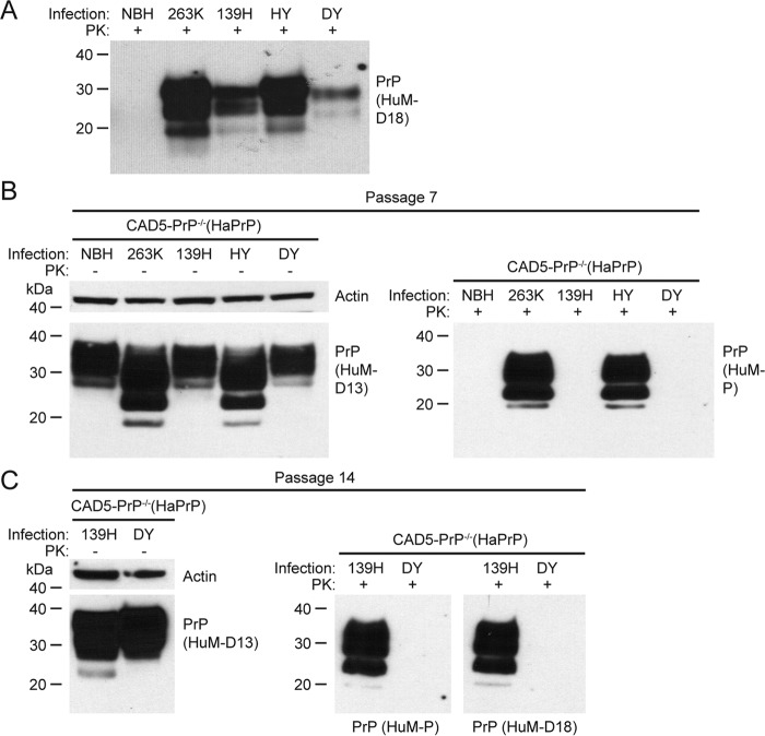
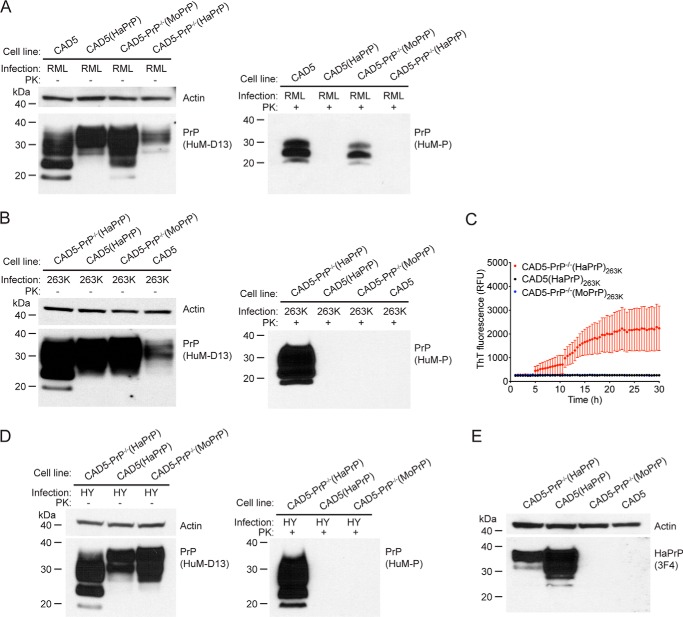
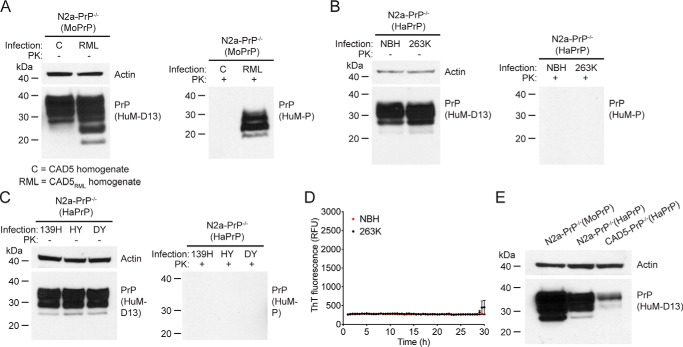
Prions are infectious protein aggregates that cause several fatal neurodegenerative diseases. Prion research has been hindered by a lack of cellular paradigms for studying the replication of prions from different species. Although hamster prions have been widely used to study prion replication in animals and within in vitro amplification systems, they have proved challenging to propagate in cultured cells. Because the murine catecholaminergic cell line CAD5 is susceptible to a diverse range of mouse prion strains, we hypothesized that it might also be capable of propagating nonmouse prions. Here, using CRISPR/Cas9-mediated genome engineering, we demonstrate that CAD5 cells lacking endogenous mouse PrP expression (CAD5-PrP-/- cells) can be chronically infected with hamster prions following stable expression of hamster PrP. When exposed to the 263K, HY, or 139H hamster prion strains, these cells stably propagated high levels of protease-resistant PrP. Hamster prion replication required absence of mouse PrP, and hamster PrP inhibited the propagation of mouse prions. Cellular homogenates from 263K-infected cells exhibited prion seeding activity in the RT-QuIC assay and were infectious to naïve cells expressing hamster PrP. Interestingly, murine N2a neuroblastoma cells ablated for endogenous PrP expression were susceptible to mouse prions, but not hamster prions upon expression of cognate PrP, suggesting that CAD5 cells either possess cellular factors that enhance or lack factors that restrict the diversity of prion strains that can be propagated. We conclude that transfected CAD5-PrP-/- cells may be a useful tool for assessing the biology of prion strains and dissecting the mechanism of prion replication.

Keywords: CRISPR/Cas; Creutzfeldt-Jakob disease; bovine spongiform encephalopathy; cell culture; heterologous system; neurodegeneration; neuron; prion; protein aggregation.


Conflict of interest statement

The authors declare that they have no conflicts of interest with the contents of this article

Figures

Figure 1.
Figure 1.
Generation of CAD5-PrP−/− cells. A, schematic of the procedure used to isolate CAD5-PrP−/− cells. Four monoclonal lines were kept for further analysis: The partial knockout clone C3 as well as CAD5-PrP−/− clones D6, E1, and A4. B, immunoblots of PrP levels in cell lysates from WT CAD5 cells, the partial knockout clone C3, and the CAD5-PrP−/− lines D6, E1, and A4. PrP was detected using the antibodies HuM-D13 (left blot) or HuM-D18 (right blot). The lower molecular weight PrP bands visible with the HuM-D18 antibody represent a physiological cleavage product termed C1 which is generated by endoproteolysis in the vicinity of PrP residue 110 (74). Blots were reprobed with an antibody to actin (20–33) to compare total protein levels. Molecular mass measurements are indicated in kDa. C, characterization of mutant Prnp alleles in CAD5-PrP−/− clone D6. The numbering is with respect to the ORF of WT Prnp. The targeting sequence of the gRNA is underlined, and the protospacer-adjacent motif (PAM) is indicated in blue. Four mutant alleles were identified: An adenine deletion (Allele 1), an adenine insertion (Allele 2), a large insertion (Allele 3), and a 10-bp deletion (Allele 4). The mutated residues present in the mutant alleles are indicated in red. D, schematic of truncated protein products predicted to be produced by the four mutant alleles. Beginning at PrP residues 34–35, frameshift mutations result in the incorporation of various amounts of non-PrP sequence (red) as well as a premature STOP codon. The domain structure of WT PrPC is shown for comparison.
Figure 2.
Figure 2.
Replication of mouse RML prions in CAD5-PrP−/−(MoPrP) cells. A, immunoblot of PK-digested cellular homogenates from either uninfected CAD5 cells (Ctrl) or RML prion–infected CAD5 cells (RML). B, infection of CAD5-PrP−/− and CAD5-PrP−/−(MoPrP) cells with RML prions. Immunoblots of either undigested (left blot) or PK-digested (right blot) lysates from cells at passage seven following exposure to homogenates from either uninfected CAD5 cells or RML prion-infected CAD5 cells. In both panels, PK-resistant PrP was visualized using the antibody HuM-P, and total (undigested) PrP was detected using the antibody HuM-D13. The undigested blot was also reprobed with an antibody to actin (20–33). Molecular mass measurements are indicated in kDa.
Figure 3.
Figure 3.
Replication of hamster 263K prions in CAD5-PrP−/−(HaPrP) cells. A, immunoblot of PK-digested NBH or 263K prion-infected brain homogenate from hamsters. PK-resistant PrP was visualized using the antibody HuM-D18. B, infection of CAD5-PrP−/− and CAD5-PrP−/−(HaPrP) cells with 263K prions. Immunoblots of either undigested (left blot) or PK-digested (right blot) lysates from cells at passage seven following exposure to NBH or 263K-infected brain homogenate. C, chronic replication of 263K prions in CAD5-PrP−/−(HaPrP) cells. Immunoblots of either undigested (left blots) or PK-digested (right blots) lysates from cells at passage 8, 12, 16, 20, or 36 following exposure to NBH or 263K-infected brain homogenate. D, immunoblots of PK-digested lysates from CAD5-PrP−/−(HaPrP) cells at passage seven following exposure to 10-fold serial dilutions of 0.2% (w/v) 263K-infected brain homogenate. Two independent replicates are shown. B–D, PK-resistant PrP was visualized using the antibody HuM-P, and total (undigested) PrP was detected using the antibody HuM-D13. The undigested blots were also reprobed with an antibody to actin (20–33). Molecular mass measurements are indicated in kDa.
Figure 4.
Figure 4.
Prion seeding activity and cellular infectivity of 263K prions passaged in CAD5-PrP−/−(HaPrP) cells. A, RT-QuIC using recombinant HaPrP(23–231) as a substrate and 100 ng of NBH (green), 263K-infected hamster brain homogenate (black), or homogenate from either 263K-exposed CAD5-PrP−/− (blue) or CAD5-PrP−/−(HaPrP) (red) cells (passage seven post infection) as a seed. Each data point represents the mean ± S.E. of quadruplicate reactions. B, RT-QuIC on 10-fold serial dilutions of CAD5-PrP−/−(HaPrP)263K homogenate (passage eight). Each data point represents the mean ± S.E. of eight independent reactions. C, RT-QuIC on 10-fold serial dilutions of 263K-infected hamster brain homogenate. Each data point represents the mean ± S.E. of four independent reactions. D, infection of CAD5-PrP−/−(HaPrP) cells with cell-passaged 263K prions. Immunoblots of either undigested (left blot) or PK-digested (right blot) lysates from cells at passage seven following exposure to homogenate from either CAD5-PrP−/−(HaPrP) cells treated with NBH (C1), CAD5-PrP−/− cells treated with 263K brain homogenate (C2), or CAD5-PrP−/−(HaPrP) cells treated with 263K brain homogenate (Sc), each at passage eight. PK-resistant PrP was visualized using the antibody HuM-P, and total (undigested) PrP was detected using the antibody HuM-D13. The undigested blot was also reprobed with an antibody to actin (20–33). Molecular mass measurements are indicated in kDa.
Figure 5.
Figure 5.
Challenge of CAD5-PrP−/−(HaPrP) cells with different hamster prion strains. A, immunoblot of PK-digested NBH or brain homogenate from hamsters infected with the 263K, 139H, Hyper (HY), or Drowsy (DY) strains. B, challenge of CAD5-PrP−/−(HaPrP) cells with various hamster prion strains. Immunoblots of either undigested (left blot) or PK-digested (right blot) lysates from cells at passage seven following exposure to NBH or hamster brain homogenate containing the indicated prion strains. C, immunoblots of either undigested (left blot) or PK-digested (right blots) lysates from cells at passage 14 following exposure to DY or 139H brain homogenate. PK-resistant PrP was visualized using the antibodies HuM-D18 (A) or HuM-P (B and C), and total (undigested) PrP was detected using the antibody HuM-D13. The undigested blots were also reprobed with an antibody to actin (20–33). Molecular mass measurements are indicated in kDa.
Figure 6.
Figure 6.
Evaluation of cross-species prion seeding and interference. A, immunoblots of either undigested (left blot) or PK-digested (right blot) lysates from CAD5, CAD5(HaPrP), CAD5-PrP−/−(MoPrP), or CAD5-PrP−/−(HaPrP) cells at passage seven following exposure to cell homogenate containing mouse RML prions. B, immunoblots of either undigested (left blot) or PK-digested (right blot) lysates from CAD5-PrP−/−(HaPrP), CAD5(HaPrP), CAD5-PrP−/−(MoPrP), or CAD5 cells at passage seven following exposure to hamster brain homogenate containing 263K prions. C, RT-QuIC using recombinant HaPrP(23–231) as a substrate and lysates (10−3 dilution, passage seven post infection) from 263K-exposed CAD5-PrP−/−(HaPrP) (red), CAD5(HaPrP) (black), or CAD5-PrP−/−(MoPrP) (blue) cells as a seed. Each data point represents the mean ± S.E. of quadruplicate reactions. D, immunoblots of either undigested (left blot) or PK-digested (right blot) lysates from CAD5-PrP−/−(HaPrP), CAD5(HaPrP), or CAD5-PrP−/−(MoPrP) cells at passage seven following exposure to hamster brain homogenate containing HY prions. E, HaPrP levels in lysates from uninfected CAD5, CAD5(HaPrP), CAD5-PrP−/−(MoPrP), or CAD5-PrP−/−(HaPrP) cells, as assessed by immunoblotting with the antibody 3F4, which recognizes HaPrP but not MoPrP. A, B, and D, PK-resistant PrP was visualized using the antibody HuM-P, and total (undigested) PrP was detected using the antibody HuM-D13. The undigested blots were also reprobed with an antibody to actin (20–33). Molecular mass measurements are indicated in kDa.
Figure 7.
Figure 7.
Challenge of transfected N2a-PrP−/− cells with prions. A, challenge of N2a-PrP−/−(MoPrP) cells with mouse RML prions. Immunoblots of either undigested (left blot) or PK-digested (right blot) lysates from cells at passage seven following exposure to homogenates from either uninfected CAD5 cells (C) or RML prion-infected CAD5 cells (RML). B, challenge of N2a-PrP−/−(HaPrP) cells with hamster 263K prions. Immunoblots of either undigested (left blot) or PK-digested (right blot) lysates from cells at passage seven following exposure to NBH or 263K-infected brain homogenate. C, challenge of N2a-PrP−/−(HaPrP) cells with hamster 139H, HY, and DY prion strains. Immunoblots of either undigested (left blot) or PK-digested (right blot) lysates from cells at passage seven following exposure to 139H-, HY-, or DY-infected brain homogenate. D, RT-QuIC using recombinant HaPrP(23–231) as a substrate and lysates (10−3 dilution, passage seven post infection) from N2a-PrP−/−(HaPrP) cells exposed to either 263K homogenate (black) or NBH (red) as a seed. Each data point represents the mean ± S.E. of quadruplicate reactions. E, PrPC levels in lysates from uninfected N2a-PrP−/−(MoPrP), N2a-PrP−/−(HaPrP), and CAD5-PrP−/−(HaPrP) cells, as detected by immunoblotting. A–C and E, PK-resistant PrP was visualized using the antibody HuM-P, and total (undigested) PrP was detected using the antibody HuM-D13. The undigested blots were also reprobed with an antibody to actin (20–33). Molecular mass measurements are indicated in kDa.

Similar articles

Cited by

References

    1. Watts J. C., Balachandran A., and Westaway D. (2006) The expanding universe of prion diseases. PLoS Pathog. 2, e26 10.1371/journal.ppat.0020026 - DOI - PMC - PubMed
    1. Oesch B., Westaway D., Wälchli M., McKinley M. P., Kent S. B. H., Aebersold R., Barry R. A., Tempst P., Teplow D. B., Hood L. E., Prusiner S. B., and Weissmann C. (1985) A cellular gene encodes scrapie PrP 27–30 protein. Cell 40, 735–746 10.1016/0092-8674(85)90333-2 - DOI - PubMed
    1. Prusiner S. B. (1982) Novel proteinaceous infectious particles cause scrapie. Science 216, 136–144 10.1126/science.6801762 - DOI - PubMed
    1. Colby D. W., and Prusiner S. B. (2011) Prions. Cold Spring Harb Perspect Biol. 3, a006833 10.1101/cshperspect.a006833 - DOI - PMC - PubMed
    1. Collinge J. (2016) Mammalian prions and their wider relevance in neurodegenerative diseases. Nature 539, 217–226 10.1038/nature20415 - DOI - PubMed

Publication types