. 2019 Apr 10;39(15):2776-2791.
doi: 10.1523/JNEUROSCI.2213-17.2019. Epub 2019 Jan 31.

Calcium-Activated Calpain Specifically Cleaves Glutamate Receptor IIA But Not IIB at the Drosophila Neuromuscular Junction

Affiliations

Calcium-Activated Calpain Specifically Cleaves Glutamate Receptor IIA But Not IIB at the Drosophila Neuromuscular Junction

Elsayed Metwally et al. J Neurosci. .

Abstract

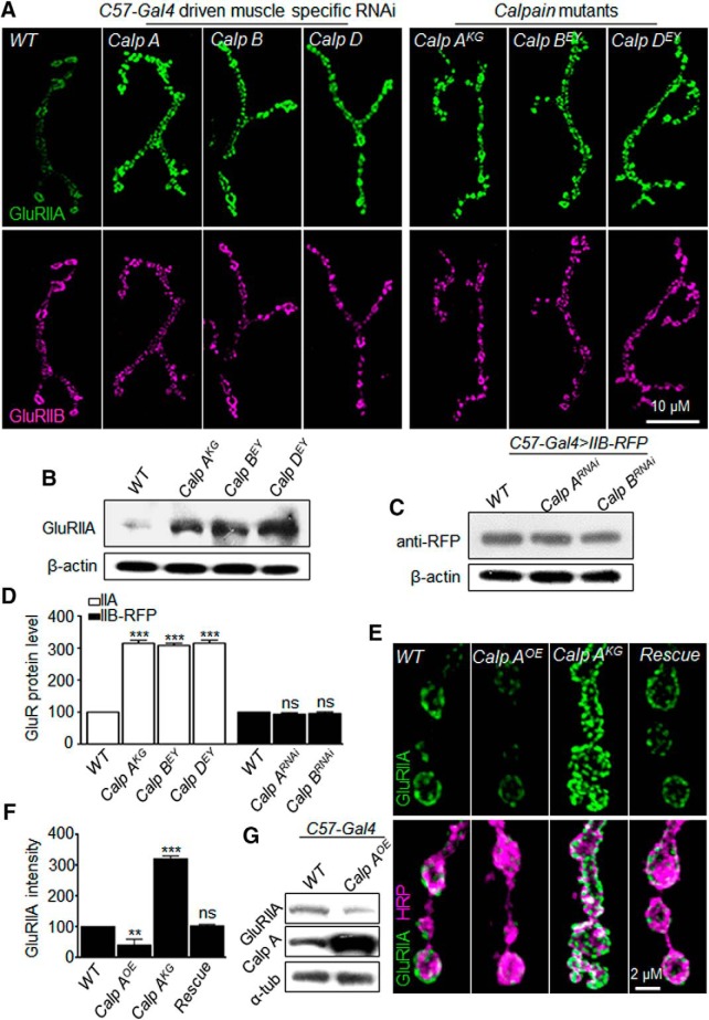
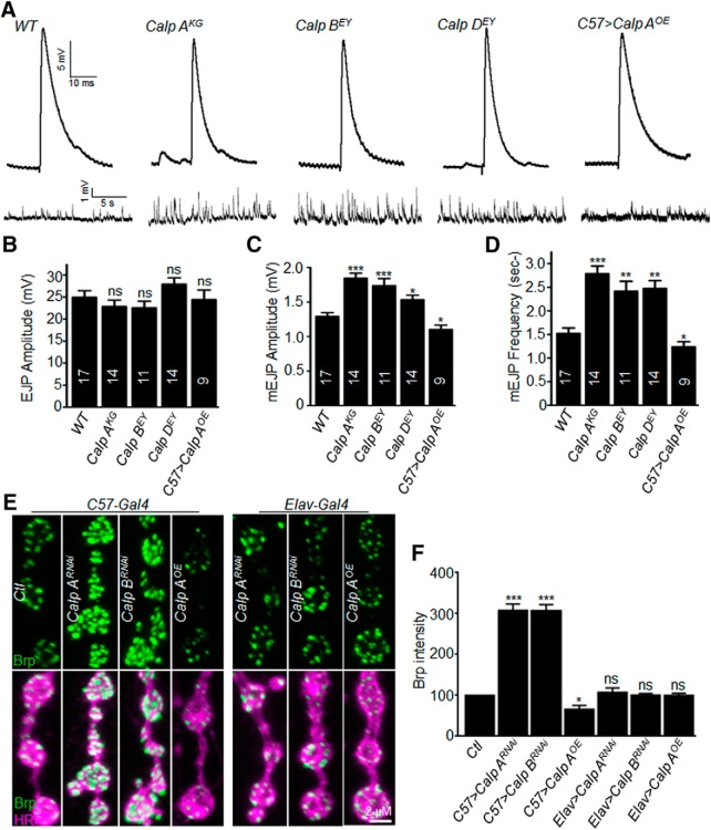
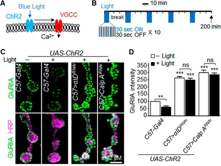
Calpains are calcium-dependent, cytosolic proteinases active at neutral pH. They do not degrade but cleave substrates at limited sites. Calpains are implicated in various pathologies, such as ischemia, injuries, muscular dystrophy, and neurodegeneration. Despite so, the physiological function of calpains remains to be clearly defined. Using the neuromuscular junction of Drosophila of both sexes as a model, we performed RNAi screening and uncovered that calpains negatively regulated protein levels of the glutamate receptor GluRIIA but not GluRIIB. We then showed that calpains enrich at the postsynaptic area, and the calcium-dependent activation of calpains induced cleavage of GluRIIA at Q788 of its C terminus. Further genetic and biochemical experiments revealed that different calpains genetically and physically interact to form a protein complex. The protein complex was required for the proteinase activation to downregulate GluRIIA. Our data provide a novel insight into the mechanisms by which different calpains act together as a complex to specifically control GluRIIA levels and consequently synaptic function.SIGNIFICANCE STATEMENT Calpain has been implicated in neural insults and neurodegeneration. However, the physiological function of calpains in the nervous system remains to be defined. Here, we show that calpain enriches at the postsynaptic area and negatively and specifically regulates GluRIIA, but not IIB, level during development. Calcium-dependent activation of calpain cleaves GluRIIA at Q788 of its C terminus. Different calpains constitute an active protease complex to cleave its target. This study reveals a critical role of calpains during development to specifically cleave GluRIIA at synapses and consequently regulate synaptic function.

Keywords: Drosophila neuromuscular junction; GluR; calcium; calpain; cytoplasmic cysteine protease; glutamate receptor.


Figures

Figure 1.
Figure 1.
Calpains negatively regulate synaptic and total levels of GluRIIA, but not GluRIIB, protein. A, Synaptic levels of GluRIIA, but not IIB, were increased in both calpain mutants and postsynaptic RNAi knockdown larvae. Representative images of NMJ4 synapse from different genotypes double-stained with anti-GluRIIA (green) and anti-GluRIIB (magenta). Scale bar, 10 μm. For a list of positive RNAi lines identified for increased GluRIIA expression at NMJ, see also Table 1-1. B, Total GluRIIA protein levels were increased in calpain mutants. Representative immunoblots detected with anti-GluRIIA in different genotypes. β-Actin was used as a loading control. C, Representative immunoblots showing RFP-tagged GluRIIB levels in different genotypes. β-Actin was used as a loading control. D, Quantitative analysis of total GluRIIA and GluRIIB-RFP protein levels. n = 3. ***p < 0.001 (one-way ANOVA with Tukey's post hoc test). Data are mean ± SEM. E, Calpain A overexpression reversed synaptic GluRIIA levels in calpain A mutants. Representative images of NMJ4 synapse from WT, calp AOE (UAS-calp A/C57-Gal4), calp AKG, and rescue (calp AKG; UAS-calp A/C57-Gal4) costained with anti-GluRIIA (green) and anti-HRP, a neuronal membrane marker (magenta). Scale bar, 2 μm. F, Quantitative analysis of fluorescence intensity of synaptic GluRIIA levels from different genotypes. n = 15 NMJs. **p < 0.01; ***p < 0.001; one-way ANOVA with Tukey's post hoc test. Data are mean ± SEM. G, Representative immunoblots showing total GluRIIA and calpain A protein levels from different genotypes. α-Tubulin was used as a loading control.
Figure 2.
Figure 2.
Temperature-sensitive temporal calpain inactivation leads to upregulation of GluRIIA at NMJ. A, Table of GluRIIA levels at NMJ synapses of C57-Gal4>Calp ARNAi+Gal80ts after temperature shift from 18°C (blue) to 30°C (orange). *p < 0.1; **p < 0.01; ***p < 0.001; one-way ANOVA with Tukey's post hoc test. B, Representative images of NMJ4 synapse from different genotypes after temperature shift from 18°C to 30°C for 1 d double-stained with anti-GluRIIA (green) and anti-HRP (magenta). Scale bar, 2 μm. C, Quantitative analysis of fluorescence intensity of synaptic GluRIIA levels from indicated genotypes. n = 8, 10, and 12 NMJs for each time point for control (C57-Gal4>Gal80ts), RNAi knockdown (C57-GAL4>Calp ARNAi), and the temperature-sensitive genotype (C57-Gal4>Calp ARNAi+Gal80ts), respectively. *p < 0.1; **p < 0.01; ***p < 0.001; one-way ANOVA with Tukey's post hoc test. Data are mean ± SEM.
Figure 3.
Figure 3.
Calpain A enriches at postsynaptic NMJ. A, Calpain A enriches at postsynaptic NMJ. Representative images of NMJ4 synapses from different genotypes, stained with anti-Dlg (magenta), anti-Calp A (green), and anti-HRP (blue). Scale bar, 2 μm. B, Representative images of WT NMJ4 synapse, stained with anti-GluRIIA (magenta) and anti-calp A (green). Scale bar, 2 μm. C, Representative immunoblots probed with anti-calp A and anti-calp B showing calpain protein level in different genotypes. β-Actin was used as a loading control. D, Quantitative analysis of the total protein level of calpains in calpain mutants and RNAi knockdown larvae. n = 3. ***p < 0.001 (one-way ANOVA with Tukey post hoc test). Data are mean ± SEM. E, A set of synaptic proteins remain unchanged at NMJs when calpain A is knocked down or overexpressed in the postsynaptic muscles. Representative images of NMJ4 synapses from indicated genotypes double-stained with antibodies against indicated synaptic markers. The intensities of DLG, dPAK, α-spectrin, β-spectrin, Futsch, and Fasciclin II levels were unchanged upon knockdown or overexpression of calpain A in postsynaptic muscles by C57-Gal4. Scale bar, 2 μm. F, Quantitative analysis of fluorescence intensity of synaptic tested proteins in indicated genotypes. n = 8. p > 0.05 by one-way ANOVA with Tukey post hoc test. Data are mean ± SEM.
Figure 4.
Figure 4.
Calpains regulate synaptic function. A, Representative recordings of EJPs and mEJPs from different genotypes. B–D, Quantitative analysis of EJP amplitudes (B), mEJP amplitudes (C), and mEJP frequencies (D) from different genotypes. Mean EJP amplitudes were normal, whereas mean mEJP amplitudes and frequencies in calpain mutants were significantly higher than WT but lower in calpain-overexpressing NMJs. n = 17, 14, 11, 14, and 9 recordings for WT, calp AKG, calp BEY, calp DEY, and calp AOE, respectively. *p < 0.05; **p < 0.01; ***p < 0.001; one-way ANOVA with Tukey post hoc test. Data are mean ± SEM. E, Postsynaptic, but not presynaptic, knockdown of calpains gives rise to upregulation of Brp at synapses. Brp intensity is increased upon calpain knockdown, whereas calpain overexpression reduces Brp intensity at NMJ. Representative confocal images of NMJ4 synapses stained with anti-Brp (green) and HRP (magenta) from different genotypes of control, calp ARNAi, calp BRNAi, and calp AOE under the control of muscle-specific C57-Gal4 or neuron-specific Elav-Gal4. Scale bar, 2 μm. F, Quantitative analysis of fluorescence intensity of synaptic Brp in indicated genotypes. n = 10 NMJ terminals for each genotype. *p < 0.05; ***p < 0.001; one-way ANOVA with Tukey post hoc test. Data are mean ± SEM.
Figure 5.
Figure 5.
Cytoplasmic calcium negatively regulates GluRIIA level via calpain. A, Diagram depicting various calcium channels on different membrane organelles. B, Intracellular calcium negatively regulates synaptic GluRIIA. Representative images of NMJ4 synapses from different genotypes costained with anti-GluRIIA (green) and anti-GluRIIB (magenta). Scale bar, 2 μm. For a list of RNAi lines for Ca2+ channels tested for GluRIIA expression at NMJ, see also Table 5-1. C, Quantitative analysis of fluorescence intensity of synaptic GluRIIA and GluRIIB in different genotypes. n = 12 NMJs. **p < 0.01; ***p < 0.001; one-way ANOVA with Tukey's post hoc test. Data are mean ± SEM. D, Calcium negatively regulates GluRIIA protein levels. Bar graph represents densitometric analysis of the relative levels of GluRIIA. n = 3. **p < 0.01; ***p < 0.001; one-way ANOVA with Tukey's post hoc test. Data are mean ± SEM. E, Representative confocal images of NMJ4 synapses from indicated genotypes double-stained with anti-GluRIIA (green) and HRP (magenta). Calpain A and calcium channels were knocked down either singly or simultaneously using the muscle-specific C57-Gal4. Scale bar, 2 μm. F, Quantitative analysis of fluorescence intensity of synaptic GluRIIA in different genotypes. n = 12 NMJs. **p < 0.01; ***p < 0.001; one-way ANOVA with Tukey's post hoc test. Data are mean ± SEM. G, Calcium treatment reduced GluRIIA abundance via calpain. Representative images of WT NMJ4 synapses costained with anti-GluRIIA (green) and anti-GluRIIB (magenta). Muscle cells were treated with vehicle dimethylsulfoxide (DMSO), calcium (10 mm), and calcium (10 mm) plus calpain inhibitor PD150606 (20 μm). Scale bar, 2 μm. H, Quantitative analysis of fluorescence intensity of synaptic GluRIIA and GluRIIB upon calcium treatment. n = 15 NMJs. ***p < 0.001 (one-way ANOVA with Tukey's post hoc test). Data are mean ± SEM.
Figure 6.
Figure 6.
Calcium influx mediated by optogenetic manipulation results in GluRIIA reduction via calpain. A, A schematic of optogenetic manipulation of calcium influx. Blue light activates ChR2, which in turn promotes calcium influx via VGCC. B, Light stimulation protocol consists of five rounds of 10 min light stimulation followed by 30 min break. During the illumination period, the light turns on and off for 30 s each for 10 times. C, Light stimulation resulted in GluRIIA reduction via calpain. Representative images of NMJ4 synapses from different genotypes costained with anti-GluRIIA (green) and anti-HRP (magenta). Scale bar, 2 μm. D, Quantitative analysis of fluorescence intensity of synaptic GluRIIA in different genotype with or without blue light illumination. n = 15 NMJs. **p < 0.01; ***p < 0.001 (one-way ANOVA with Tukey's post hoc test). Data are mean ± SEM.
Figure 7.
Figure 7.
Calcium-dependent cleavage of GluRIIA at C terminus by calpain. A, B, Calpain specifically cleaves GluRIIA at its C terminus. Diagram showing location of Myc tags at N termini. Arrow indicates putative cleavage site. Black bar at the C-terminal of GluRIIA represents anti-GluRIIA epitope. Muscle cells were treated with vehicle (DMSO), calcium, and calcium plus the calpain inhibitor PD150606. Lysates were subjected to IP with anti-Myc followed by immunoblotting with anti-Myc. Black arrowheads indicate full-length GluRIIA (A) or GluRIIB (B). Open arrowheads indicate truncated N-terminal band of GluRIIA. C, Calpain autolysis-associated cleavage of GluRIIA. S2 cells expressing Flag-GluRIIA were treated with vehicle (DMSO), calcium at the indicated concentrations, and calcium plus PD150606. Black arrowheads indicate full-length. Open arrowheads indicate N-terminal fragment of GluRIIA. Arrows indicate autolytic bands of calpains. D, Concentration dependency of calcium-induced truncation of GluRIIA and calpain autolysis. Quantitative analysis of the level of GluRIIA, calpain A, and calpain B fragments in arbitrary units (a.u.). n = 3. *p < 0.05; **p < 0.01; ***p < 0.001; one-way ANOVA with Tukey's post hoc test. Data are mean ± SEM. E, F, Calcium-dependent interaction of calpain with GluRIIA. S2 cells coexpressing Flag-IIA and calp A-His or calp B-His were treated with vehicle (DMSO) and calcium (10 mm). Lysates were IPed with anti-Flag antibody or mouse IgG as a control, followed by IB with anti-His and anti-Flag (E). F, Lysates were IPed with anti-calp A or rabbit IgG as a control, followed by Western analysis with anti-Flag and anti-calp A. E, F, Black arrowheads indicate GluRIIA/calpain interaction. White arrowheads indicate GluRIIA fragments. Arrows indicate calpain autolysis fragments.
Figure 8.
Figure 8.
GluRIIA is cleaved by calpain at Q788. A, Bioinformatics analysis of a peptide from G785 to F840 of GluRIIA for putative calpain cleavage sites. An analysis using CaMPDA (www.calpain.org/prediction_view.rb) showed Q788, M790, K791, and N797 as calpain cleavage sites, whereas an analysis using GPS-CCD 1.0 (http://ccd.biocuckoo.org) identified Q788 and K791 as putative calpain cleavage sites. B, HNE induces cleavage of GluRIIA by calpain in a concentration-dependent manner. S2 cells expressing Flag-GluRIIA were treated with vehicle (DMSO) and HNE at the indicated concentrations. Black arrow indicates the N-terminal fragment of GluRIIA at 105 kDa. Gray arrow indicates an unspecific band. β-Actin was used as a loading control. C, S2 cells expressing WT full-length Flag-IIA, single or double amino acid mutant GluRIIA at Q788 and K791 were analyzed by immunoblotting with anti-GluIIA. Black arrow indicates the N-terminal fragments of GluRIIA. β-Actin was used as a loading control. Red arrow indicates the calpain cleavage site in GluRIIA (based on Accession No. NP_523484), but not GluRIIB (based on Accession No. AAF52269).
Figure 9.
Figure 9.
Different calpains form a protein complex to downregulate GluRIIA. A, Different calpains interact genetically to downregulate IIA at NMJs. Representative images of NMJ synapses from different genotypes costained with anti-GluRIIA (green) and anti-GluRIIB (magenta). Scale bar, 2 μm. B, Quantitative analysis of fluorescence intensity of synaptic GluRIIA from indicated genotypes. n = 12. ***p < 0.001 (one-way ANOVA with Tukey's post hoc test). Data are mean ± SEM. C, Calpain A interacts physically with calpain B in vivo. Lysates were extracted from WT and calp A-GFP. Muscle lysates were IPed with anti-GFP and detected with indicated antibodies on Western blots. *IgG heavy chain at 55 kDa. D, E, Calpains interact with each other in S2 cells. S2 cells expressing tagged proteins were IPed with anti-calp A (D) or anti-Flag (E), followed by immune-blotting (IB) analysis with the indicated antibodies. *IgG heavy chain at 55 kDa. F, Interaction of calpain A and B is not affected by calcium. The lysates were IPed with anti-calp B or rabbit IgG as a control and subsequently detected by Western analysis using the indicated antibodies. G, Representative images of calp A-GFP-expressing muscles costained with anti-GFP (green) and anti-calp B (magenta). Bottom, A higher magnification of the inset. Scale bar, 5 μm. H, HA-tagged calpain A and His-tagged other calpains were substantially colocalized in S2 cells. S2 cells were stained with anti-HA (green) and anti-His (magenta). Scale bar, 5 μm.
Figure 10.
Figure 10.
Calpain activity, but not protein level, depends on the integrity of calpain complex. The expression of calpain A and B is not dependent on each other. A, B, Representative images of NMJ4 synapses from indicated genotypes costained with GluRIIA (green) and calp A (magenta) (A) or calp B (magenta) (B). Scale bar, 2 μm. C, Quantitative analysis of fluorescence intensities of synaptic GluRIIA, calpain A, and calpain B in different genotypes. n = 11 NMJs. ***p < 0.001 (one-way ANOVA with Tukey post hoc test). Data are mean ± SEM. D, Representative immunoblots showing the total protein levels of calpains in different genotypes. β-Actin was used as a loading control. E, Muscle lysates from different genotypes were subjected to Western blotting with anti-calp A or anti-calp B. α-Tubulin was used as a loading control. Black arrowhead indicates full-length calpain. Open arrowhead indicates autolytic fragments of calpains in WT larvae. Arrows indicate autolytic calpain fragments in calpain mutants. F, Different calpains act together, probably as a protein complex, to control GluR levels during development. In the absence of one calpain, the activity of other calpains is compromised, suggesting that the full activity of calpains requires the integrity of the calpain complex.

Similar articles

Cited by

References

    1. Abe K, Takeichi M (2007) NMDA-receptor activation induces calpain-mediated beta-catenin cleavages for triggering gene expression. Neuron 53:387–397. 10.1016/j.neuron.2007.01.016 - DOI - PubMed
    1. Albin SD, Davis GW (2004) Coordinating structural and functional synapse development: postsynaptic p21-activated kinase independently specifies glutamate receptor abundance and postsynaptic morphology. J Neurosci 24:6871–6879. 10.1523/JNEUROSCI.1538-04.2004 - DOI - PMC - PubMed
    1. Anggono V, Huganir RL (2012) Regulation of AMPA receptor trafficking and synaptic plasticity. Curr Opin Neurobiol 22:461–469. 10.1016/j.conb.2011.12.006 - DOI - PMC - PubMed
    1. Bano D, Young KW, Guerin CJ, Lefeuvre R, Rothwell NJ, Naldini L, Rizzuto R, Carafoli E, Nicotera P (2005) Cleavage of the plasma membrane Na+/Ca2+ exchanger in excitotoxicity. Cell 120:275–285. 10.1016/j.cell.2004.11.049 - DOI - PubMed
    1. Baudry M, Bi X (2016) Calpain-1 and calpain-2: the yin and yang of synaptic plasticity and neurodegeneration. Trends Neurosci 39:235–245. 10.1016/j.tins.2016.01.007 - DOI - PMC - PubMed

Publication types

MeSH terms

LinkOut - more resources