Review
. 2019 Jan 31;5(1):9.
doi: 10.1038/s41572-019-0060-9.

Kaposi sarcoma

Affiliations
Review

Kaposi sarcoma

Ethel Cesarman et al. Nat Rev Dis Primers. .

Abstract

Kaposi sarcoma (KS) gained public attention as an AIDS-defining malignancy; its appearance on the skin was a highly stigmatizing sign of HIV infection during the height of the AIDS epidemic. The widespread introduction of effective antiretrovirals to control HIV by restoring immunocompetence reduced the prevalence of AIDS-related KS, although KS does occur in individuals with well-controlled HIV infection. KS also presents in individuals without HIV infection in older men (classic KS), in sub-Saharan Africa (endemic KS) and in transplant recipients (iatrogenic KS). The aetiologic agent of KS is KS herpesvirus (KSHV; also known as human herpesvirus-8), and viral proteins can induce KS-associated cellular changes that enable the virus to evade the host immune system and allow the infected cell to survive and proliferate despite viral infection. Currently, most cases of KS occur in sub-Saharan Africa, where KSHV infection is prevalent owing to transmission by saliva in childhood compounded by the ongoing AIDS epidemic. Treatment for early AIDS-related KS in previously untreated patients should start with the control of HIV with antiretrovirals, which frequently results in KS regression. In advanced-stage KS, chemotherapy with pegylated liposomal doxorubicin or paclitaxel is the most common treatment, although it is seldom curative. In sub-Saharan Africa, KS continues to have a poor prognosis. Newer treatments for KS based on the mechanisms of its pathogenesis are being explored.


Figures
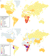
Fig. 1 ∣
Fig. 1 ∣. Geographical prevalence of KS and seroprevalence of KSHV.
a ∣ The age-standardized incidence rate of Kaposi sarcoma (KS) per 100,000 males is depicted, and rates (apart from for the USA) were obtained from the International Agency for Research on Cancer (IARC) Cancer Incidence in Five Continents Volume X1 and ‘Cancer Today’ Global Cancer Observatory resources,. The rate provided for the USA is an average for 2000–2015 (0.7 affected individuals per 100,000 males) and rates are from Surveillance, Epidemiology, and End Results (SEER). However, rates in some regions based on the population reported are higher than others, ranging from 1.7 affected individuals per 100,000 males (for Atlanta) to 0.1 affected individuals per 100,000 males (for Iowa and Utah). Overall rates in the USA show racial disparities: among non-Hispanic white individuals, white Hispanics and black individuals, the incidence rate is 0.4, 0.7 and 1 affected individual per 100,000 males, respectively. b ∣ Seroprevalence rates were compiled from multiple studies,,,-. The seroprevalence of KS herpesvirus (KSHV) infection in northern Europe, Asia and the USA is <10%, but in most of sub-Saharan Africa, overall seroprevalence is >40%. The Mediterranean region (that is, Italy, Sicily and Sardinia) has intermediate seroprevalence rates of 10–30%. Figure adapted from reF., Springer Nature Limited.
Fig. 2 ∣
Fig. 2 ∣. The viral life cycle of KSHV.
The Kaposi sarcoma herpesvirus (KSHV) virion binds to receptors present on the cell surface (such as integrins, the cystine–glutamate transporter (xCT), CD98 and heparan sulfate) via glycoproteins (such as gpK8.1, gB, gM–gN and gH–gL) on its envelope; this binding, in most cases, results in the endocytosis of the virion into the cell. The virion uncoats itself in the cell cytoplasm, and the capsid containing the viral genome traverses to the nucleus. The viral genome enters the nucleus, where it can remain latent as a circular episome tethered to host chromosomes via its latency-associated nuclear protein (LANA), or it can enter the lytic cycle where the viral genomes are replicated and new virions are produced through a complex mechanism of envelopment and ultimately released from the cell via budding. Note that KSHV proteins can increase host signalling through the phosphoinositide 3-kinase (PI3K), mitogen-activated protein kinase (MAPK) and nuclear factor-κB (NF-κB) signalling pathways. RTA, replication and transcription activator.
Fig. 3 ∣
Fig. 3 ∣. Modulation of host signalling pathways by KSHV-encoded proteins.
A graphic representation illustrates how Kaposi sarcoma herpesvirus (KSHV) representative viral proteins (in orange) modulate host cellular signalling proteins (in blue) to promote cellular survival and the inhibition of apoptosis. Viral proteins including KSHV K15, K1, viral G protein-coupled receptor (vGPCR) and viral IL-6 (vIL-6) can activate the phosphoinositide 3-kinase (PI3K)–protein kinase B (AKT)–mechanistic target of rapamycin (mTOR) pathway, thereby increasing cell survival and promoting protein synthesis. KSHV ORF36 acts further downstream and directly phosphorylates cellular ribosomal protein S6, which in turn leads to increased global protein synthesis. KSHV ORF36 also phosphorylates Jun N-terminal kinase (JNK). Some of the KSHV viral proteins, such as vGPCR, K1 and K15, also activate the extracellular-signal-regulated kinase 1 (ERK1)–ERK2 pathway to promote protein synthesis through the activation of ribosomal S6 kinase (RSK; which in turn phosphorylates S6), and KSHV ORF45 directly binds to RSK, leading to its activation. Note that there is a certain degree of crosstalk between both the PI3K–AKT–mTOR and ERK1–ERK2 pathways. Finally, KSHV viral FLICE inhibitory protein (vFLIP) potently activates nuclear factor-κB (NF-κB) signalling by directly binding NF-κB essential modulator (NEMO). This signalling leads to degradation of the inhibitor of NF-κB, IκBα, and translocation of the NF-κB transcription factors into the nucleus. Dashed lines indicate indirect activation. 4EBP1, eukaryotic translation initiation factor 4E-binding protein 1; eIF4B, eukaryotic translation initiation factor 4B; gp130, membrane glycoprotein 130; MEK, MAPK/ERK kinase; mTORC1, mechanistic TOR complex 1; PLCγ1, phospholipase C, γ1; SRK, Syk-related tyrosine kinase; S6KB1, ribosomal protein S6 kinase.
Fig. 4 ∣
Fig. 4 ∣. Modulation of innate host immunity by KSHV.
Kaposi sarcoma herpesvirus (KSHV) encodes an arsenal of viral proteins that modulate multiple innate immune pathways. KSHV viral interferon regulatory factor 1 (vIRF1), viral G protein-coupled receptor (vGPCR) and replication and transcription activator (RTA) can downregulate Toll-like receptor 2 (TLR2) and TLR4, and TRIF (also known as TIR domain-containing adaptor molecule 1 (TICAM1)), a TLR adaptor protein, is further inhibited by KSHV RTA. Inhibition of these TLR signalling pathways results in the downregulation of the type I interferon response. Ubiquitylation of the RNA sensor, retinoic acid-inducible gene I protein (RIG-I), which is crucial for its activation, is inhibited by KSHV ORF64, resulting in the dampening of RIG-I activity and inhibition of the interferon response. KSHV vIRFs and ORF45 can inhibit activation of cellular IRFs such as IRF3 and IRF7. Cyclic GMP-AMP synthase (cGAS)–stimulator of interferon genes protein (STING) is a cytosolic DNA sensing pathway, and cGAS activation is inhibited by KSHV ORF52 and KSHV latency-associated nuclear protein (LANA), whereas KSHV vIRF1 prevents STING association with the serine/threonine-protein kinase TBK1, thereby inhibiting interferon production through the pathway mediated by IRF3 and IRF7. The NOD-, LRR- and pyrin domain-containing 1 (NLRP1) inflammasome, which comprises NLRP1, apoptosis-associated speck-like protein containing a CARD (ASC) and pro-caspase-1, is inhibited by KSHV ORF63, resulting in inhibition of IL-1β and IL-18 activation. 5'ppp, 5'-triphosphate; dsRNA, double-stranded RNA; MAVS, mitochondrial antiviral-signalling protein; MDA5, melanoma differentiation-associated protein 5; MyD88, myeloid differentiation primary response protein MyD88; NF-κB, nuclear factor-κB; NLR, Nod-like receptor; TRAF3, TNF receptor-associated factor 3; Ub, ubiquitin.
Fig. 5 ∣
Fig. 5 ∣. Clinical manifestations of KS.
Different manifestations of Kaposi sarcoma (KS) include (part a) macular lesions on the back and nodules on the arm; (part b) extensive KS plaques on the legs with tumour-associated oedema; (part c) exophytic KS lesions on the foot; (part d) extensive gingival KS nodules; and (part e) flat, violaceous lesions on the hard palate. The image in part c is of a patient with classic KS; all other images are of patients with AIDS-related KS.
Fig. 6 ∣
Fig. 6 ∣. Histopathology of KS.
a ∣ Haematoxylin and eosin (H&E) staining of a skin biopsy sample showing characteristic features of Kaposi sarcoma (KS) is shown. Note the presence of spindle cells replacing dermal collagen, vascular spaces containing red blood cells, extravasated red blood cells and haemosiderin in macrophages (that is, siderophages). The original magnification is 20× (left panel) and 60× (right panels). b ∣ H&E staining showing sheets of spindle cells in a biopsy sample of a nodular KS lesion. Original magnification 60×. c ∣ A biopsy sample of a patch-stage KS lesion, stained with H&E, which has a sparse cellular infiltrate with many inflammatory cells (seen as dark, small cells). Original magnification is 20×. d ∣ Immunohistochemistry for KS herpesvirus (KSHV) latency-associated nuclear protein (LANA) in a skin biopsy sample of patch-stage KS, in which cells with brown nuclei line vascular spaces. e ∣ H&E staining of a nodular-stage KS lesion is shown at low power (4×), with a large nodule formed by swirling fascicles of spindle cells. Original magnification 20×. f ∣ Immunohistochemistry for KSHV LANA in an area of a nodular KS lesion biopsy sample, showing immunoreactivity indicated by brown staining with a characteristic punctate pattern in the nuclei of spindle cells. Original magnification 60×

Similar articles

Cited by

References

    1. Kaposi M Idiopatisches multiples pigmentsarkom der haut [German]. Arch. Dermatol. Syph. 4, 265–273 (1872).
    1. Montpellier J & Mussini-Montpellier J Le Cancer en France d’outre-mer: Considérations Pathogéniques (Libraire Ferraris, 1947).
    1. D’Oliveira JJ & Torres FO Kaposi’s sarcoma in the Bantu of Mozambique. Cancer 30, 553–561 (1972). - PubMed
    1. Thijs A L’angiosarcomatose de Kaposi au Congo Beige et au Ruanda-Urundi [French]. Ann. Soc. Belg. Med. Trop 37, 295–307 (1957). - PubMed
    1. Gottlieb GJ et al. A preliminary communication on extensively disseminated Kaposi’s sarcoma in young homosexual men. Am. J. Dermatopathol. 3, 111–114 (1981).

      This paper presents the first report of KS in MSM as a harbinger of the AIDS epidemic.

Publication types

MeSH terms

Supplementary concepts