. 2019 Jan 31;9(1):1022.
doi: 10.1038/s41598-018-37395-9.

Memory-like Liver Natural Killer Cells are Responsible for Islet Destruction in Secondary Islet Transplantation

Affiliations

Memory-like Liver Natural Killer Cells are Responsible for Islet Destruction in Secondary Islet Transplantation

Y Saeki et al. Sci Rep. .

Abstract

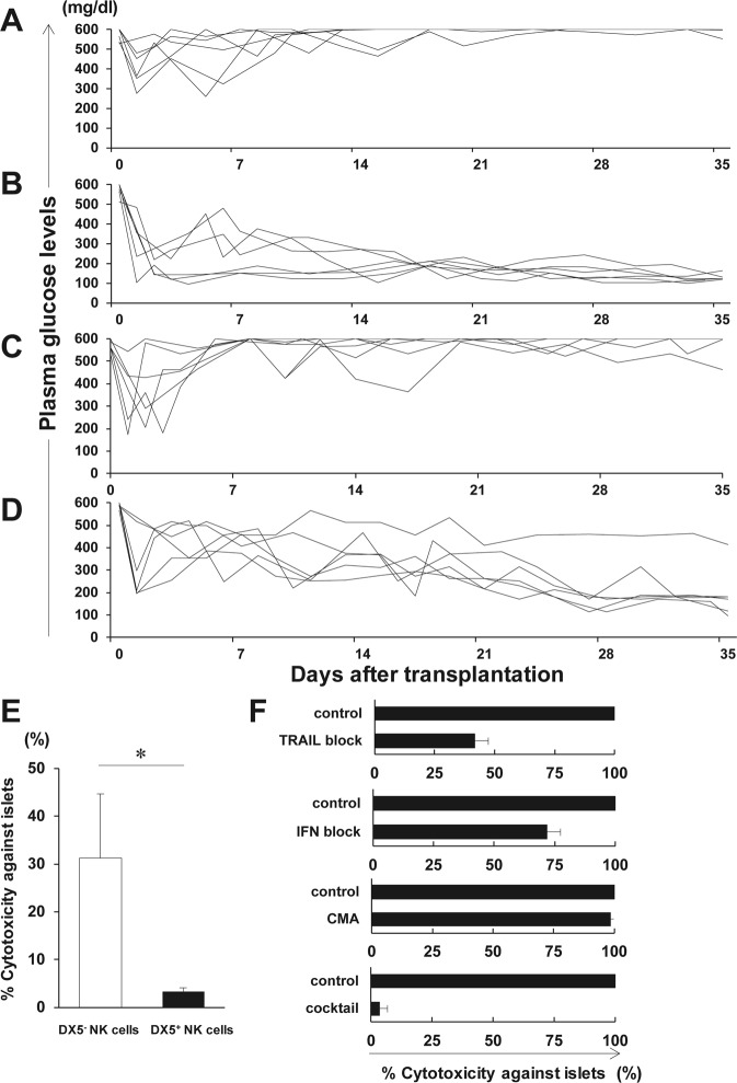
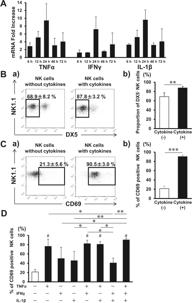
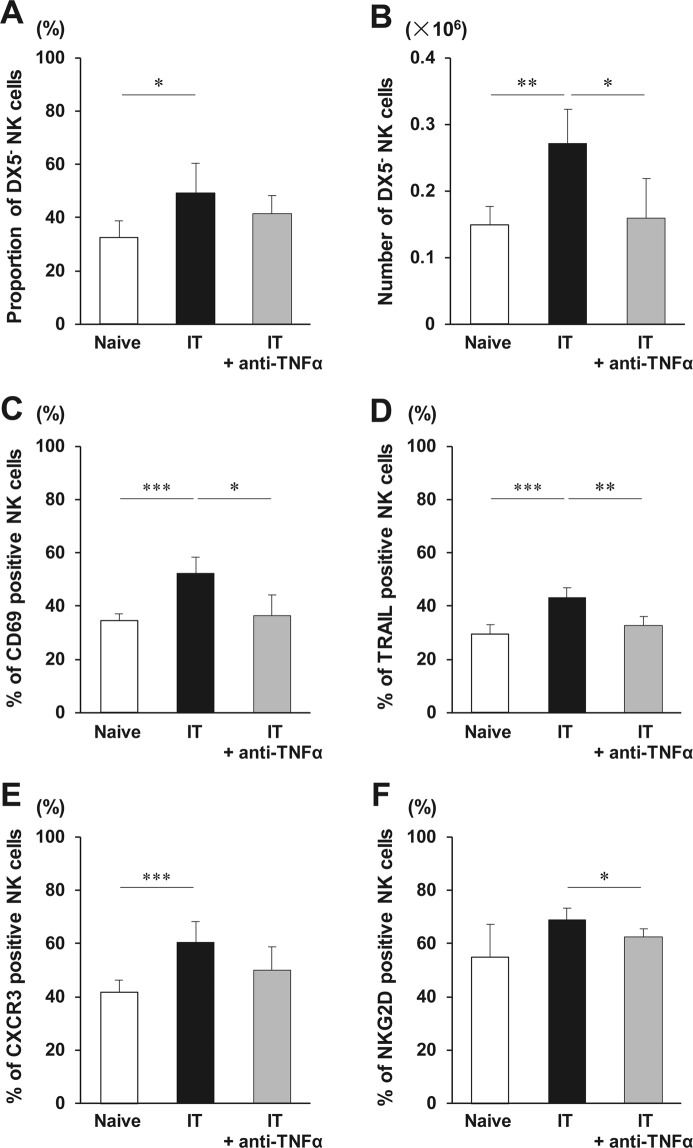
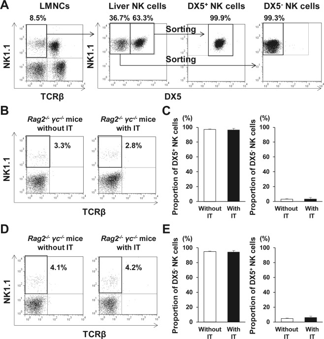
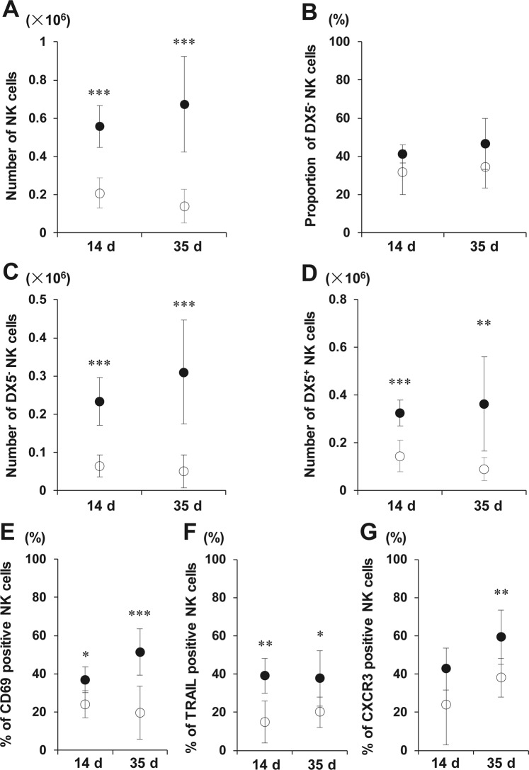
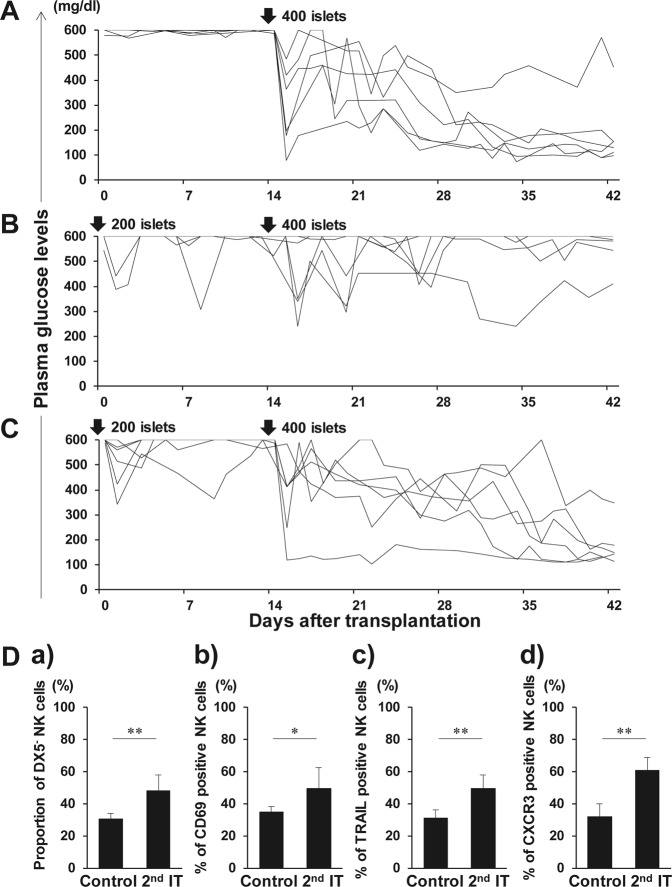
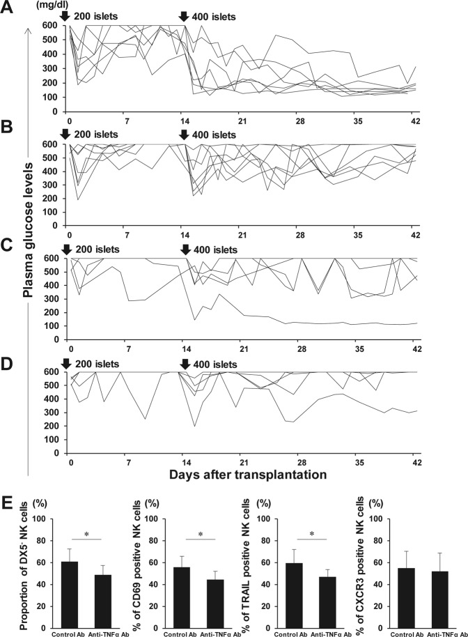
We previously demonstrated the pivotal role of natural killer (NK) cells in islet graft loss during the early phase after intraportal syngeneic islet transplantation (IT). Liver-resident DX5- NK cells were reported to possess memory-like properties, distinguishing them from conventional DX5+ NK cells. Here, we investigated the impact of primary IT-induced liver DX5- NK cells on the engraftment of secondary-transplanted islets in mice. The culture of liver NK cells isolated from naive mice with TNF-α, IFN-γ, and IL-lβ, mimicking instant blood-mediated inflammatory reaction, led to significantly increased DX5- NK cell percentage among total liver NK cells. Consistently, the prolonged expansion of DX5- CD69+ TRAIL+ CXCR3+ NK cells was observed after intraportal IT of 300 syngeneic islets (marginal mass). In most diabetic mice, 400 syngeneic islets of primary IT were sufficient to achieve normoglycaemia, whereas the same mass after secondary IT failed to induce normoglycaemia in mice that received 200 syngeneic islets during primary IT. These findings indicated that liver-resident DX5- NK cells significantly expanded even after syngeneic IT, and that these memory-like NK cells may target both originally engrafted and secondary-transplanted islets. Furthermore, anti-TNF-α treatment suppressed the expansion of liver-resident DX5- NK cells, resulting in successful islet engraftment after sequential ITs.


Conflict of interest statement

The authors declare no competing interests.

Figures

Figure 1
Figure 1
Involvement of liver DX5 natural killer (NK) cells in islet destruction after islet transplantation (IT). (A,B) Syngeneic islet graft survival in diabetic C57BL/6 (B6) wild-type (WT) or B6 CXCR3−/− mice was monitored by measuring the non-fasting plasma glucose level after intraportal IT. Glucose levels less than 200 mg/dL indicated diabetes reversal. Blood glucose levels of the control diabetic B6 WT or B6 CXCR3−/− mice that were intraportally transplanted with 200 syngeneic islets (n = 6). Data were collected from 5 independent experiments. (C,D) Syngeneic islet graft survival in diabetic B6 WT or B6 Tbx21−/− mice was monitored by measuring the non-fasting plasma glucose level after intraportal IT. Glucose levels less than 200 mg/dL indicated diabetes reversal. Blood glucose levels of the control diabetic B6 WT or B6 Tbx21−/− mice that were intraportally transplanted with 200 syngeneic islets (n = 6). Data were collected from 4 independent experiments. (E) The cytotoxicity of NK cells from B6 mice was tested using islets isolated from B6 mice as targets. NK cells were isolated from the liver of naive B6 mice and were used as effector cells at an E:T ratio of 105:1. The cytotoxicity of NK cells is shown in bar graphs (DX5 NK cells, open bar; DX5+ NK cells, solid bar) as the mean ± SD of 3 independent experiments (n = 3, pooled liver mononuclear cells were from 18 mice per experiment). *p < 0.05. (F) The cytotoxicity of DX5 NK cells with or without anti-TRAIL mAb, anti-IFN-γ mAb, or concanamycin A (CMA) is shown in bar graphs as the mean ± SD (n = 3 per mAb). The experiments were repeated 12 times (n = 17 mice per experiment). Data were collected from 7 independent experiments.
Figure 2
Figure 2
Liver natural killer (NK) cells are activated during instant blood-mediated inflammatory reaction (IBMIR). (A) Livers were harvested from C57BL/6J (B6) mice that received 300 syngeneic islets 6, 12, 24, 48, and 72 h after islet transplantation. Time course of intrahepatic mRNA for TNF-α, IFN-γ, and IL-lβ in islet transplant recipients after transplantation were compared with that of non-treated wild-type B6 mice (naive livers) as quantified by real-time RT-PCR. The relative fold increase was calculated using the delta-delta Ct method. Data are presented as the means ± standard deviation (SD) (n = 3–4). (B) Liver TCRβ NK1.1+ NK cells were separated from liver mononuclear cells and cultured without or with TNF-α, IFN-γ, and IL-1β. Cells were harvested after 24 h and analysed with flow cytometry. Representative flow cytometry plots of NK1.1 and DX5 in isolated NK cells after the incubation without or with three cytokines. The proportions of DX5 NK cells among total NK cells are shown (n = 5). The data in bar graphs are presented as the means ± SD of 4 independent experiments. **p < 0.01. (C) Representative flow cytometry plots of NK1.1 and CD69 in isolated NK cells after their incubation without or with three cytokines. The percentages of cells expressing CD69 among total liver NK cells are shown in bar graphs as the means ± SD of 4 independent experiments (n = 5). ***p < 0.001. (D) Liver NK cells treated with the cytokine combinations for 24 h (n = 4–5). The data in bar graphs are presented as the means ± SD of 4 independent experiments. *p < 0.05; **p < 0.01. #p < 0.001, compared with the results of liver NK cells without cytokines.
Figure 3
Figure 3
Phenotypic alterations of liver natural killer (NK) cells early after islet transplantation (IT). C57BL/6 wild-type mice administered PBS or anti-TNF-α antibody were treated with 300 syngeneic islets. Phenotypic alterations of NK cells in the liver were analysed 24 h after intraportal IT. (A,B) Proportion of TCRβ NK1.1+ DX5 NK cells in total NK cells and the absolute number of DX5 NK cells obtained from the liver of mice that received 300 syngeneic islets, and that were treated or not with anti-TNF-α antibody (naive group, open bar; group that received transplantation, solid bar; group that received islet and anti-TNF-α antibody treatment, gray bar) (n = 5–7). The data in bar graphs are shown as the means ± standard deviation (SD) of 5 independent experiments. *p < 0.05. **p < 0.01. (CF) Percentages of CD69-, TRAIL-, CXCR3-, or NKG2D positive NK cells in the liver after intraportal IT were analysed with flow cytometry (naive group, open bar; group that received transplantation, solid bar; group that received islet and anti-TNF-α antibody treatment, gray bar) (n = 5–7). The data in bar graphs are shown as the means ± SD of 5 independent experiments. *p < 0.05. **p < 0.01. ***p < 0.001.
Figure 4
Figure 4
Propagation of liver DX5 natural killer (NK) cells in response to islet transplantation (IT). (A) TCRβ NK1.1+ DX5+ NK cells and DX5 NK cells were isolated from liver mononuclear cells (LMNCs) of C57BL/6 (B6) wild-type mice. Representative dot plots show the isolated DX5+ NK cells and DX5 NK cells. (B) Isolated DX5+ NK cells were transferred into B6 Rag-2−/− γ chain−/− mice, which was followed by IT, after which LMNCs from the recipient mice were analysed. Representative dot plots show the gated TCRβ NK1.1+ NK cells and their percentage in total liver NK cells from DX5+ NK cell-transferred recipients with/without IT. (C) Bar graph, representing the mean percentage ± standard deviation (SD) of each subset of NK cells in total NK cells isolated from B6 Rag-2−/− γ chain−/− mice that received DX5+ NK cells, followed or not by IT (n = 5). Data were collected from of 2 independent experiments. (D) Isolated DX5 NK cells were transferred into B6 Rag-2−/− γ chain−/− mice, which was followed by IT, after which LMNCs from the recipient mice were analysed. Representative dot plots show the gated TCRβ NK1.1+ NK cells and their percentage in total liver NK cells from DX5 NK cell-transferred recipient with/without IT. (E) Bar graph, representing the mean percentage ± SD of each subset of NK cells in total NK cells isolated from B6 Rag-2−/− γ chain−/− mice that received DX5 NK cells, followed or not by IT (n = 5). Data were collected from of 2 independent experiments.
Figure 5
Figure 5
Expansion of liver DX5 natural killer (NK) cells at the late phase after islet transplantation (IT). Phenotypic alteration of TCRβ NK1.1+ NK cells in the liver from diabetic C57BL/6 (B6) mice was analysed 14 days (n = 6, 3 independent experiments) or 35 days (n = 8, 6 independent experiments) after intraportal IT of syngeneic 300 islets. The data from transplanted diabetic mice are compared with the data from control diabetic B6 mice (open circles; control diabetic B6 mice, closed circles; transplanted diabetic mice). Diabetic B6 mice treated with streptozotocin 7 days before were used as recipients. (A) The absolute number of NK cells obtained from the whole livers of diabetic mice that were transplanted with 300 islets. ***p < 0.001, compared with control. (B,C) Proportion of DX5 NK cells in total NK cells and the absolute number of DX5 NK cells obtained from the whole liver of diabetic mice that were transplanted with 300 islets. ***p < 0.001, compared with control. (D) The absolute number of DX5+ NK cells obtained from the whole liver of diabetic mice that were transplanted with 300 islets. **p < 0.01; ***p < 0.001, compared with control. (EG) The proportion of liver NK cells positive for markers at the indicated time points. *p < 0.05; **p < 0.01; ***p < 0.001, compared with control.
Figure 6
Figure 6
Graft survival after secondary islet transplantation (IT). Syngeneic islet graft survival in diabetic C57BL/6 (B6) mice was monitored by measuring the non-fasting plasma glucose level after intraportal IT. Glucose levels less than 200 mg/dL indicated diabetes reversal. Liver TCRβ NK1.1+ natural killer (NK) cells were harvested at day 42, and analysed with flow cytometry. (A) Blood glucose levels of the control diabetic B6 mice that were intraportally transplanted with 400 syngeneic islets (n = 6). Data were collected from 4 independent experiments. (B) Blood glucose levels of diabetic B6 mice that were intraportally transplanted with 400 syngeneic islets 14 days after the primary transplantation of 200 syngeneic islets (n = 6). Data were collected from 4 independent experiments. (C) Blood glucose levels of anti-asialo GM1-treated diabetic B6 mice that were intraportally transplanted with 400 syngeneic islets 14 days after the primary transplantation of 200 syngeneic islets (n = 6). Diabetic B6 mice were treated with intraperitoneal injection of rabbit anti-asialo GM1 serum on days −3, 0, and 14 of the secondary IT. Data were collected from 4 independent experiments. (D) Proportion of DX5 NK cells in total NK cells obtained from the control and secondary IT mice. Expression of CD69, TRAIL, and CXCR3 on these cells was analysed. Data are presented as the means ± standard deviation of 4 independent experiments. *p < 0.05; **p < 0.01.
Figure 7
Figure 7
Graft survival after secondary islet transplantation (IT) with anti-TNF-α antibodies. Syngeneic islet graft survival in diabetic C57BL/6 (B6) mice that were intraportally transplanted with 400 syngeneic islets 14 days after the primary transplantation of 200 syngeneic islets was monitored by measuring the non-fasting plasma glucose level after intraportal IT. Glucose levels less than 200 mg/dL indicated diabetes reversal. Liver TCRβ NK1.1+ natural killer (NK) cells were harvested at day 42 and analysed with flow cytometry. (A) Diabetic B6 mice were treated with intraperitoneal injection of anti-TNF-α antibodies on days 0, 3, 7, and 10 of each IT (n = 7). Data were collected from 5 independent experiments. (B) Diabetic B6 mice were treated with intraperitoneal injection of normal goat IgG antibodies on days 0, 3, 7, and 10 of each IT (n = 7). Data were collected from 5 independent experiments. (C) Diabetic B6 mice were treated with intraperitoneal injection of anti-TNF-α antibodies on days 0, 3, 7, and 10 of the secondary IT (n = 5). Data were collected from 4 independent experiments. (D) Diabetic B6 mice were treated with intraperitoneal injection of normal goat IgG antibodies on days 0, 3, 7, and 10 of the secondary IT (n = 5). Data were collected from 4 independent experiments. (E) Proportion of DX5 NK cells in total NK cells obtained from IT recipients together with the control or anti-TNF-α antibodies at primary and secondary IT (n = 7). Expression of CD69, TRAIL, and CXCR3 on these cells was analysed. Data are presented as the means ± standard deviation of 5 independent experiments. *p < 0.05.

Similar articles

Cited by

References

    1. Shapiro AM, et al. International trial of the Edmonton protocol for islet transplantation. New Eng. J. Med. 2006;355:1318–1330. doi: 10.1056/NEJMoa061267. - DOI - PubMed
    1. Barton FB, et al. Improvement in outcomes of clinical islet transplantation: 1999-2010. Diabetes Care. 2012;35:1436–1445. doi: 10.2337/dc12-0063. - DOI - PMC - PubMed
    1. Hering BJ, et al. Phase 3 Trial of Transplantation of Human Islets in Type 1 Diabetes Complicated by Severe Hypoglycemia. Diabetes Care. 2016;39:1230–1240. doi: 10.2337/dc15-1988. - DOI - PMC - PubMed
    1. Bellin MD, et al. Potent induction immunotherapy promotes long-term insulin independence after islet transplantation in type 1 diabetes. Am. J. Transplant. 2012;12:1576–1583. doi: 10.1111/j.1600-6143.2011.03977.x. - DOI - PMC - PubMed
    1. Olsson R, Olerud J, Pettersson U, Carlsson PO. Increased numbers of low-oxygenated pancreatic islets after intraportal islet transplantation. Diabetes. 2011;60:2350–2353. doi: 10.2337/db09-0490. - DOI - PMC - PubMed

Publication types

MeSH terms