. 2019 Jan 15:9:3167.
doi: 10.3389/fimmu.2018.03167. eCollection 2018.

Sex and Pubertal Differences in the Type 1 Interferon Pathway Associate With Both X Chromosome Number and Serum Sex Hormone Concentration

Affiliations

Sex and Pubertal Differences in the Type 1 Interferon Pathway Associate With Both X Chromosome Number and Serum Sex Hormone Concentration

Kate Webb et al. Front Immunol. .

Abstract

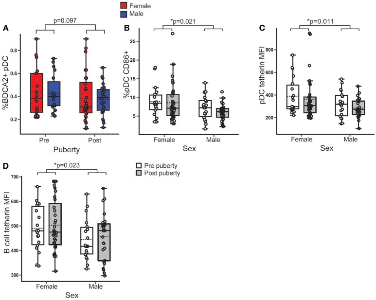
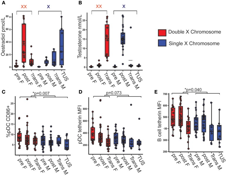
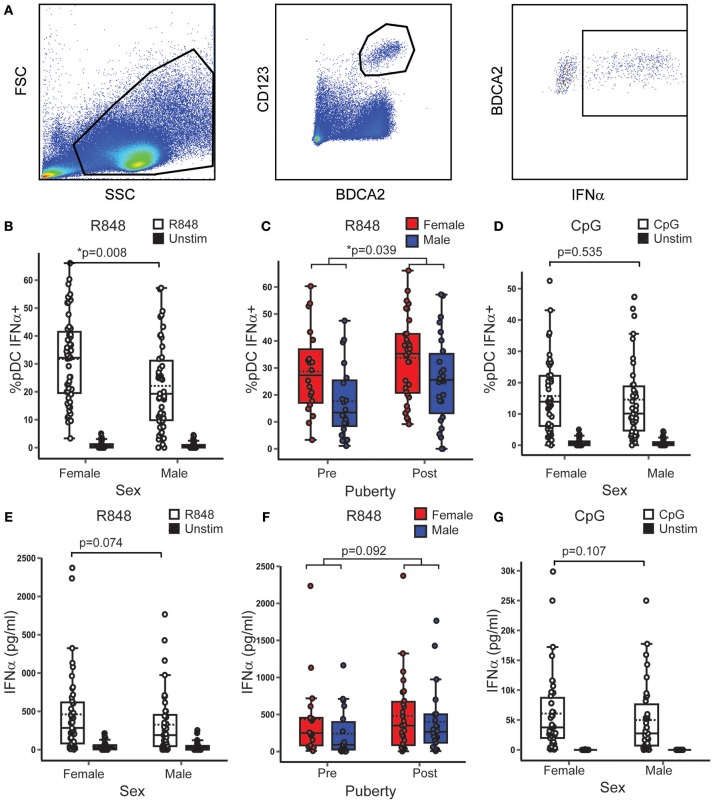
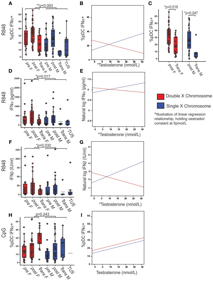
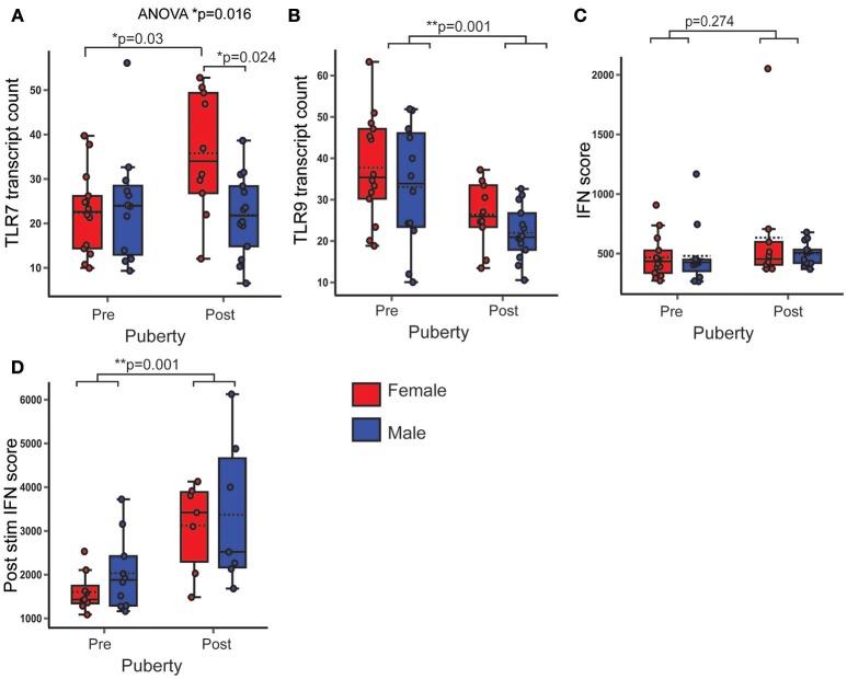
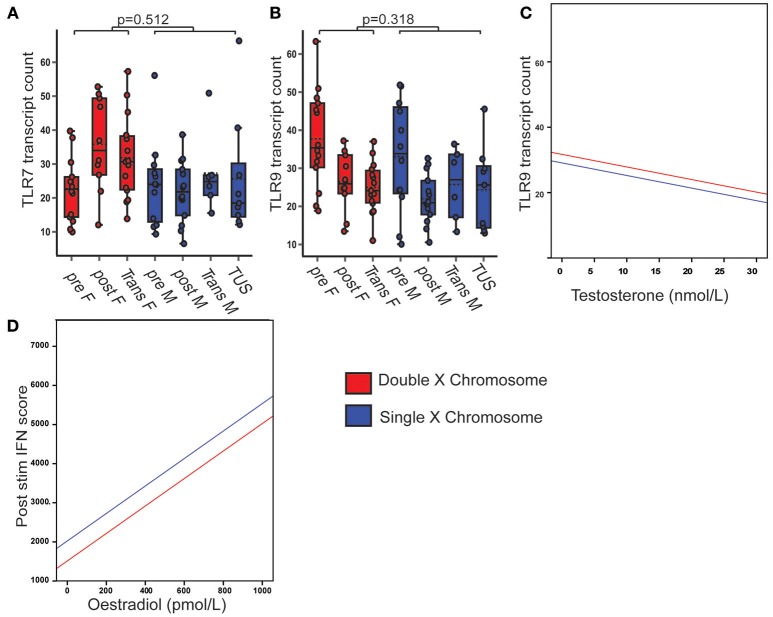
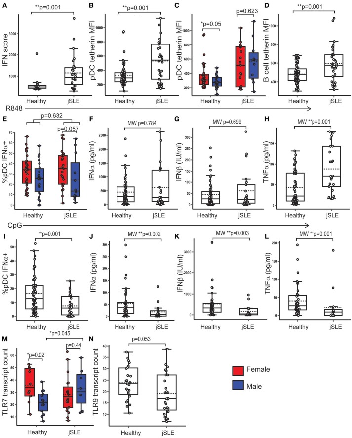
Type 1 interferons (IFN) are an antiviral cytokine family, important in juvenile onset systemic lupus erythematosus (jSLE) which is more common in females, around puberty. We report that plasmacytoid dendritic cells (pDC) from healthy females produced more type 1 IFN after toll like receptor (TLR) 7 signaling than males, even before puberty, but that puberty itself associated with increased production of type 1 IFN. A unique human model allows us to show that this was related to X chromosome number, and serum testosterone concentration, in a manner which differed depending on the number of X chromosomes present. In addition, we have showed that pDC were more activated in females overall, and immune cell TLR7 gene expression was higher in females after puberty. Therefore, sex hormones and X chromosome number were associated individually and interactively with the type 1 IFN response, which contributes to our understanding of why females are more likely to develop an IFN mediated disease like jSLE after puberty.

Keywords: SLE; TLR7; X Chromosome; immunity; interferon; puberty; sex; sex hormone.


Figures

Figure 1
Figure 1
Female pDC expressed more surface CD86 and tetherin than males, regardless of puberty. Healthy volunteer PBMC were assessed by flow cytometry (n = 106). p-values represent the significance of the coefficient the variable shown as estimated by linear regression after correcting for the other variable in the model (sex or puberty). (A) %BDCA2+ pDC did not differ with pubertal phase (p = 0.097). (B) In females, a higher % pDC expressed CD86 than in males (p = 0.021). (C,D) Females had a higher tetherin expression in pDC (p = 0.011) and B cells (p = 0.023) than males. *p < 0.05.
Figure 2
Figure 2
pDC CD86 and tetherin expression were higher when two X chromosomes were present, regardless of serum testosterone and oestradiol concentration. (A,B) Serum hormone levels in pre- and post-pubertal healthy, transgender and TUS volunteers allowed for a full spectrum of hormone concentration upon the background of one or two X chromosomes (n = 135). p-values represent the significance of the coefficient the variable shown as estimated by linear regression after correcting for the other variables in the model (X chromosome number, serum testosterone, and oestradiol). (C) If two X chromosomes were present, there was a higher %pDC expressing CD86 (p = 0.007). (D) The presence of two X chromosomes showed a non-significant trend toward a higher pDC tetherin expression (p = 0.073). (E) B cell tetherin expression was higher in the presence of two X chromosomes (p = 0.040). pre F, Pre- pubertal female; post F, Post-pubertal female; Trans F, Transgender birth female; pre M, Pre-pubertal male; post M, Post-pubertal male; Trans M, transgender birth male; TUS, turners syndrome female. *p < 0.05.
Figure 3
Figure 3
In healthy volunteers, TLR7, and not TLR9 induced pDC IFNα production was higher in females and after puberty. (A) After stimulation with R848 or CpG, PBMC from healthy volunteers were assessed by flow cytometry as shown. p-values represent the significance of the coefficient the variable shown as estimated by linear regression after correcting for the other variable in the model (sex or puberty). (B,C) Upon R848 stimulation, females had a higher % pDC IFNα+ (p = 0.008) than males, and post-pubertal volunteers had a higher % pDC IFNα+ (p = 0.039), than pre-pubertal volunteers (n = 109). (D) After CpG stimulation, there was no sex difference in the % pDC IFNα+ (p = 0.535, n = 90). (E,F) After R848 stimulation, the same trend toward higher PBMC production of IFNα in females (p = 0.074) and post-pubertal volunteers (p = 0.092) was seen as in the pDC specific experiment, although this did not reach significance (n = 86). (G) No sex differences were seen in PBMC IFNα production after CpG stimulation (p = 0.107, n = 74). *p < 0.05.
Figure 4
Figure 4
X chromosome number and serum testosterone concentration was associated with TLR7 induced IFNα production. Pre- and post-pubertal healthy, transgender, and TUS volunteers were analyzed (n = 128), p-values represent the significance of the coefficient the variable shown as estimated by linear regression after correcting for the other variables in the model (X chromosome number, serum testosterone, and oestradiol). (A) If two X chromosomes were present, a higher % pDC produced IFNα after R848 stimulation (p = 0.003). (B) % pDC IFNα+ after R848 stimulation associated with serum testosterone (p = 0.008) with a significant interaction term between X chromosome number and serum testosterone (p = 0.002) as illustrated, holding oestradiol constant at 5pmol/L. (C) Transgender birth females (p = 0.018) and males (p = 0.047) both had a lower %pDC IFNα+ after R848 stimulation than their chromosomal counterparts when analyzed by ANOVA with post hoc analysis. (D–G) After R848 stimulation, if two X chromosomes were present, PBMC produced more IFNα (p = 0.017) and IFNβ (p = 0.03) with a significant interaction between testosterone and X chromosome number (p = 0.028; p = 0.019 respectively). (H,I) After CpG stimulation, % pDC IFNα+ did not associate with X chromosome number (p = 0.243) but did associate with serum testosterone concentration (p = 0.007). pre F, Pre-pubertal female; post F, Post- pubertal female; Trans F, Transgender birth female; pre M, Pre-pubertal male; post M, Post-pubertal male; Trans M, transgender birth male; TUS, turners syndrome female. *p < 0.05; **p < 0.005.
Figure 5
Figure 5
PBMC gene expression of TLR7 was higher in post-pubertal females. In healthy volunteers, PBMC gene expression was assessed by Nanostring and normalized gene transcript counts are shown (n = 50). p-values represent significance of the coefficient for the variables shown after correcting for the other variable (sex or puberty). (A) There was an overall difference in TLR7 expression (p = 0.016). After post hoc testing with Bonferroni correction, post-pubertal females had a higher TLR7 expression than post-pubertal males (p = 0.024) and pre-pubertal females (p = 0.03) (B) PBMC TLR9 gene expression was lower in post-pubertal volunteers. (C) There were no pubertal (p = 0.274) differences in the PBMC IFN score. (D) After stimulation with IFNα, the IFN score (post stim IFN score) was significantly higher in post pubertal volunteers (p = 0.001, n = 33). *p < 0.05; **p < 0.005.
Figure 6
Figure 6
PBMC TLR7 gene expression did not associate with X chromosome number or serum sex hormone. Pre- and post-pubertal healthy, transgender and TUS volunteers were included (n = 77), p-values represent the significance of the coefficient the variable shown as estimated by linear regression after correcting for the other variables in the model (X chromosome number, serum testosterone, and oestradiol). (A) PBMC TLR7 gene expression did not differ with X chromosome number (p = 0.512). (B,C) PBMC TLR 9 gene expression did not differ with X chromosome number (p = 0.318), but was negatively associated with serum testosterone concentration (p = 0.014). (D) After cells were pre-stimulated with IFNα, IFN score was significantly associated with serum oestradiol (p = 0.009, n = 58).
Figure 7
Figure 7
PBMC TLR7 gene expression was higher in males with jSLE. Healthy volunteers and young people with jSLE were included. p-values represent t-tests unless data was not normally distributed, when a Mann Whitney U test was done as indicated by “MW.” (A) PBMC IFN score was higher in jSLE (p = 0.001, n = 79). (B) pDC tetherin expression was higher in jSLE (p = 0.001, n = 136). (C) The sex difference in pDC tetherin expression seen in healthy volunteers (p = 0.05) was lost in those with jSLE (p = 0.623). (D) B cell tetherin expression was higher in jSLE (p = 0.004, n = 136). (E) After R848 stimulation, there was no significant difference in the %pDC producing IFNα in jSLE (p = 0.632, n = 138). Females with jSLE had a trend toward a higher % pDC producing IFNα than males with jSLE (p = 0.057). (F–H) There were no differences in jSLE in the amount of IFNα (p = 0.784) or IFNβ (p = 0.699) produced by PBMC in supernatant after R848 stimulation, but there was more TNFα produced in jSLE (p = 0.001, n = 108). (I–L) After stimulation with CpG, there was a reduction in the production of all cytokines in patients with jSLE (n = 92). (M) PBMC TLR7 gene expression was not different overall in jSLE (p = 0.636, n = 79) although the sex difference was lost in jSLE (p = 0.440). Males with jSLE had a higher PBMC TLR7 gene expression (p = 0.045) than healthy males. (N) There was a trend toward decreased PBMC gene expression of TLR9 in jSLE (p = 0.053, n = 43). *p < 0.05; **p < 0.005.

Similar articles

Cited by

References

    1. Flanagan KL, Fink AL, Plebanski M, Klein SL. Sex and gender differences in the outcomes of vaccination over the life course. Annu Rev Cell Dev Biol. (2017) 33:577–99. 10.1146/annurev-cellbio-100616-060718 - DOI - PubMed
    1. Klein SL, Flanagan KL. Sex differences in immune responses. Nat Rev Immunol. (2016) 16:626–38. 10.1038/nri.2016.90 - DOI - PubMed
    1. Ziegler SM, Altfeld M. Human Immunodeficiency Virus 1 and Type I Interferons—where sex makes a difference. Front Immunol. (2017) 8:1224. 10.3389/fimmu.2017.01224 - DOI - PMC - PubMed
    1. Whitacre CC. Sex differences in autoimmune disease. Nat Immunol. (2001) 2:777–780. 10.1038/ni0901-777 - DOI - PubMed
    1. Bennett L, Palucka AK, Arce E, Cantrell V, Borvak J, Banchereau J, et al. . Interferon and granulopoiesis signatures in systemic lupus erythematosus blood. J Exp Med. (2003) 197:711–23. 10.1084/jem.20021553 - DOI - PMC - PubMed

Publication types

MeSH terms