

SGK1-dependent stimulation of vascular smooth muscle cell osteo-/chondrogenic transdifferentiation by interleukin-18
- PMID: 30706178
- PMCID: PMC6533237
- DOI: 10.1007/s00424-019-02256-5
SGK1-dependent stimulation of vascular smooth muscle cell osteo-/chondrogenic transdifferentiation by interleukin-18
Abstract
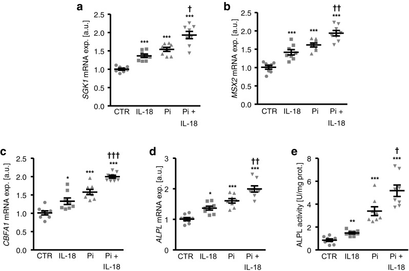
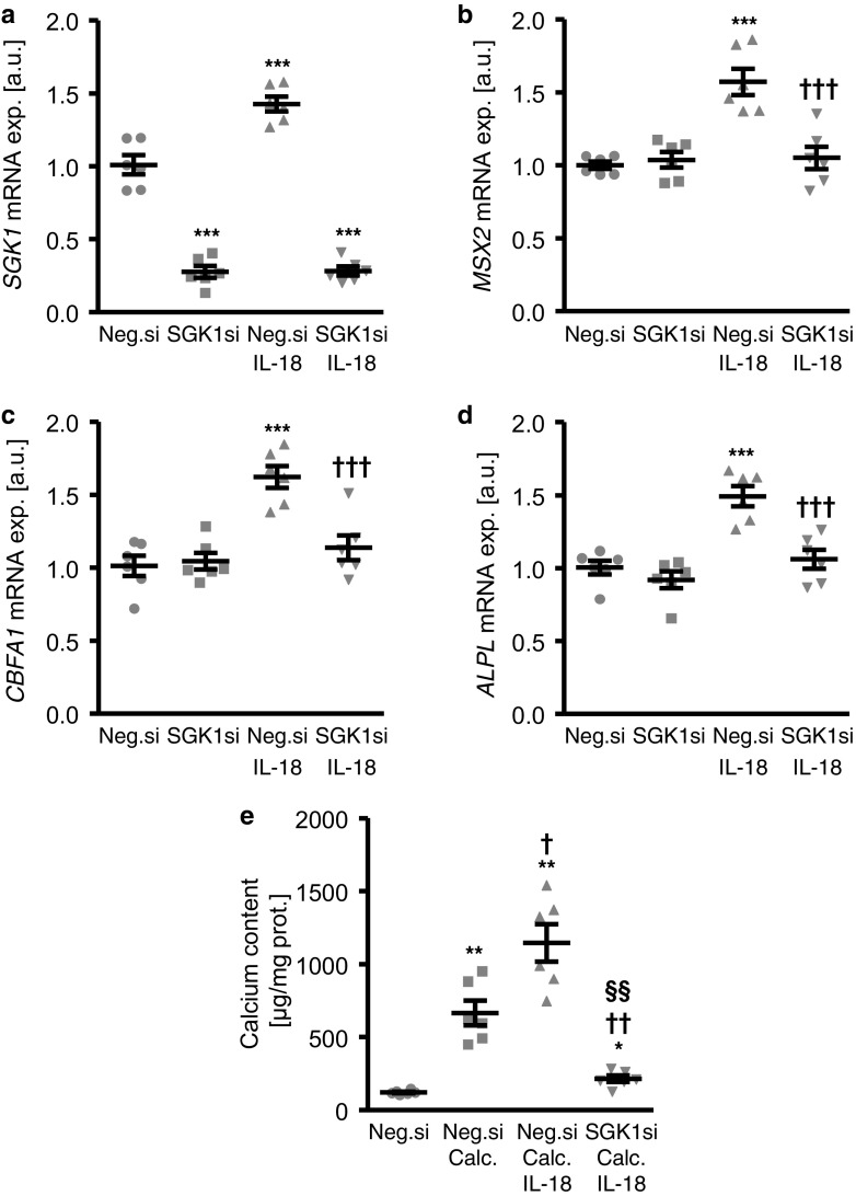
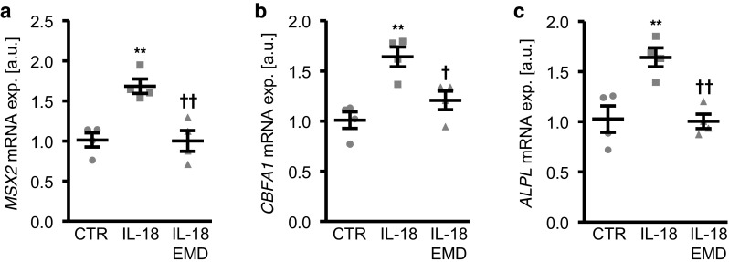
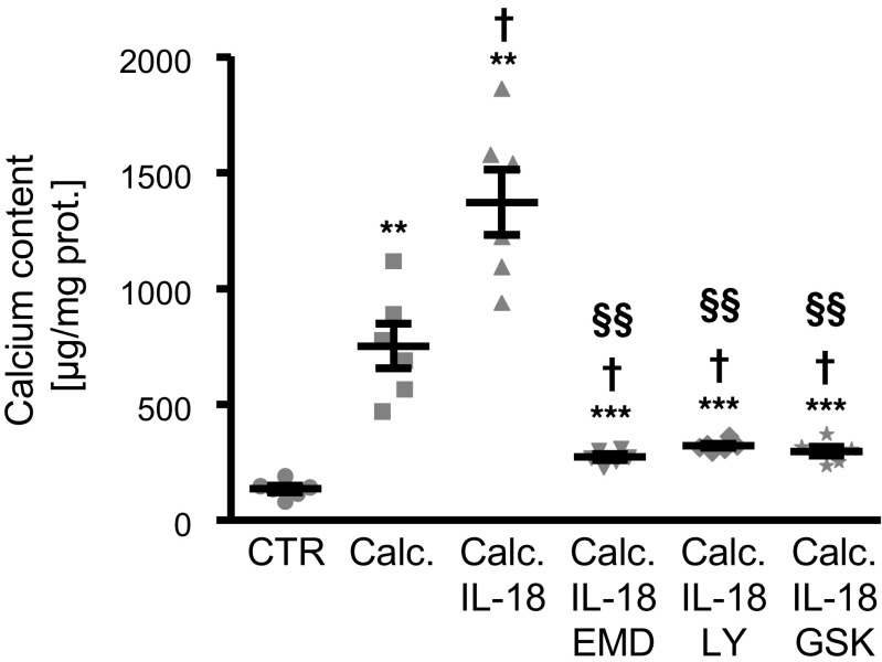
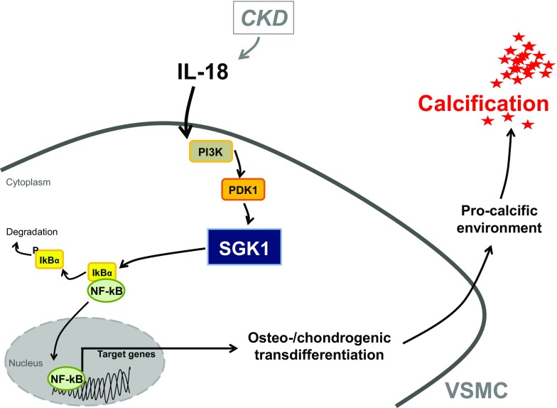
The serum- and glucocorticoid-inducible kinase 1 (SGK1) is a key regulator of osteo-/chondrogenic transdifferentiation and subsequent calcification of vascular smooth muscle cells (VSMCs). The phenotypical transdifferentiation of VSMCs is associated with increased interleukin-18 (IL-18) levels and generalized inflammation. Therefore, the present study investigated the possible involvement of SGK1 in IL-18-induced vascular calcification. Experiments were performed in primary human aortic smooth muscle cells (HAoSMCs) treated with recombinant human IL-18 protein in control or high phosphate conditions and following SGK1 knockdown by siRNA or pharmacological inhibition of SGK1, PI3K, and PDK1. As a result, IL-18 treatment increased SGK1 mRNA and protein expression in HAoSMCs. IL-18 upregulated SGK1 mRNA expression in a dose-dependent manner. This effect was paralleled by upregulation of the mRNA expression of MSX2 and CBFA1, osteogenic transcription factors, and of tissue-nonspecific alkaline phosphatase (ALPL), an osteogenic enzyme, as markers of increased osteo-/chondrogenic transdifferentiation. Phosphate treatment increased SGK1 and osteogenic markers mRNA expression as well as ALPL activity and induced calcification of HAoSMCs, all effects significantly augmented by additional treatment with IL-18. Conversely, silencing of SGK1 or cotreatment with the SGK1 inhibitor EMD638683 blunted the effects of IL-18 on osteo-/chondrogenic transdifferentiation and calcification of HAoSMCs. The procalcific effects of IL-18 were similarly suppressed in the presence of PI3K or PDK1 inhibitors. In conclusion, SGK1 expression is upregulated by IL-18 in VSMCs and SGK1 participates in the intracellular signaling of IL-18-induced osteo-/chondrogenic transdifferentiation of VSMCs. Thus, SGK1 may serve as therapeutic target to limit the progression of medial vascular calcification during vascular inflammation.
Keywords: Osteo-/chondrogenic signaling; PI3K; SGK1, interleukin-18; Vascular calcification; Vascular smooth muscle cells.
Conflict of interest statement
The authors declare that they have no conflict of interest.
Figures


Similar articles
-
SGK1 induces vascular smooth muscle cell calcification through NF-κB signaling.J Clin Invest. 2018 Jul 2;128(7):3024-3040. doi: 10.1172/JCI96477. Epub 2018 Jun 11. J Clin Invest. 2018. PMID: 29889103 Free PMC article.
-
Phosphate-induced ORAI1 expression and store-operated Ca2+ entry in aortic smooth muscle cells.J Mol Med (Berl). 2019 Oct;97(10):1465-1475. doi: 10.1007/s00109-019-01824-7. Epub 2019 Aug 5. J Mol Med (Berl). 2019. PMID: 31385016
-
Role of Cytosolic Serine Hydroxymethyl Transferase 1 (SHMT1) in Phosphate-Induced Vascular Smooth Muscle Cell Calcification.Kidney Blood Press Res. 2018;43(4):1212-1221. doi: 10.1159/000492248. Epub 2018 Aug 2. Kidney Blood Press Res. 2018. PMID: 30071536
-
Signaling pathways involved in vascular smooth muscle cell calcification during hyperphosphatemia.Cell Mol Life Sci. 2019 Jun;76(11):2077-2091. doi: 10.1007/s00018-019-03054-z. Epub 2019 Mar 18. Cell Mol Life Sci. 2019. PMID: 30887097 Free PMC article. Review.
-
Therapeutic Interference With Vascular Calcification-Lessons From Klotho-Hypomorphic Mice and Beyond.Front Endocrinol (Lausanne). 2018 May 4;9:207. doi: 10.3389/fendo.2018.00207. eCollection 2018. Front Endocrinol (Lausanne). 2018. PMID: 29780355 Free PMC article. Review.
Cited by
-
Inflammation: a putative link between phosphate metabolism and cardiovascular disease.Clin Sci (Lond). 2021 Jan 15;135(1):201-227. doi: 10.1042/CS20190895. Clin Sci (Lond). 2021. PMID: 33416083 Free PMC article. Review.
-
Impact of β-glycerophosphate on the bioenergetic profile of vascular smooth muscle cells.J Mol Med (Berl). 2020 Jul;98(7):985-997. doi: 10.1007/s00109-020-01925-8. Epub 2020 Jun 2. J Mol Med (Berl). 2020. PMID: 32488546 Free PMC article.
-
Stress, Vascular Smooth Muscle Cell Phenotype and Atherosclerosis: Novel Insight into Smooth Muscle Cell Phenotypic Transition in Atherosclerosis.Curr Atheroscler Rep. 2024 Aug;26(8):411-425. doi: 10.1007/s11883-024-01220-8. Epub 2024 May 30. Curr Atheroscler Rep. 2024. PMID: 38814419 Review.
-
From Cells to Plaques: The Molecular Pathways of Coronary Artery Calcification and Disease.J Clin Med. 2024 Oct 23;13(21):6352. doi: 10.3390/jcm13216352. J Clin Med. 2024. PMID: 39518492 Free PMC article. Review.
-
Similarities and Differences of Vascular Calcification in Diabetes and Chronic Kidney Disease.Diabetes Metab Syndr Obes. 2024 Jan 10;17:165-192. doi: 10.2147/DMSO.S438618. eCollection 2024. Diabetes Metab Syndr Obes. 2024. PMID: 38222032 Free PMC article. Review.
References
-
- Alesutan I, Feger M, Tuffaha R, Castor T, Musculus K, Buehling SS, Heine CL, Kuro OM, Pieske B, Schmidt K, Tomaschitz A, Maerz W, Pilz S, Meinitzer A, Voelkl J, Lang F. Augmentation of phosphate-induced osteo-/chondrogenic transformation of vascular smooth muscle cells by homoarginine. Cardiovasc Res. 2016;110:408–418. doi: 10.1093/cvr/cvw062. - DOI - PubMed
-
- Alesutan I, Voelkl J, Feger M, Kratschmar DV, Castor T, Mia S, Sacherer M, Viereck R, Borst O, Leibrock C, Gawaz M, Kuro OM, Pilz S, Tomaschitz A, Odermatt A, Pieske B, Wagner CA, Lang F. Involvement of vascular aldosterone synthase in phosphate-induced osteogenic transformation of vascular smooth muscle cells. Sci Rep. 2017;7:2059. doi: 10.1038/s41598-017-01882-2. - DOI - PMC - PubMed
Publication types
MeSH terms
Substances
LinkOut - more resources
Full Text Sources
Research Materials
Miscellaneous
