

Levels Propagation Approach to Image Segmentation: Application to Breast MR Images
- PMID: 30706211
- PMCID: PMC6499864
- DOI: 10.1007/s10278-018-00171-2
Levels Propagation Approach to Image Segmentation: Application to Breast MR Images
Abstract
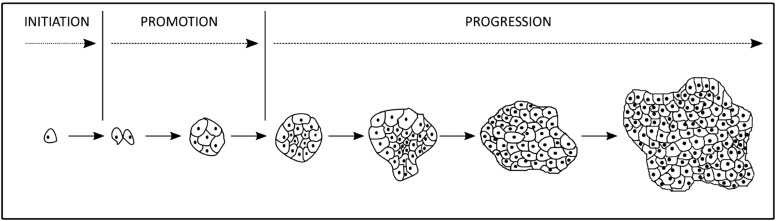
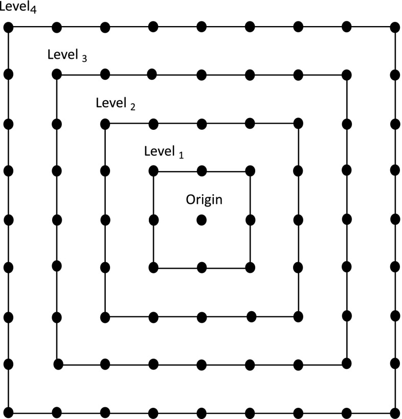
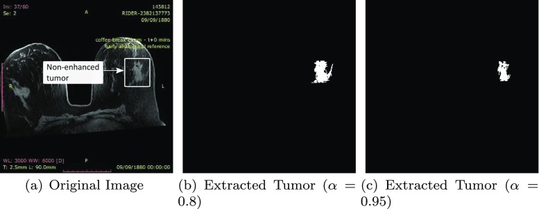
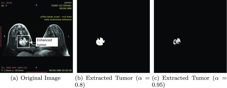
Accurate segmentation of a breast tumor region is fundamental for treatment. Magnetic resonance imaging (MRI) is a widely used diagnostic tool. In this paper, a new semi-automatic segmentation approach for MRI breast tumor segmentation called Levels Propagation Approach (LPA) is introduced. The introduced segmentation approach takes inspiration from tumor propagation and relies on a finite set of nested and non-overlapped levels. LPA has several features: it is highly suitable to parallelization and offers a simple and dynamic possibility to automate the threshold selection. Furthermore, it allows stopping of the segmentation at any desired limit. Particularly, it allows to avoid to reach the breast skin-line region which is known as a significant issue that reduces the precision and the effectiveness of the breast tumor segmentation. The proposed approach have been tested on two clinical datasets, namely RIDER breast tumor dataset and CMH-LIMED breast tumor dataset. The experimental evaluations have shown that LPA has produced competitive results to some state-of-the-art methods and has acceptable computation complexity.
Keywords: Breast; Image segmentation; Levels; MRI; Propagation; Tumor.
Figures


Similar articles
-
Multistage processing procedure for 4D breast MRI segmentation.Annu Int Conf IEEE Eng Med Biol Soc. 2008;2008:3036-9. doi: 10.1109/IEMBS.2008.4649843. Annu Int Conf IEEE Eng Med Biol Soc. 2008. PMID: 19163346
-
Image manifold revealing for breast lesion segmentation in DCE-MRI.Biomed Mater Eng. 2015;26 Suppl 1:S1353-60. doi: 10.3233/BME-151433. Biomed Mater Eng. 2015. PMID: 26405896
-
Semi-automated brain tumor segmentation on multi-parametric MRI using regularized non-negative matrix factorization.BMC Med Imaging. 2017 May 4;17(1):29. doi: 10.1186/s12880-017-0198-4. BMC Med Imaging. 2017. PMID: 28472943 Free PMC article.
-
Principles and methods for automatic and semi-automatic tissue segmentation in MRI data.MAGMA. 2016 Apr;29(2):95-110. doi: 10.1007/s10334-015-0520-5. Epub 2016 Jan 11. MAGMA. 2016. PMID: 26755062 Review.
-
State of the art survey on MRI brain tumor segmentation.Magn Reson Imaging. 2013 Oct;31(8):1426-38. doi: 10.1016/j.mri.2013.05.002. Epub 2013 Jun 20. Magn Reson Imaging. 2013. PMID: 23790354 Review.
References
-
- World Health Organization (2018) Available at http://www.who.int/en/. Accessed on October 20th
-
- Mughal B, Sharif M. Automated detection of breast tumor in different imaging modalities: A Review. Current Medical Imaging Reviews. 2017;13:121–139. doi: 10.2174/1573405612666160901121802. - DOI
MeSH terms
LinkOut - more resources
Full Text Sources
Medical
Miscellaneous
