

Foot-and-mouth disease virus O/ME-SA/Ind 2001 lineage outbreak in vaccinated Holstein Friesian cattle in Saudi Arabia in 2016
- PMID: 30706772
- PMCID: PMC6831000
- DOI: 10.1080/01652176.2018.1539568
Foot-and-mouth disease virus O/ME-SA/Ind 2001 lineage outbreak in vaccinated Holstein Friesian cattle in Saudi Arabia in 2016
Abstract
Background: Foot-and-mouth disease virus (FMDV) is a highly contagious viral infection of large ruminants. Despite the massive application of vaccines against FMDV, several outbreaks are still being reported in Africa and Asia.
Aim: To perform molecular characterization of FMDV in an outbreak among a cattle herd Saudi Arabia in 2016. This herd had been vaccinated with a polyvalent FMDV vaccine.
Methods: To investigate this outbreak, we collected specimens from 77 animals showing typical clinical signs of FMDV. Specimens including sera, nasal swabs, and tissues (tongue, coronary bands, hooves, and hearts) were collected. We tested the collected cattle sera for the presence of FMDV antibodies with commercial ELISA kits. In addition, we tested the swabs for the presence of the most common FMDV strains (O, A, Asia-1 and SAT-2) with RT-PCR using serotype-specific oligonucleotides.
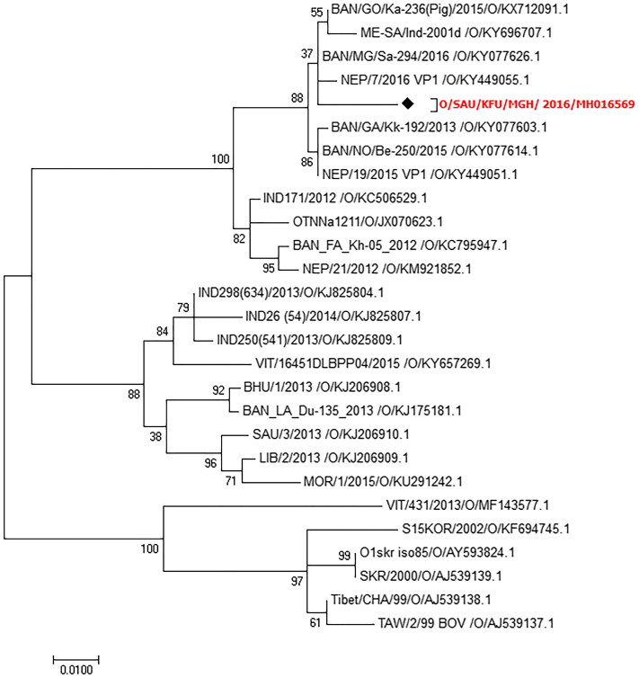
Results: Serology showed that 22% of the tested sera were positive. Molecular testing of the examined swabs confirmed that 24% of the tested animals were positive. Our sequencing analysis confirmed that the circulating strains of FMDV belonged to FMDV serotype O. The phylogenetic tree based on the FMDV-VP-1 gene revealed high nucleotide identity between the circulating strains and the Bangladesh strain (99%). These strains were distinct (shared 89% nucleotide identity) from the FMDV-O strains used for the preparation of the vaccine administered to the animals in this herd. Moreover, they had 7% nucleotide difference between the FMDV-O strains reported in Saudi Arabian in 2013.
Conclusion: More in-depth molecular characterization of these FMDV strains is warranted.
Keywords: Cattle; PCR; Saudi Arabia; foot-and-mouth disease; pathology; serotype O.
Figures


Similar articles
-
Characterization of foot-and-mouth disease viruses (FMDVs) from Ugandan cattle outbreaks during 2012-2013: evidence for circulation of multiple serotypes.PLoS One. 2015 Feb 9;10(2):e0114811. doi: 10.1371/journal.pone.0114811. eCollection 2015. PLoS One. 2015. PMID: 25664876 Free PMC article.
-
Emergence of an exotic strain of serotype O foot-and-mouth disease virus O/ME-SA/Ind-2001d in South-East Asia in 2015.Transbound Emerg Dis. 2018 Feb;65(1):e104-e112. doi: 10.1111/tbed.12687. Epub 2017 Aug 30. Transbound Emerg Dis. 2018. PMID: 28856846
-
Outbreak investigations and molecular characterization of foot-and-mouth disease viruses circulating in south-west Niger.Transbound Emerg Dis. 2018 Feb;65(1):146-157. doi: 10.1111/tbed.12642. Epub 2017 Mar 27. Transbound Emerg Dis. 2018. PMID: 28345819
-
Review of the Global Distribution of Foot-and-Mouth Disease Virus from 2007 to 2014.Transbound Emerg Dis. 2017 Apr;64(2):316-332. doi: 10.1111/tbed.12373. Epub 2015 May 20. Transbound Emerg Dis. 2017. PMID: 25996568 Review.
-
Foot-and-mouth disease status in India during the second decade of the twenty-first century (2011-2020).Vet Res Commun. 2022 Dec;46(4):1011-1022. doi: 10.1007/s11259-022-10010-z. Epub 2022 Oct 3. Vet Res Commun. 2022. PMID: 36190601 Free PMC article. Review.
Cited by
-
Determinants of Foot and Mouth Virus in Eastern Algeria.Trop Anim Health Prod. 2025 Apr 11;57(3):167. doi: 10.1007/s11250-025-04413-8. Trop Anim Health Prod. 2025. PMID: 40214836
-
Epidemiology and economics of foot-and-mouth disease: current understanding and knowledge gaps.Vet Res. 2025 Jul 7;56(1):141. doi: 10.1186/s13567-025-01561-5. Vet Res. 2025. PMID: 40624570 Free PMC article. Review.
-
A review of foot-and-mouth disease in Ethiopia: epidemiological aspects, economic implications, and control strategies.Virol J. 2023 Dec 15;20(1):299. doi: 10.1186/s12985-023-02263-0. Virol J. 2023. PMID: 38102688 Free PMC article. Review.
-
Foot-and-Mouth Disease Surveillance Using Pooled Milk on a Large-Scale Dairy Farm in an Endemic Setting.Front Vet Sci. 2020 May 27;7:264. doi: 10.3389/fvets.2020.00264. eCollection 2020. Front Vet Sci. 2020. PMID: 32537458 Free PMC article.
-
Current trends and challenges in the management of foot and mouth disease in Saudi Arabia: A review.Open Vet J. 2025 May;15(5):1907-1933. doi: 10.5455/OVJ.2025.v15.i5.6. Epub 2025 May 31. Open Vet J. 2025. PMID: 40557100 Free PMC article. Review.
References
-
- Abd El-Rahim IH, Asghar AH, Mohamed AM, Fat'hi SM. 2016. The impact of importation of live ruminants on the epizootiology of foot and mouth disease in Saudi Arabia. Rev Off Int Epizoot. 35(3):769–778. - PubMed
-
- Al-Afaleq AI, Abu Elzein EM, Mousa SM, Abbas AM. 2003. A retrospective study of Rift Valley fever in Saudi Arabia. Rev Off Int Epizoot. 22(3):867–871. - PubMed
-
- Alexandersen S, Zhang Z, Donaldson AI, Garland AJ. 2003. The pathogenesis and diagnosis of foot-and-mouth disease. J Comp Pathol. 129(1):1–36. - PubMed
-
- Ali W, Habib M, Khan RSA, Zia MA, Farooq M, Sajid S, Shah M. 2018. Molecular investigation of foot-and-mouth disease virus circulating in Pakistan during 2014–17. Arch Virol. 163(7):1733–1743. - PubMed
-
- Arzt J, Juleff N, Zhang Z, Rodriguez LL. 2011. The pathogenesis of foot-and-mouth disease I: viral pathways in cattle. Transbound Emerg Dis. 58(4):291–304. - PubMed
MeSH terms
Substances
LinkOut - more resources
Full Text Sources
Research Materials