Review
. 2019 Feb;25(1):14-33.
doi: 10.1212/CON.0000000000000700.

Late-onset Alzheimer Disease

Review

Late-onset Alzheimer Disease

Gil D Rabinovici. Continuum (Minneap Minn). 2019 Feb.

Abstract

Purpose of review: Alzheimer disease (AD) is the most common cause of late-onset dementia. This article describes the epidemiology, genetic and environmental risk factors, clinical diagnosis, biomarkers, and treatment of late-onset AD, defined by age of onset of 65 years or older.

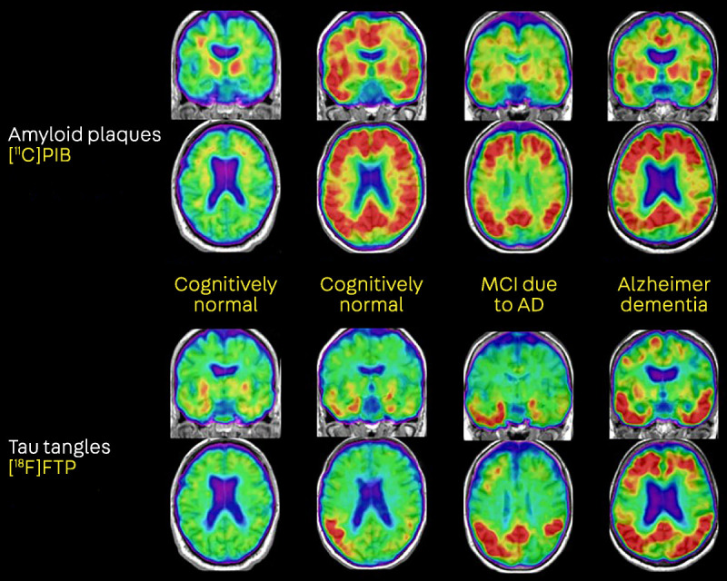
Recent findings: An estimated 5.7 million Americans are living with AD dementia, with the number of affected individuals growing rapidly because of an aging population. Vascular risk factors, sleep disorders, and traumatic brain injury are associated with an increased risk of AD, while increased cognitive and physical activity throughout the lifespan reduce the risk of disease. The primary genetic risk factor for late-onset AD is the apolipoprotein E (APOE) ε4 allele. AD typically presents with early and prominent episodic memory loss, although this clinical syndrome is neither sensitive nor specific for underlying AD neuropathology. Emerging CSF and imaging biomarkers can now detect the key neuropathologic features of the disease (amyloid plaques, neurofibrillary tangles, and neurodegeneration) in living people, allowing for characterization of patients based on biological measures. A comprehensive treatment plan for AD includes use of symptomatic medications, optimal treatment of comorbid conditions and neuropsychiatric symptoms, counseling about safety and future planning, and referrals to community resources.

Summary: AD is very common in older neurologic patients. Neurologists should set the standard for the diagnosis and care of patients with AD and should be familiar with emerging biomarkers that have transformed AD research and are primed to enter the clinical arena.


Figures

FIGURE 1-1
FIGURE 1-1
MRI findings in Alzheimer disease (AD). Axial (A), sagittal (B), and coronal (C) T1-weighted images demonstrate prominent hippocampal and medial temporal lobe atrophy, moderate diffuse cortical atrophy, and ventricular enlargement in an 81-year-old with AD dementia, subsequently confirmed at autopsy. D, Fluid-attenuated inversion recovery (FLAIR) sequence demonstrates subcortical and periventricular white matter hyperintensities in a 78-year-old with a clinical diagnosis of AD, likely representing comorbid small vessel ischemic disease. E, Hallmarks of cerebral amyloid angiopathy, including scattered microbleeds and superficial siderosis, are revealed on susceptibility-weighted imaging (SWI) in a 75-year-old with acute altered mental status superimposed on progressive memory and executive dysfunction. F, Confluent white matter hyperintensities on FLAIR in a 75-year-old with pathology-proven severe cerebral amyloid angiopathy and AD neuropathology. In all panels, key findings are highlighted by arrows. L = left; R = right.
FIGURE 1-2
FIGURE 1-2
Neuropathologic features of Alzheimer disease (AD). A, Gross neuropathologic features of AD include cortical atrophy (arrow), ventricular enlargement (dashed arrow) and hippocampal atrophy (arrowhead). B, Low-power photomicrograph (1x) of hippocampus stained with anti–phosphorylated tau (p-tau) antibody (CP13), demonstrating an abundance of p-tau deposition, most prominent in CA1 (arrow). C, Thioflavin S staining identifies senile amyloid plaques (arrowhead) and tau neurofibrillary tangles (arrow), the pathologic hallmarks of AD. D, High-power photomicrographs (40x) of senile plaques (arrow) identified via anti–amyloid-β immunohistochemistry (1-16, clone DE2, mouse, 1:500). E, High-power photomicrograph (40x) of anti–p-tau (CP13) immunohistochemistry illustrates neurofibrillary tangles (arrow) and dystrophic neurites (arrowhead). F, Low-power photomicrograph (4x) of anti–amyloid-β immunostaining of cerebellum reveals amyloid deposition in blood vessel walls (arrow), the pathologic hallmark of cerebral amyloid angiopathy. Bars represent 25 microns.
FIGURE 1-3
FIGURE 1-3
Amyloid positron emission tomography (PET) with [18F]florbetapir. A, Example of a positive scan in a 72-year-old with amnestic mild cognitive impairment, as defined by absent gray-white matter contrast and intense gray matter binding that exceeds the binding in adjacent white matter. B, Example of a negative scan in an 80-year-old with amnestic mild cognitive impairment, as defined by greater tracer binding in white matter compared with gray matter, creating clear gray-white contrast.
FIGURE 1-4
FIGURE 1-4
Amyloid and tau positron emission tomography (PET) of the Alzheimer disease (AD) continuum. The first column shows a cognitively normal individual with negative amyloid PET (no cortical tracer retention) and tau PET uptake restricted to the medial temporal lobes. The second column shows a cognitively normal individual with evidence of both cortical amyloid and spread of tau into temporal and parietal cortices. The third and fourth columns, representing patients with mild cognitive impairment (MCI) and dementia, show a plateau of amyloid PET signal but increased spread of tau with increasing clinical stages of AD. [18F]FTP = [18F]flortaucipir, a radiotracer selective for paired helical filaments of tau; [11C]PIB = [11C]Pittsburgh Compound B, an amyloid-β–specific tracer.

References

    1. Alzheimer A. Uber eine eigenaritage, schweren Erkrankung der Hirnrinde. Neurol Zbl 1907;25:1134.
    1. Alzheimer’s Association. 2018 Alzheimer’s disease facts and figures. Alzheimers Dement 2018;14(3):367–429. doi:10.1016/j.jalz.2018.02.001. - PubMed
    1. Petersen RC, Lopez O, Armstrong MJ, et al. Practice guideline update summary: mild cognitive impairment: report of the Guideline Development, Dissemination, and Implementation Subcommittee of the American Academy of Neurology. Neurology 2018;90(3):126–135. doi:10.1212/WNL.0000000000004826. - PMC - PubMed
    1. Hebert LE, Beckett LA, Scherr PA, Evans DA. Annual incidence of Alzheimer disease in the United States projected to the years 2000 through 2050. Alzheimer Dis Assoc Disord 2001;15(4):169–173. - PubMed
    1. Hebert LE, Weuve J, Scherr PA, Evans DA. Alzheimer disease in the United States (2010–2050) estimated using the 2010 census. Neurology 2013;80(19):1778–1783. doi:10.1212/WNL.0b013e31828726f5. - PMC - PubMed

Publication types

MeSH terms