Review
. 2019 Feb;25(1):147-164.
doi: 10.1212/CON.0000000000000684.

Vascular Cognitive Impairment

Review

Vascular Cognitive Impairment

Jonathan Graff-Radford. Continuum (Minneap Minn). 2019 Feb.

Abstract

Purpose of review: This article provides an overview of vascular cognitive impairment; discusses its epidemiology, subtypes, and associations with other neurodegenerative diseases; and reviews the diagnostic evaluation and management of these disorders.

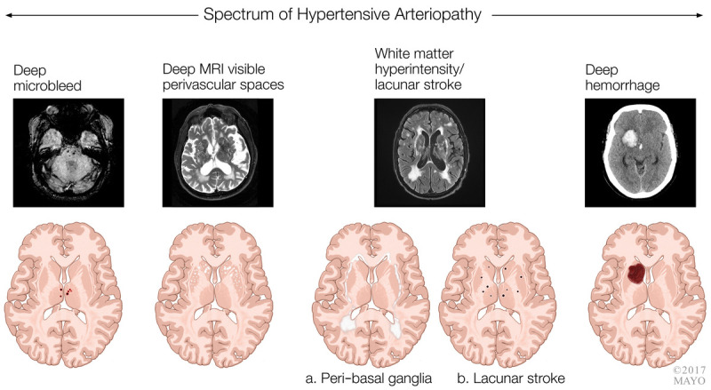
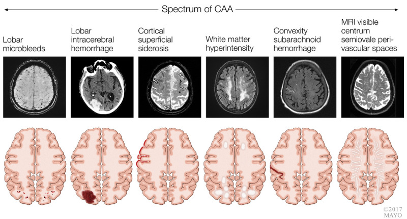
Recent findings: Cerebrovascular disease is a common cause of dementia and frequently coexists with neurodegenerative causes. The heterogeneity of mechanisms leading to vascular cognitive impairment makes developing unifying clinical and research criteria difficult. Recognizing the neuroimaging hallmarks of different forms of vascular cognitive impairment can allow for individualized treatment and management. In individuals with mild vascular cognitive impairment, aerobic exercise appears to be a promising treatment but requires further investigation.

Summary: Vascular cognitive impairment can be caused by several mechanisms. While treating vascular risk factors is rational to prevent worsening of cognitive impairment, well-designed studies are needed to demonstrate efficacy.


Figures

FIGURE 7-1
FIGURE 7-1
Spectrum of imaging changes with hypertensive arteriopathy. MRI = magnetic resonance imaging.
FIGURE 7-2
FIGURE 7-2
Spectrum of imaging changes associated with cerebral amyloid angiopathy (CAA). MRI = magnetic resonance imaging.
FIGURE 7-3
FIGURE 7-3
Imaging of the patient in case 7-1. Axial diffusion-weighted MRI shows an acute ischemic infarct in the anterior nucleus of the thalamus (arrow).
FIGURE 7-4
FIGURE 7-4
Imaging of the patient in case 7-2. Axial diffusion-weighted MRI shows an acute infarction in the left posterior cerebral artery distribution involving the left hippocampus and the left ventral visual stream (occipitotemporal area), resulting in dementia related to a strategic infarct.
FIGURE 7-5
FIGURE 7-5
Imaging of the patient in case 7-4. A, Axial T2-weighted MRI shows an infarct involving the right caudate. B, Fludeoxyglucose positron emission tomography (FDG-PET) statistical map shows regions of significant hypometabolism relative to age-matched controls. Hypometabolism is present in areas functionally connected to the caudate, including the right medial prefrontal cortex, dorsolateral prefrontal cortex, and right anterior cingulate cortex. Contralateral cerebellar hypometabolism is also seen. Reprinted with permission from Graff-Radford J, et al, Neurology. © 2017 American Academy of Neurology.
FIGURE 7-6
FIGURE 7-6
Embolic disease and large vessel disease. Multiple cortical infarcts (left) due to cardiogenic emboli and watershed infarct (right) related to hypoperfusion in the setting of a significant carotid stenosis. ACA = anterior cerebral artery; MCA = middle cerebral artery; PCA = posterior cerebral artery.
FIGURE 7-7
FIGURE 7-7
Imaging of the patient in case 7-6. Axial fluid-attenuated inversion recovery (FLAIR) MRI shows subcortical ischemic infarcts (A, B) in addition to white-matter T2 hyperintensities with notable involvement of the anterior temporal lobes (C) and external capsule (A) characteristic of cerebral autosomal dominant arteriopathy with subcortical infarcts and leukoencephalopathy (CADASIL).

References

    1. Hachinski VC, Lassen NA, Marshall J. Multi-infarct dementia. A cause of mental deterioration in the elderly. Lancet 1974;2(7874):207–210. doi:10.1016/S0140-6736(74)91496-2. - PubMed
    1. O’Brien JT, Erkinjuntti T, Reisberg B, et al. Vascular cognitive impairment. Lancet Neurol 2003;2(2):89–98. doi:10.1016/S1474-4422(03)00305-3. - PubMed
    1. Román GC, Tatemichi TK, Erkinjuntti T, et al. Vascular dementia: diagnostic criteria for research studies. Report of the NINDS-AIREN International Workshop. Neurology 1993;43(2):250–260. doi:10.1212/WNL.43.2.250. - PubMed
    1. Gold G, Bouras C, Canuto A, et al. Clinicopathological validation study of four sets of clinical criteria for vascular dementia. Am J Psychiatry 2002;159(1):82–87. doi:10.1176/appi.ajp.159.1.82. - PubMed
    1. Gorelick PB, Scuteri A, Black SE, et al. Vascular contributions to cognitive impairment and dementia: a statement for healthcare professionals from the American Heart Association/American Stroke Association. Stroke 2011;42(9):2672–2713. doi:10.1161/STR.0b013e3182299496. - PMC - PubMed

Publication types

MeSH terms