

Electrochemistry at the Synapse
- PMID: 30707593
- PMCID: PMC6989097
- DOI: 10.1146/annurev-anchem-061318-115434
Electrochemistry at the Synapse
Abstract
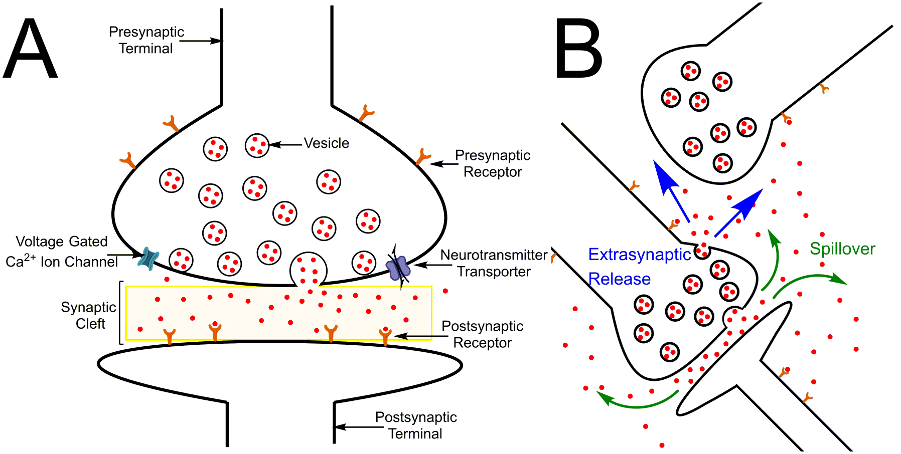
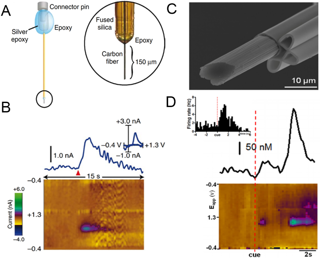
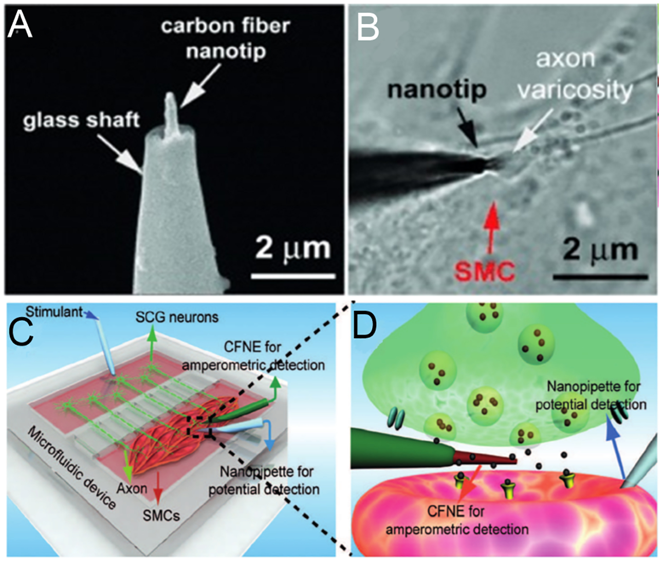
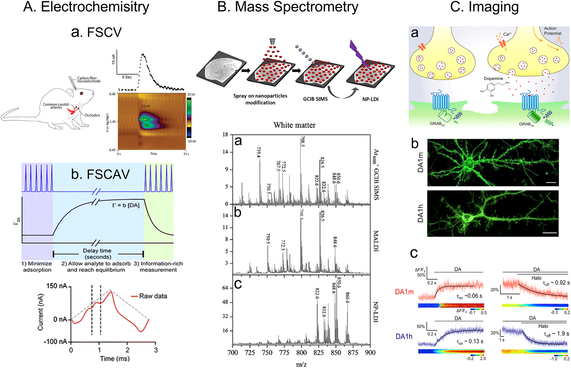
Electrochemical measurements of neurotransmitters provide insight into the dynamics of neurotransmission. In this review, we describe the development of electrochemical measurements of neurotransmitters and how they started with extrasynaptic measurements but now are pushing toward synaptic measurements. Traditionally, biosensors or fast-scan cyclic voltammetry have monitored extrasynaptic levels of neurotransmitters, such as dopamine, serotonin, adenosine, glutamate, and acetylcholine. Amperometry and electrochemical cytometry techniques have revealed mechanisms of exocytosis, suggesting partial release. Advances in nanoelectrodes now allow spatially resolved, electrochemical measurements in a synapse, which is only 20-100 nm wide. Synaptic measurements of dopamine and acetylcholine have been made. In this article, electrochemical measurements are also compared to optical imaging and mass spectrometry measurements, and while these other techniques provide enhanced spatial or chemical information, electrochemistry is best at monitoring real-time neurotransmission. Future challenges include combining electrochemistry with these other techniques in order to facilitate multisite and multianalyte monitoring.
Keywords: amperometry; dopamine; glutamate; microelectrode; nanoelectrodes; voltammetry.
Figures
Similar articles
-
Electrochemical Analysis of Neurotransmitters.Annu Rev Anal Chem (Palo Alto Calif). 2015;8:239-61. doi: 10.1146/annurev-anchem-071114-040426. Epub 2015 May 4. Annu Rev Anal Chem (Palo Alto Calif). 2015. PMID: 25939038 Free PMC article. Review.
-
Nanoelectrode for amperometric monitoring of individual vesicular exocytosis inside single synapses.Angew Chem Int Ed Engl. 2014 Nov 10;53(46):12456-60. doi: 10.1002/anie.201404744. Epub 2014 Jul 24. Angew Chem Int Ed Engl. 2014. PMID: 25060546
-
Wireless Instantaneous Neurotransmitter Concentration System-based amperometric detection of dopamine, adenosine, and glutamate for intraoperative neurochemical monitoring.J Neurosurg. 2009 Oct;111(4):701-11. doi: 10.3171/2009.3.JNS0990. J Neurosurg. 2009. PMID: 19425899 Free PMC article.
-
A test potential booster for fast-scan cyclic voltammetry with an electrophysiological amplifier.Anal Biochem. 2020 Dec 1;610:113934. doi: 10.1016/j.ab.2020.113934. Epub 2020 Sep 3. Anal Biochem. 2020. PMID: 32891595
-
Electrochemical techniques for subsecond neurotransmitter detection in live rodents.Comp Med. 2014 Aug;64(4):249-55. Comp Med. 2014. PMID: 25296011 Free PMC article. Review.
Cited by
-
Accelerating the development of implantable neurochemical biosensors by using existing clinically applied depth electrodes.Anal Bioanal Chem. 2023 Mar;415(6):1137-1147. doi: 10.1007/s00216-022-04445-1. Epub 2022 Dec 2. Anal Bioanal Chem. 2023. PMID: 36456747 Free PMC article.
-
Carbon nanospike coated nanoelectrodes for measurements of neurotransmitters.Faraday Discuss. 2022 Apr 5;233(0):303-314. doi: 10.1039/d1fd00053e. Faraday Discuss. 2022. PMID: 34889344 Free PMC article.
-
Parkinson's Disease: Cells Succumbing to Lifelong Dopamine-Related Oxidative Stress and Other Bioenergetic Challenges.Int J Mol Sci. 2024 Feb 7;25(4):2009. doi: 10.3390/ijms25042009. Int J Mol Sci. 2024. PMID: 38396687 Free PMC article. Review.
-
Expression of Genes Involved in Axon Guidance: How Much Have We Learned?Int J Mol Sci. 2020 May 18;21(10):3566. doi: 10.3390/ijms21103566. Int J Mol Sci. 2020. PMID: 32443632 Free PMC article. Review.
-
Electrochemical treatment in KOH renews and activates carbon fiber microelectrode surfaces.Anal Bioanal Chem. 2021 Nov;413(27):6737-6746. doi: 10.1007/s00216-021-03539-6. Epub 2021 Jul 23. Anal Bioanal Chem. 2021. PMID: 34302181 Free PMC article.
References
Publication types
MeSH terms
Substances
Grants and funding
LinkOut - more resources
Full Text Sources
