. 2019 Apr 1;316(4):H862-H872.
doi: 10.1152/ajpheart.00696.2018. Epub 2019 Feb 1.

Sympathoexcitation in response to cardiac and pulmonary afferent stimulation of TRPA1 channels is attenuated in rats with chronic heart failure

Affiliations

Sympathoexcitation in response to cardiac and pulmonary afferent stimulation of TRPA1 channels is attenuated in rats with chronic heart failure

Ryan J Adam et al. Am J Physiol Heart Circ Physiol. .

Abstract

Excessive sympathoexcitation characterizes the chronic heart failure (CHF) state. An exaggerated cardiac sympathetic afferent reflex (CSAR) contributes to this sympathoexcitation. Prior studies have demonstrated that the CSAR to capsaicin [transient receptor potential (TRP) vanilloid 1 agonist] is exaggerated in CHF animal models. We recently discovered that capsaicin application to the lung visceral pleura in anesthetized, vagotomized, open-chested rats increases mean arterial pressure (MAP), heart rate (HR), and renal sympathetic nerve activity (RSNA). We named this response the pulmonary spinal afferent reflex (PSAR). Due to the similarities between TRP vanilloid 1 and TRP ankyrin 1 (TRPA1) channels as well as the excessive sympathoexcitation of CHF, we hypothesized that stimulation of the CSAR and PSAR with a specific TRPA1 agonist would result in an augmented response in CHF rats (coronary ligation model) compared with sham control rats. In response to a TRPA1 agonist, both CSAR and PSAR in sham rats resulted in biphasic changes in MAP and increases in HR and RSNA 10-12 wk postmyocardial infarction (post-MI). These effects were blunted in CHF rats. Assessment of TRPA1 expression levels in cardiopulmonary spinal afferents by immunofluorescence, quantitative RT-PCR, and Western blot analysis 10-12 wk post-MI all indicates reduced expression in CHF rats but no reduction at earlier time points. TRPA1 protein was reduced in a dorsal root ganglia cell culture model of inflammation and simulated tissue ischemia, raising the possibility that the in vivo reduction of TRPA1 expression was, in part, caused by CHF-related tissue ischemia and inflammation. These data provide evidence that reflex responses to cardiopulmonary spinal afferent TRPA1 stimulation may be attenuated in CHF rather than enhanced. NEW & NOTEWORTHY Excessive sympathoexcitation characterizes chronic heart failure (CHF). The contribution of transient receptor potential ankyrin 1 (TRPA1) channel-mediated reflexes to this sympathoexcitation is unknown. We found that application of TRPA1 agonist to the heart and lung surface resulted in increased heart rate and sympathetic output and a biphasic change in mean arterial pressure in control rats. These effects were attenuated in CHF rats, decreasing the likelihood that TRPA1 channels contribute to cardiopulmonary afferent sensitization in CHF.

Keywords: autonomic dysfunction; cardiovascular reflexes; heart failure; sensory afferents; sympathoexcitation.


Conflict of interest statement

No conflicts of interest, financial or otherwise, are declared by the authors.

Figures
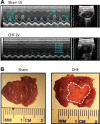
Fig. 1.
Fig. 1.
Echocardiography and necropsy evaluation. A: M-mode, long-axis (left), and two-dimensional (right) echocardiography images of the left ventricle (LV) from a sham rat and chronic heart failure (CHF) rat. B: photo of the dissected LV at necropsy from one sham rat and one CHF rat. The outline marks the scar area in CHF.
Fig. 2.
Fig. 2.
Epicardial application of ally isothiocyanate (AITC). A and B: representative tracings of a sham (A) and chronic heart failure (CHF; B) rat’s reflex to application of 30 mM AITC to the epicardium. C−E: corresponding changes in mean arterial pressure (ΔMAP; C), heart rate (ΔHR; D), and renal sympathetic nerve activity (ΔRSNA; E) for 50 and 300 mM doses. Data are means ± SE. Veh, vehicle; P, pressor response; DP, depressor response; BPM, beats/min; BL, baseline. Animals were 10–12 wk postmyocardial infarction (MI)/sham surgery. *P < 0.05 comparing sham and CHF groups by a Mann-Whitney rank-sum test; #P < 0.05 compared with BL by a Wilcoxon matched-pair signed-rank test.
Fig. 3.
Fig. 3.
Lung visceral pleura application of ally isothiocyanate (AITC). A and B: representative tracings of a sham (A) and chronic heart failure (CHF; B) rat’s reflex to application of 30 mM AITC to the lung visceral pleura. C−E: corresponding changes in mean arterial pressure (ΔMAP; C), heart rate (ΔHR; D), and renal sympathetic nerve activity (ΔRSNA; E) for 50 and 300 mM doses. Data are means ± SE. Veh, vehicle; P, pressor response; DP, depressor response; BPM, beats/min; BL, baseline. Animals were 10–12 wk postmyocardial infarction (MI)/sham surgery. *P < 0.05 comparing sham and CHF groups by a Mann-Whitney rank-sum test; #P < 0.05 compared with BL by a Wilcoxon matched-pair signed-rank test.
Fig. 4.
Fig. 4.
T1−T4 dorsal root ganglia (DRG) transient receptor potential ankyrin 1 (TRPA1) expression in sham and chronic heart failure (CHF) rats. A: immunofluorescence images of T1−T4 DRG in a sham and CHF rat 10 wk postmyocardial infarction (MI)/sham surgery. Arrows in the merged image mark colocalization of TRPA1 and IB4 in two fibers. Triangles mark colocalization of TRPA1 and NF200 in two fibers. B: sham-normalized Western blots for TRPA1 in T1−T4 DRG in sham and MI rats from 1, 3–4, 6–8, and 10–12 wk post-MI. MI size is listed below as a percentage of left ventricular surface ± SE. C: sham-normalized TRPA1/β-actin mRNA levels in T1−T4 DRG. *P < 0.05 by a Mann-Whitney rank-sum test.
Fig. 5.
Fig. 5.
Transient receptor potential ankyrin 1 (TRPA1) protein in dorsal root ganglia (DRG) cell culture models of inflammation. AD: Western blot analysis for TRPA1 from a cell culture of DRG cells (50B11 rat line) under normal conditions (control) and with exposure to the proinflammatory cytokines IL-6 (A), IL-1β (B), and TNF-α (C) or a cocktail of all three combined (D). *P > 0.05 between control and the respective cytokine group for all four cell culture experiments by a Mann-Whitney rank-sum test.
Fig. 6.
Fig. 6.
Transient receptor potential ankyrin 1 (TRPA1) protein in a dorsal root ganglia (DRG) cell culture model of tissue ischemia. A and B: hypoxia-inducible factor (HIF)-1α (A) and TRPA1 (B) protein expression in a DRG cell culture (50B11 DRG rat cell line) grown under normoxic and simulated tissue ischemia (hypoxic) conditions. *P < 0.05 by a Mann-Whitney rank-sum test.

References

    1. Bandell M, Story GM, Hwang SW, Viswanath V, Eid SR, Petrus MJ, Earley TJ, Patapoutian A. Noxious cold ion channel TRPA1 is activated by pungent compounds and bradykinin. Neuron 41: 849–857, 2004. doi:10.1016/S0896-6273(04)00150-3. - DOI - PubMed
    1. Barretto AC, Santos AC, Munhoz R, Rondon MU, Franco FG, Trombetta IC, Roveda F, de Matos LN, Braga AM, Middlekauff HR, Negrão CE. Increased muscle sympathetic nerve activity predicts mortality in heart failure patients. Int J Cardiol 135: 302–307, 2009. doi:10.1016/j.ijcard.2008.03.056. - DOI - PubMed
    1. Bautista DM, Jordt SE, Nikai T, Tsuruda PR, Read AJ, Poblete J, Yamoah EN, Basbaum AI, Julius D. TRPA1 mediates the inflammatory actions of environmental irritants and proalgesic agents. Cell 124: 1269–1282, 2006. doi:10.1016/j.cell.2006.02.023. - DOI - PubMed
    1. Chang RB, Strochlic DE, Williams EK, Umans BD, Liberles SD. Vagal sensory neuron subtypes that differentially control breathing. Cell 161: 622–633, 2015. doi:10.1016/j.cell.2015.03.022. - DOI - PMC - PubMed
    1. Chen W, Mi R, Haughey N, Oz M, Höke A. Immortalization and characterization of a nociceptive dorsal root ganglion sensory neuronal line. J Peripher Nerv Syst 12: 121–130, 2007. doi:10.1111/j.1529-8027.2007.00131.x. - DOI - PMC - PubMed

Publication types