. 2019 Feb 1;14(2):e0211739.
doi: 10.1371/journal.pone.0211739. eCollection 2019.

Identification of the X-linked germ cell specific miRNAs (XmiRs) and their functions

Affiliations

Identification of the X-linked germ cell specific miRNAs (XmiRs) and their functions

Hiromitsu Ota et al. PLoS One. .

Abstract

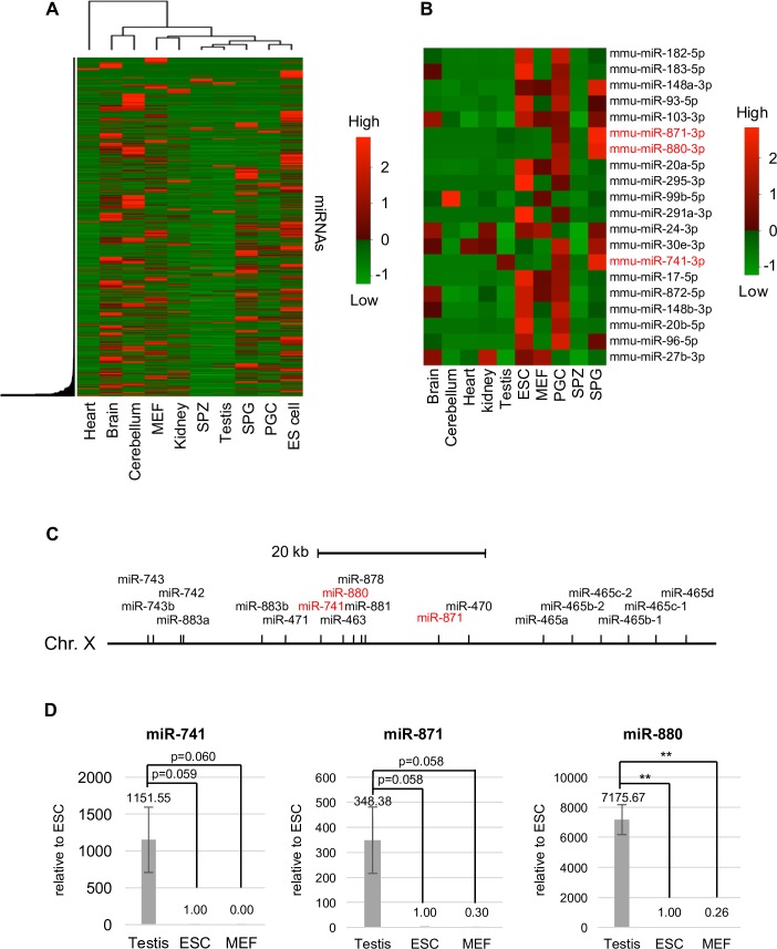
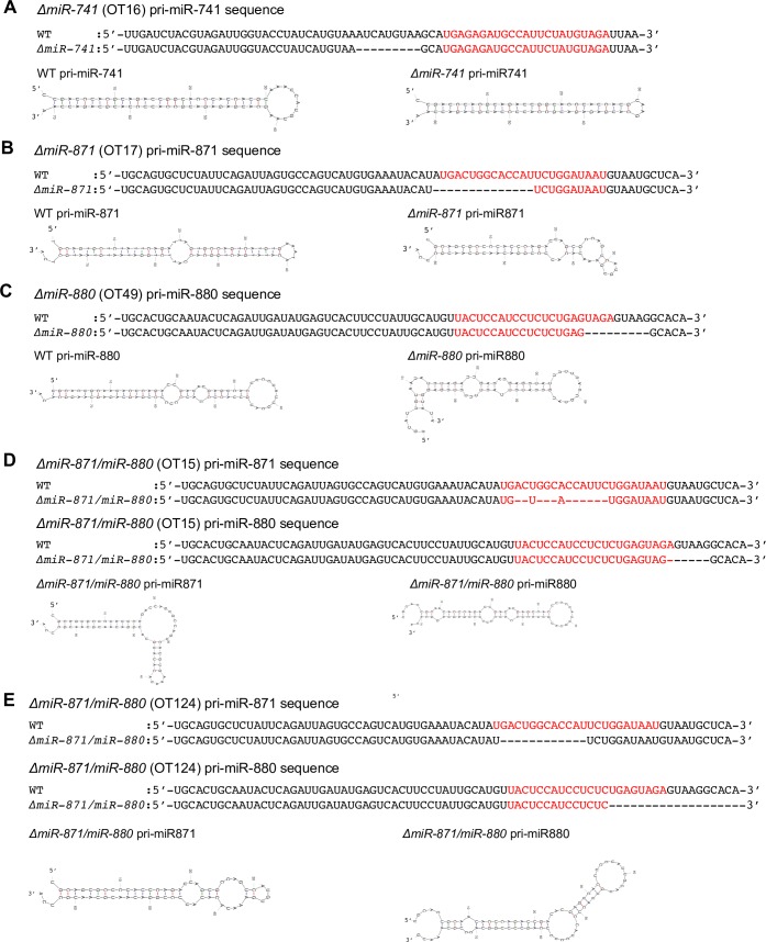
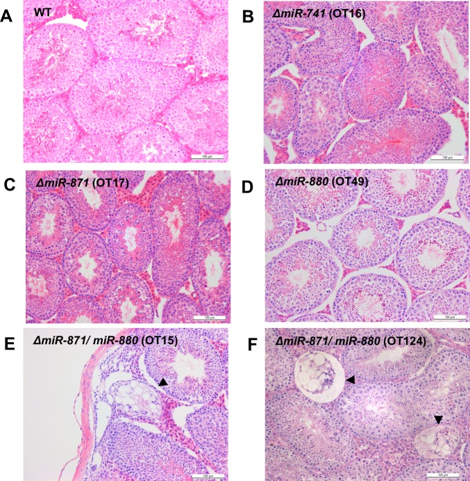
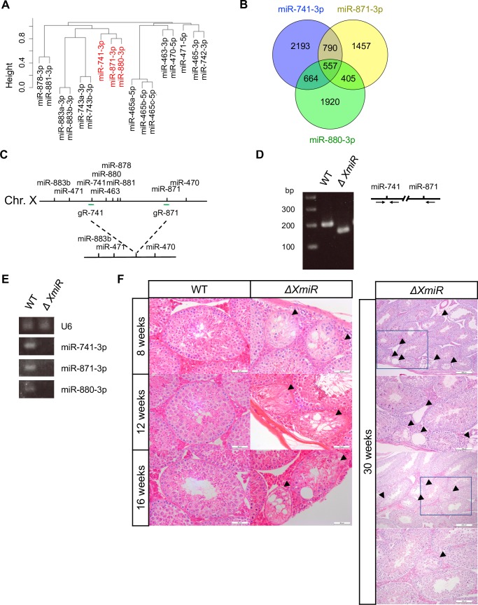
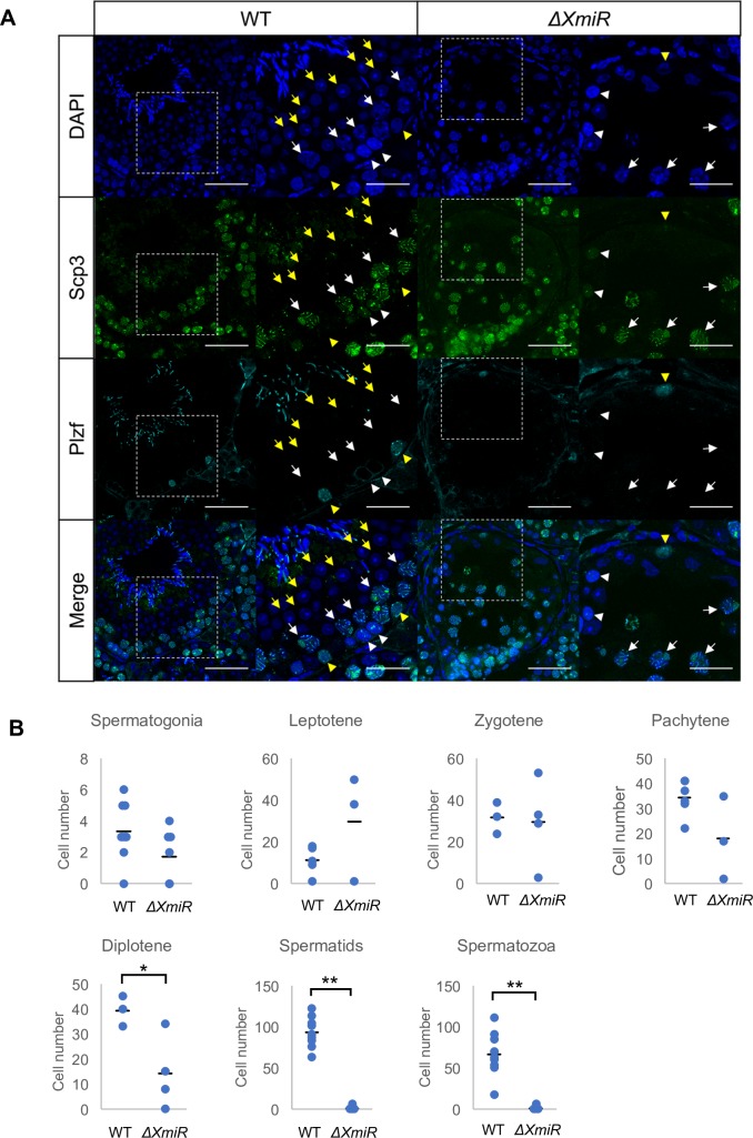
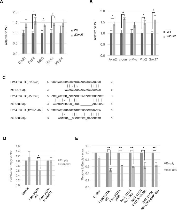
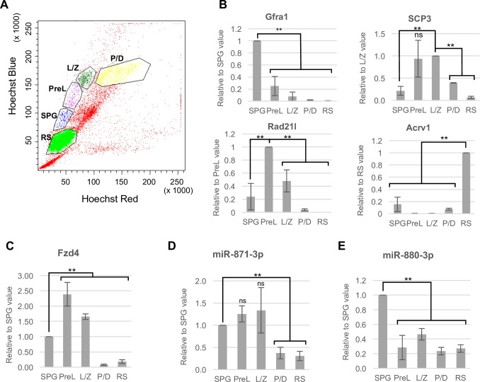
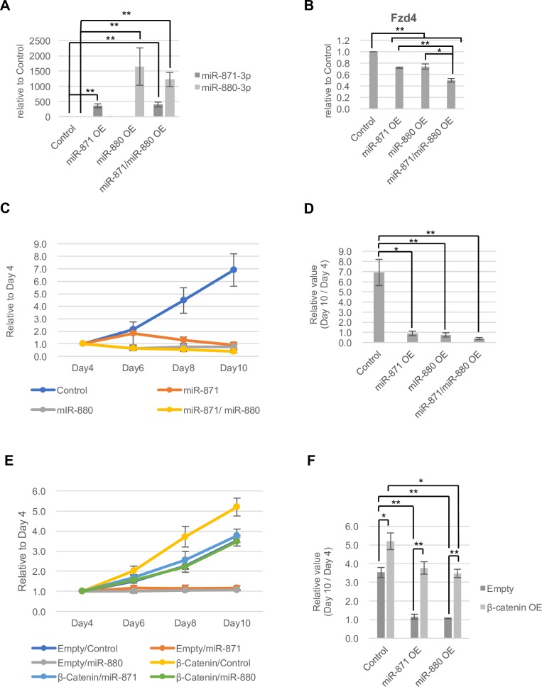
MicroRNAs (miRNAs) play a critical role in multiple aspects of biology. Dicer, an RNase III endonuclease, is essential for the biogenesis of miRNAs, and the germ cell-specific Dicer1 knockout mouse shows severe defects in gametogenesis. How miRNAs regulate germ cell development is still not fully understood. In this study, we identified germ cell-specific miRNAs (miR-741-3p, miR-871-3p, miR-880-3p) by analyzing published RNA-seq data of mouse. These miRNA genes are contiguously located on the X chromosome near other miRNA genes. We named them X chromosome-linked miRNAs (XmiRs). To elucidate the functions of XmiRs, we generated knockout mice of these miRNA genes using the CRISPR/Cas9-mediated genome editing system. Although no histological abnormalities were observed in testes of F0 mice in which each miRNA gene was disrupted, a deletion covering miR-871 and miR-880 or covering all XmiRs (ΔXmiRs) resulted in arrested spermatogenesis in meiosis in a few seminiferous tubules, indicating their redundant functions in spermatogenesis. Among candidate targets of XmiRs, we found increased expression of a gene encoding a WNT receptor, FZD4, in ΔXmiRs testis compared with that in wildtype testis. miR-871-3p and miR-880-3p repressed the expression of Fzd4 via the 3'-untranslated region of its mRNA. In addition, downstream genes of the WNT/β-catenin pathway were upregulated in ΔXmiRs testis. We also found that miR-871, miR-880, and Fzd4 were expressed in spermatogonia, spermatocytes and spermatids, and overexpression of miR-871 and miR-880 in germ stem cells in culture repressed their increase in number and Fzd4 expression. Previous studies indicated that the WNT/β-catenin pathway enhances and represses proliferation and differentiation of spermatogonia, respectively, and our results consistently showed that stable β-catenin enhanced GSC number. In addition, stable β-catenin partially rescued reduced GSC number by overexpression of miR-871 and miR-880. The results together suggest that miR-871 and miR-880 cooperatively regulate the WNT/β-catenin pathway during testicular germ cell development.


Conflict of interest statement

The authors have declared that no competing interests exist.

Figures

Fig 1
Fig 1. The expression profile of miRNAs in various tissues and cell lines.
(A) A heat map of hierarchical clustering of miRNAs detected in small RNA-seq data used in this study. (B) A heat map of 20 miRNAs highly expressed in PGCs. Relative miRNA expression is described according to the color scale. Red and green indicate high and low expression, respectively. Mouse embryonic fibroblasts (MEFs), embryonic stem (ES) cells, primordial germ cells (PGCs), spermatogonia (SPG), spermatozoa (SPZ). (C) The locus of XmiR genes on the X chromosome. (D) The expression of XmiRs in testes, ES cells, and MEFs determined by quantitative RT-PCR. Each expression level was normalized to the expression of U6 snRNA. The expression in ES cells was set as 1.0. Error bars show standard errors of three biological replicates. **P < 0.01.
Fig 2
Fig 2. Predicted altered secondary structures of XmiR precursors by genome editing.
Secondary structures of predicted miRNA precursors of (A) miR-741 WT (left) and ΔmiR-741 (OT16) (right), (B) miR-871 WT (left) and ΔmiR-871 (OT17) (right), (C) miR-880 WT (left) and ΔmiR-880 (OT49) (right), and (D, E) miR-871 (top) and miR-880 (bottom) in ΔmiR-871/ΔmiR-880 in two different mice, OT15 (D) and OT124 (E).
Fig 3
Fig 3. Spermatogenesis in mice with mutations in miR-741, miR-871, and miR-880.
Hematoxylin-eosin (HE)-stained sections of seminiferous tubules in the testes of (A) WT, (B) ΔmiR-741 (OT16), (C) ΔmiR-871 (OT17), (D) ΔmiR-880 (OT49), and ΔmiR-871/ΔmiR-880 (E) (OT15) mice at 8 weeks of age, and (F) ΔmiR-871/ΔmiR-880 (OT124) mouse at 24 weeks of age. The arrowheads show abnormal seminiferous tubules. Scale bars = 100 μm.
Fig 4
Fig 4. Relationship between target mRNAs of XmiRs and generation of ΔXmiRs mice.
(A) A dendrogram of hierarchical clustering analysis of target mRNAs of XmiRs and their neighboring miRNAs. (B) Venn diagram showing the relationship among putative target mRNAs of miR-741-3p, miR-871-3p, and miR-880-3p. Corresponding gene lists are shown in S3 Table. (C) A schematic presentation of the WT and ΔXmiRs locus. gR-741 and gR-871 represent positions of guide RNAs used for genome editing. (D) Representative PCR for genotyping of WT and ΔXmiRs (OT84) mice. Arrows in the right panel represent primers used for PCR. (E) Expression of XmiRs in WT and a ΔXmiRs testes (F2 of OT84) determined with semi-quantitative RT-PCR analysis. U6 snRNA was used as an internal control. (F) HE-stained sections of seminiferous tubules in WT (left) and ΔXmiR (F2 of OT100) (right) testes at 8, 12, 16, and 30 weeks of age. The second and fourth panels for 30 weeks show higher magnification views corresponding to the rectangular area in the first and third panels. Lower two panels show mildly affected seminiferous tubules. Arrowheads show abnormal seminiferous tubules. Scale bar = 50 μm (8, 12, 16 weeks), 200 μm (30 weeks, lower magnification), 100 μm (30 weeks, higher magnification).
Fig 5
Fig 5. Spermatogenesis was arrested at meiotic prophase in abnormal seminiferous tubules in ΔXmiR testes.
(A) Testis sections were co-stained with anti-SCP3 (green) and anti-PLZF (cyan) antibodies in WT and ΔXmiRs (F2 generation of the OT100 line) mice at 12 weeks of age. The second and fourth column show higher magnification views corresponding to the rectangular area in the pictures in the first and third columns. Mildly affected seminiferous tubules in ΔXmiR testis shown in Fig 4F are presented. Spermatogonia (yellow arrowheads), leptotene spermatocytes (white arrowheads), pachytene spermatocytes (white arrows), and round spermatids (yellow arrows) are indicated. Scale bars = 50 μm (the first and third columns) and 25 μm (the second, and fourth columns). (B) Number of cells at different spermatogenic stages determined by staining for Scp3 and Plzf in WT and mildly affected seminiferous tubules in ΔXmiR testis. Cells in two sections in a single mouse of WT and ΔXmiR testes were counted. Vertical lines in the graphs indicate means. *P < 0.05 and **P < 0.01.
Fig 6
Fig 6. Upregulation of WNT/β-catenin signaling genes in testes of ΔXmiRs mice, and repression of Fzd4 by XmiRs.
(A, B) Relative expression of the putative common target genes of miR-871-3p and miR-880-3p (A) and of the downstream genes of WNT/β-catenin signaling (B) in the testes of WT and ΔXmiRs (F2 of the OT84 line) mice at 12 weeks of age was determined by quantitative RT-PCR analysis. The expression in WT testis was set as 1. (C) Potential target sites of miR-871-3p and miR-880-3p in Fzd4-3′-UTR. (D, E) Relative luciferase activity from the reporter vectors with Fzd4-3′-UTR with or without expression vectors for miR-871 (D) or miR-880 (E). Fzd4-3′-UTR ΔmiR-871; Fzd4-3′-UTR with deleted miR-871-3p target site (D). Fzd4-3′-UTR 1–927 or 927–2263 with or without ΔmiR-880; 1–927 or 927–2263 bp fragment of Fzd4-3′-UTR with or without deleted miR-880-3p target sites (E). Luciferase activity was measured 48 h after transfection. Luciferase activity with an empty expression vector was set as 1. Error bars represent standard errors of three biological replicates. *P < 0.05 and **P < 0.01.
Fig 7
Fig 7. Expression of XmiRs and Fzd4 in spermatogenic cells.
(A) Scatter plot of FACS for testicular cells stained with Hoechst 33342. Spermatogonia (SPG), pre-leptotene (PreL), leptotene-zygotene (L/Z), pachytene-diplotene (P/D), round spermatids (RS). (B-E) Relative expression of stage-specific germ cell marker genes (B), Fzd4 (C), miR-871-3p (D), and miR-880-3p (E) in FACS-purified spermatogenic cells. Gfra1 for spermatogonia, Scp3 and Rad21l for spermatocytes, Acrv1 for spermatids. Gene expression was determined by RT-qPCR. Error bars represent standard errors of three biological replicates. **P < 0.01.
Fig 8
Fig 8. Roles of miR-871, miR-880 and stable β-catenin in GSCs.
(A, B) Relative expression of miR-871 and miR-880 (A) and Fzd4 (B) in GSCs overexpressing XmiRs at 8 days after infection with the lenti-virus vectors. Gene expression was determined with RT-qPCR. (C, D) The effect of XmiRs overexpression in GSCs in culture. The pLKO1 empty vector was used as the control. Cell number at day 4 to day 10 after infection with the lenti-virus vector (C), and ratios of GSC number at day10 compared with that at day4 (D) are shown. (E, F) The effect of co-overexpression of stable β-catenin and miR-871 or miR-880. The pLKO1 empty vector and CSII-EF-MCS vector were used as the control. Cell number at day4 to day10 after infection with the indicated lenti-virus vectors (E), and ratios of GSC number at day10 compared with that at day4 (F) are shown. Error bars represent standard errors of three biological replicates. *P < 0.05 and **P < 0.01.

References

    1. Ginsburg M, Snow MH, McLaren A. Primordial germ cells in the mouse embryo during gastrulation. Development. 1990;110(2):521–8. - PubMed
    1. Richardson BE, Lehmann R. Mechanisms guiding primordial germ cell migration: strategies from different organisms. Nat Rev Mol Cell Biol. 2010;11(1):37–49. 10.1038/nrm2815 - DOI - PMC - PubMed
    1. McLaren A. Meiosis and differentiation of mouse germ cells. Symp Soc Exp Biol. 1984;38:7–23. - PubMed
    1. Western PS, Miles DC, van den Bergen JA, Burton M, Sinclair AH. Dynamic regulation of mitotic arrest in fetal male germ cells. Stem Cells. 2008;26(2):339–47. 10.1634/stemcells.2007-0622 - DOI - PubMed
    1. de Rooij DG. Proliferation and differentiation of spermatogonial stem cells. Reproduction. 2001;121(3):347–54. - PubMed

Publication types